. 2024 Feb;13(4):e2302656.
doi: 10.1002/adhm.202302656. Epub 2023 Nov 27.

A pH-Responsive Virus-Like Particle as a Protein Cage for a Targeted Delivery

Affiliations

A pH-Responsive Virus-Like Particle as a Protein Cage for a Targeted Delivery

Kwan-Jip Kim et al. Adv Healthc Mater. 2024 Feb.

Abstract

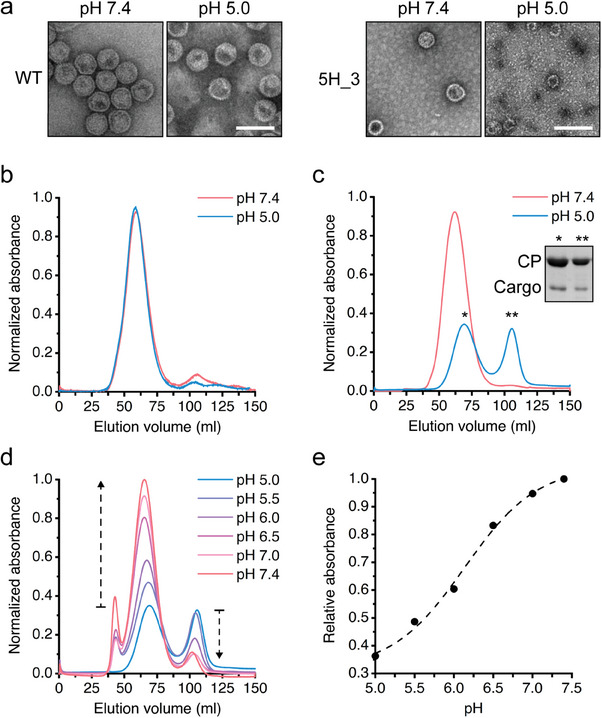
A stimuli-responsive protein self-assembly offers promising utility as a protein nanocage for biotechnological and medical applications. Herein, the development of a virus-like particle (VLP) that undergoes a transition between assembly and disassembly under a neutral and acidic pH, respectively, for a targeted delivery is reported. The structure of the bacteriophage P22 coat protein is used for the computational design of coat subunits that self-assemble into a pH-responsive VLP. Subunit designs are generated through iterative computational cycles of histidine substitutions and evaluation of the interaction energies among the subunits under an acidic and neutral pH. The top subunit designs are tested and one that is assembled into a VLP showing the highest pH-dependent structural transition is selected. The cryo-EM structure of the VLP is determined, and the structural basis of a pH-triggered disassembly is delineated. The utility of the designed VLP is exemplified through the targeted delivery of a cytotoxic protein cargo into tumor cells in a pH-dependent manner. These results provide strategies for the development of self-assembling protein architectures with new functionality for diverse applications.

Keywords: computational design; pH-responsive assembly; targeted delivery; virus-like particle.


Conflict of interest statement

The authors declare no conflict of interest.

Figures

Figure 1
Figure 1
Development of a pH‐responsive VLP through a computational approach. a) Schematic illustration of the pH‐dependent assembly and disassembly of a VLP. b) Cryo‐EM structure of bacteriophage P22 protein coat (PDB: 5UU5). Dashed line indicates an asymmetric unit (ASU) comprising seven identical subunits. c) Iterative computational cycles for the design of a subunit that assembles into a pH‐responsive VLP: Step 1) Calculate the interaction energies for the ASU at pH 7.4 and 5.0 (ΔG IE 7.4 and ΔG IE 5.0) as well as their differences (ΔΔ GIE 5.0–7.4) using FoldX as single histidine mutations are introduced. Predict the subunit sites that satisfy the criteria. Step 2) Introduce a single histidine mutation into the predicted sites and select the ASU models with higher differences in the interaction energies. Step 3) Repeat step 2 until the differences in the interaction energies no longer increase as single histidine mutations are introduced into the selected ASU models. Step 4) Identify key sites at a subunit for histidine substitutions, introduce multiple histidine mutations, and select the top‐10 subunit designs based on the interaction energies. The locations of the nine finally selected key sites are shown in the subunit in bottom‐right panel.
Figure 2
Figure 2
Assembly and disassembly of respective VLPs from ten subunit designs in response to pH changes. a) Conservation of key sites in the ten subunit designs. b) Schematic illustration of a VLP containing a cargo‐fused scaffolding protein. eGFP was used as a protein cargo and genetically fused to the N‐terminus of a truncated scaffolding protein. c) Each of the ten subunit designs and the wild‐type (48 kDa) were co‐expressed with an eGFP‐fused scaffolding protein (eGFP‐SP, 37 kDa) in E. coli, and the pellet was analyzed on SDS/PAGE after ultracentrifugation. Sol, supernatants after sonication; Sup, supernatants after ultracentrifugation; and Pel, pellet after ultracentrifugation. CP indicates a coat protein. d) Representative SDS/PAGE analyses of five pH‐responsive VLPs after ultracentrifugation. The wild‐type VLP was used as control. Bf, supernatants before ultracentrifugation; Af, supernatants after ultracentrifugation. e) The remaining amounts of coat proteins in the supernatants of VLPs at pH 7.4 and pH 5.0 after ultracentrifugation. The error bars show the standard deviations in triplicate experiments.
Figure 3
Figure 3
The pH‐dependent structural transition of 5H_3 VLP. a) TEM images of the wild‐type (left) and 5H_3 (right) VLPs encapsulating eGFP‐SP at pH 7.4 and 5.0. The scale bar represents 100 nm. b) Elution profiles of wild‐type VLP at pH 7.4 and 5.0. c) Elution profiles of the 5H_3 VLP at pH 7.4 and 5.0. d) Elution profiles of partially disassembled 5H_3 VLPs based on size exclusion chromatography with a gradual shift in pH from 5.0 to 7.4. The buffers used are as follows: 0.1 m citrate‐phosphate (pH 5.0), 0.1 m citrate (pH 5.5), 0.1 m phosphate (pH 6.0), 0.1 m phosphate (pH 6.5), 0.1 m phosphate (pH 7.0), and 0.1 m phosphate (pH 7.4). e) Relative portions of the reassembled 5H_3 VLP were estimated at each pH based on the elution profiles in (d) and plotted against the pH.
Figure 4
Figure 4
Cryo‐EM structure of 5H_3 VLP. a) A representative cryo‐EM micrograph of the 5H_3 VLP. b) Cryo‐EM map of the 5H_3 VLP at a resolution of 4.02 Å. c) An entire model of the 5H_3 VLP (PDB 8GN5). d) Location of substituted histidine residues at the subunits of the 5H_3 VLP (middle). Subunit interfaces near His43 (right) and His54, His153, and His287 (left) are illustrated, respectively. The interface around e) His43 is outlined in green, and the interfaces around f) His54, g) His153, and h) His287 are in pink. The histidine residues are colored in sky blue. The distances between the histidine and the amino acid residues that are presumably involved in a pH‐dependent disassembly of the 5H_3 are indicated by the dotted lines.
Figure 5
Figure 5
The pH‐dependent delivery of a protein cargo through receptor‐mediated endocytosis into tumor cell using the 5H_3 VLP. a) Schematic illustration of the 5H_3 VLP encapsulating a targeting moiety‐fused cargo. b) Schematics for the tumor microenvironment‐targeted delivery of a cargo through receptor‐mediated endocytosis using the 5H_3 VLP encapsulating a targeting moiety‐fused cargo. c) SDS/PAGE analyses of the wild‐type and 5H_3 VLPs encapsulating rEGFR‐eGFP‐SP. Here, rEGFR indicates an EGFR‐specific protein binder. CP indicates a coat protein. d) TEM images of the wild‐type and 5H_3 VLPs at pH 7.4 and 5.0. The scale bar represents 100 nm. Fluorescent microscopic images of e) high EGFR‐expressing MDA‐MB‐468 cells and f) low EGFR‐expressing MCF7 cells when treated with the wild‐type and 5H_3 VLPs encapsulating rEGFR‐eGFP‐SP at pH 7.4 and 6.5. Free rEGFR‐eGFP‐SP was used as a control. Equal concentration of cargos was treated. Blue, DAPI; green, rEGFR‐eGFP‐SP. The scale bar represents 20 nm.
Figure 6
Figure 6
Cell cytotoxicity of 5H_3 VLP encapsulating a cytotoxic cargo in a pH‐dependent manner. a) Schematics for the 5H_3 VLP encapsulating an exotoxin A from Pseudomonas aeruginosa (PE24). b) SDS/PAGE analyses of the wild‐type and 5H_3 VLPs encapsulating rEGFR‐PE24‐SP. CP indicates a coat protein. Dose‐dependent cytotoxic effects on c) high EGFR‐expressing MDA‐MB‐468 cells and d) low EGFR‐expressing MCF7 cells using the wild‐type and 5H_3 VLPs encapsulating rEGFR‐PE24‐SP at pH 7.4 and pH 6.5. Non‐targeted indicates the use of an off‐target protein binder (rOff) instead of rEGFR. Free rEGFR‐PE24‐SP was tested under the same conditions. The error bars represent standard deviations in triplicate experiments.

Similar articles

Cited by

References

    1. Lee E. J., Lee N. K., Kim I.‐S., Adv. Drug Delivery Rev. 2016, 106, 157. - PubMed
    1. Hu H., Steinmetz N. F., Biotechnol. J. 2020, 15, 2000077. - PMC - PubMed
    1. Estrada L. H., Chu S., Champion J. A., J. Pharm. Sci. 2014, 103, 1863. - PubMed
    1. Herrera Estrada L. P., Champion J. A., Biomater. Sci. 2015, 3, 787. - PubMed
    1. Edwardson T. G. W., Mori T., Hilvert D., J. Am. Chem. Soc. 2018, 140, 10439. - PubMed

Publication types

MeSH terms

Substances

LinkOut - more resources