. 2023 Nov 28;42(11):113449.
doi: 10.1016/j.celrep.2023.113449. Epub 2023 Nov 15.

Keratinocyte FABP5-VCP complex mediates recruitment of neutrophils in psoriasis

Affiliations

Keratinocyte FABP5-VCP complex mediates recruitment of neutrophils in psoriasis

Jiaqing Hao et al. Cell Rep. .

Abstract

One of the hallmarks of intractable psoriasis is neutrophil infiltration in skin lesions. However, detailed molecular mechanisms of neutrophil chemotaxis and activation remain unclear. Here, we demonstrate a significant upregulation of epidermal fatty acid binding protein (E-FABP, FABP5) in the skin of human psoriasis and psoriatic mouse models. Genetic deletion of FABP5 in mice by global knockout and keratinocyte conditional (Krt6a-Cre) knockout, but not myeloid cell conditional (LysM-Cre) knockout, attenuates psoriatic symptoms. Immunophenotypic analysis shows that FABP5 deficiency specifically reduces skin recruitment of Ly6G+ neutrophils. Mechanistically, activated keratinocytes produce chemokines and cytokines that trigger neutrophil chemotaxis and activation in an FABP5-dependent manner. Proteomic analysis further identifies that FABP5 interacts with valosin-containing protein (VCP), a key player in NF-κB signaling activation. Silencing of FABP5, VCP, or both inhibits NF-κB/neutrophil chemotaxis signaling. Collectively, these data demonstrate dysregulated FABP5 as a molecular mechanism promoting NF-κB signaling and neutrophil infiltration in psoriasis pathogenesis.

Keywords: CP: Immunology; FABP5; NF-κB signaling; chemotaxis; chronic inflammation; epidermal fatty acid binding protein; keratinocytes; macrophages; neutrophil; psoriasis; valosin-containing protein.


Conflict of interest statement

Declaration of interests The authors declare no competing interests.

Figures

Figure 1.
Figure 1.. The increasing levels of FABP5 are associated with psoriasis.
(A) FABP5 levels in skin samples from healthy individuals and patients with psoriasis. Data were analyzed by publicly accessing database GSE14905 from the Gene Expression Omnibus (GEO). The database of GSE14905 contained 21 healthy individuals and 33 psoriasis patients. (B) FABP5 levels in skin samples from healthy individuals and patients with psoriasis. Data were analyzed by publicly accessing GEO database GSE13355, which contained 64 healthy individuals and 58 psoriasis patients. (C) Alexa Fluor 488-labeled FABP5 expression profile in human skin tissue collected from psoriasis patients (lower panels) and healthy individuals (upper panels). H&E staining (left panels) showed the structure of skin tissue from psoriasis patients and healthy individuals. (D) Fabp5 mRNA levels in skin tissues from imiquimod (IMQ)-induced psoriasis mice and normal mice (n = 8/group). (E) H&E staining (left panels) and immunohistochemistry staining (right panels) showed the structure of skin tissue and Fabp5 levels from IMQ-induced psoriasis mice and control mice, respectively. Data are shown as mean ± SD in (A, B, and D) (**p ≤ 0.01, ****p ≤ 0.0001 as compared with healthy or control groups, unpaired Student’s t test). See also Figure S1.
Figure 2.
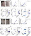
Figure 2.. Fabp5 deficiency attenuated IMQ-induced psoriasis progression with less neutrophil infiltration.
(A) Representative images of skin from Fabp5 global knockout and WT mice after treatment with IMQ for 7 days. On day 7 and WT controls. (B) The score of erythema, induration, and desquamation on skin were evaluated from the first day on both Fabp5 global knockout mice and WT controls. The cumulative score was calculated in a sum of the score of erythema, induration, and desquamation at the indicated time point (n = 5/group). (C) Ear thickness was measured daily for both strains, and its change was calculated by subtracting the first day value (n = 5/group). (D and E) H&E staining for ear (D) and skin (E) from both Fabp5 global knockout mice and WT controls. (F–H) Neutrophil infiltration and ratio were analyzed from ear (F), dermis (G), and PBMC (H) using flow cytometry at the endpoint of treatment for both Fabp5 global knockout mice and WT controls. In each figure, left panel: representative flow plot with gate on neutrophils (Ly6G+) from Fabp5 global knockout mice and WT controls. The parent population is CD45+Zombie; right panel: histogram represents neutrophil ratio in different skin tissues (n = 5/group). Data are shown as mean ± SD in (B, C, and F–H) (*p ≤ 0.05, **p ≤ 0.01, ***p ≤ 0.001, ****p ≤ 0.0001 as compared with healthy or control groups, unpaired Student’s t test). See also Figure S2.
Figure 3.
Figure 3.. Fabp5 deficiency in keratinocyte but not in myeloid cells attenuated neutrophil infiltration.
(A) Representative images of the skin from Fabp5f/f Krt6A-Cre+ and Fabp5f/f Krt6A-Cre mice after treatment with IMQ for 7 days. (B) The cumulative score (a sum score of erythema, induration, and desquamation on skin) was calculated from the first day on both Fabp5f/f Krt6A-Cre+ and Fabp5f/f Krt6A-Cre mice (n = 10/group). (C) Ear thickness was measured daily from Fabp5f/f Krt6A-Cre+ and Fabp5f/f Krt6A-Cre, and its change was calculated by subtracting the first day value (n = 10/group). (D) Neutrophil infiltration and its ratio were analyzed from the ear using flow cytometry at the endpoint of treatment for both Fabp5f/f Krt6A-Cre+ and Fabp5f/f Krt6A-Cre mice. In each figure, left panel: representative flow plot with gate on neutrophils (Ly6G+) from Fabp5 global knockout mice and WT controls. The parent population is CD45+Zombie; right panel: histogram represents neutrophil ratio in different tissues (n = 6/group). (E and F) Neutrophils infiltration and ratio were analyzed from the ear (D), dermis (E), and PBMC (F) using flow cytometry at the endpoint of treatment for both Fabp5f/f Krt6A-Cre+ and Fabp5f/f Krt6A-Cre mice. In each figure, left is the flow plot with gate on neutrophils (Ly6G+) in Fabp5 global knockout and WT mice. The parent population is CD45+Zombie; right is the histogram showing neutrophil ratio in individual tissues (n = 8/group). (G) Images were taken from skin with IMQ-induced psoriasis on day 7 from Fabp5f/f LysM-Cre+ and Fabp5f/f LysM-Cre. (H) Ear thickness was measured daily from Fabp5f/f LysM-Cre+ and Fabp5f/f LysM-Cre, and its change was calculated by subtracting the first day value (n = 6/group). (I) The cumulative score (a sum score of erythema, induration, and desquamation on skin) was calculated from the first day on both Fabp5f/f LysM-Cre+ and Fabp5f/f LysM-Cre mice (n = 6/group). (J–L) Neutrophils infiltration and its ratio was analyzed from the ear (J), dermis (K), and PBMC (L) using flow cytometry at the endpoint of treatment for both Fabp5f/f LysM-Cre+ and Fabp5f/f LysM-Cre mice. In each panel, flow cytometric plot (left) showed neutrophils (Ly6G+) population in Fabp5 global knockout and WT mice; histogram (right) represented neutrophil ratio in CD45+ immune cells in individual tissues (n = 7/group). Data are shown as mean ± SD in (D–F, K, and L) (*p ≤ 0.05, **p ≤ 0.01, ***p ≤ 0.001; ns, not significant compared with control groups, unpaired Student’s t test). See also Figures S3 and S4.
Figure 4.
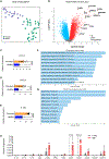
Figure 4.. Human psoriasis is associated with NFκB-mediated chemokine signaling.
(A) The Uniform Manifold Approximation and Projection showed two different clusters, which are healthy samples (green, n = 21) and psoriasis samples (blue, n = 33). (B) The differentially expressed genes (DEGs) from volcano plot with an adjusted p value cutoff of 0.05 (red dots are upregulated genes, blue dots are downregulated genes, gray dots are genes with no significance). (C–E) Relative expression of CXCL1 (C), CXCL8 (D), and IL-36 γ (E) in skin samples from healthy individuals and lesional skin samples from patients with psoriasis. Data were analyzed by publicly accessing the GSE14905 database. This database contains 21 healthy individuals and 33 psoriasis patients. (F) The analysis of gene ontology biological process (version 2021) from top hits cytokines indicated the most important signaling pathways. (G) The most relevant transcription factors were predicted by the analysis of transcriptional regulatory relationships unraveled by sentence-based text mining (TRRUST). (H) Real-time PCR for cytokine expression in skin tissue on both IMQ-treated mice and control mice. Data were analyzed by two-way ANOVA followed by Bonferroni’s multiple comparison test (n = 3/group). Data are shown as mean ± SD in (C–E and H) (****p < 0.0001 compared with control groups, unpaired Student’s t test). See also Figure S5.
Figure 5.
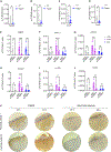
Figure 5.. Silencing FABP5 in HaCaT cells inhibits TNF-α-induced neutrophil chemotaxis.
(A–D) Real-time PCR showed downregulation of Fabp5 (A), IL-36γ (B), Cxcl1 (C), and Cxcl2 (D) with IMQ-treated skin tissue for 24 h in Fabp5 global knockout mice compared with WT controls (n = 3/group). (E–J) Real-time PCR showed the levels of FABP5 (E), CXCL1 (F), CXCL2 (G), CXCL8 (H), IL-36γ (I), and KLK6 (J) were downregulated in FABP5-deficient HaCaT cells transfected with 40 nM siRNA compared with siNC controls with 10 ng/mL TNF-α, 50 μg/mL IMQ, and DMSO as control treatment for 6 h (n = 5/group). (K) Immunohistochemistry staining on skin tissues from two representative psoriasis patients showed FABP5 expression (left) and neutrophil elastase expression (right). Data are shown as mean ± SD in (A–J) (*p ≤ 0.05, **p ≤ 0.01, ***p ≤ 0.001; ns, not significant compared with control groups, unpaired Student’s t test). See also Figure S6.
Figure 6.
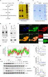
Figure 6.. FABP5-VCP interaction promotes the TNF-α-induced NF-κB signaling pathway.
(A) The workflow on immunoprecipitation with anti-FABP5 antibody in HaCaT cells. (B and C) Silver staining (B) and Coomassie blue staining (C) on SDS-PAGE gel for immunoprecipitated complexes. Blue arrow shows the band for FABP5, and orange arrow shows the band for VCP. (D and E) Validation of FABP5-VCP interaction using immunoprecipitation and western blot. HaCaT lysates were immunoprecipitated (IP) with anti-FABP5 antibody (D) and anti-VCP antibody (E), separately. Immunoblots were performed with anti-VCP antibody and anti-FABP5 antibody as indicated in the figure. (F and G) Images of HaCaT cells show co-localization of FABP5 and VCP using confocal analysis (F). The straight line indicates the region of interest (ROI) utilized to measure the fluorescence intensity in both VCP and FABP5 (G). FABP5 (green), VCP (red), Hochest33342 (blue). Scale bars, 50 μm. (H) Detection of NF-κB signaling activation using western blot. Transient silencing FABP5 with RNAi for 24 h in HaCaT cells and controls were treated with 10 ng/mL TNF-α for the indicated time points. FABP5, VCP, p-IκBα(S32), and p-NFκB(S536) were detected. β-Actin was used as internal control. (I–K) The levels of p-IκBα(S32), p-NFκB p65 (S536), and FABP5 were quantified using ImageJ from three independent western blots experiments (*p < 0.05). Data are shown as mean ± SD in (I–K) (*p ≤ 0.05 compared with the siNC control group, unpaired Student’s t test). See also Figure S7.

Similar articles

Cited by

References

    1. Griffiths CE, and Barker JN (2007). Pathogenesis and clinical features of psoriasis. Lancet 370, 263–271. - PubMed
    1. Gangwar RS, Gudjonsson JE, and Ward NL (2022). Mouse Models of Psoriasis: A Comprehensive Review. J. Invest. Dermatol. 142, 884–897. - PMC - PubMed
    1. van der Fits L, Mourits S, Voerman JSA, Kant M, Boon L, Laman JD, Cornelissen F, Mus AM, Florencia E, Prens EP, and Lubberts E (2009). Imiquimod-induced psoriasis-like skin inflammation in mice is mediated via the IL-23/IL-17 axis. J. Immunol. 182, 5836–5845. - PubMed
    1. Machado Á, and Torres T (2018). Guselkumab for the Treatment of Psoriasis. BioDrugs 32, 119–128. - PubMed
    1. Singh S, Kroe-Barrett RR, Canada KA, Zhu X, Sepulveda E, Wu H, He Y, Raymond EL, Ahlberg J, Frego LE, et al. (2015). Selective targeting of the IL23 pathway: Generation and characterization of a novel high-affinity humanized anti-IL23A antibody. mAbs 7, 778–791. - PMC - PubMed

Publication types