. 2023 Nov 17;9(46):eadi5764.
doi: 10.1126/sciadv.adi5764. Epub 2023 Nov 15.

Centromere innovations within a mouse species

Affiliations

Centromere innovations within a mouse species

Craig W Gambogi et al. Sci Adv. .

Abstract

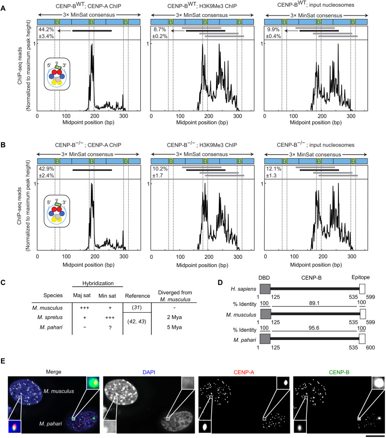
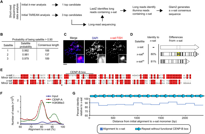
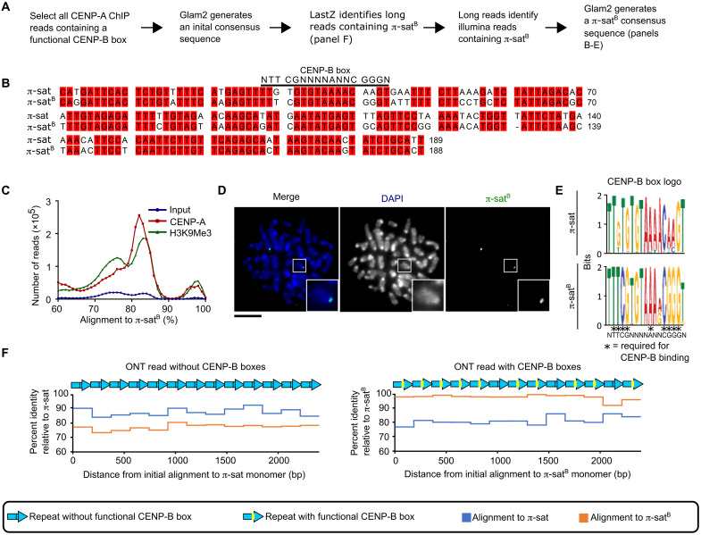
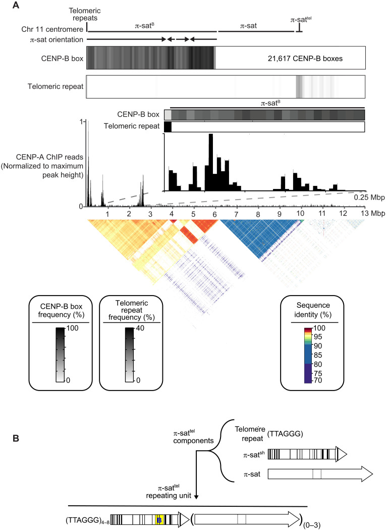
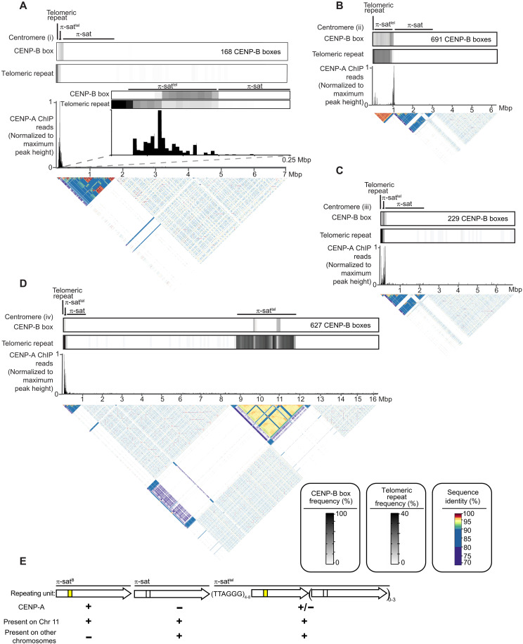
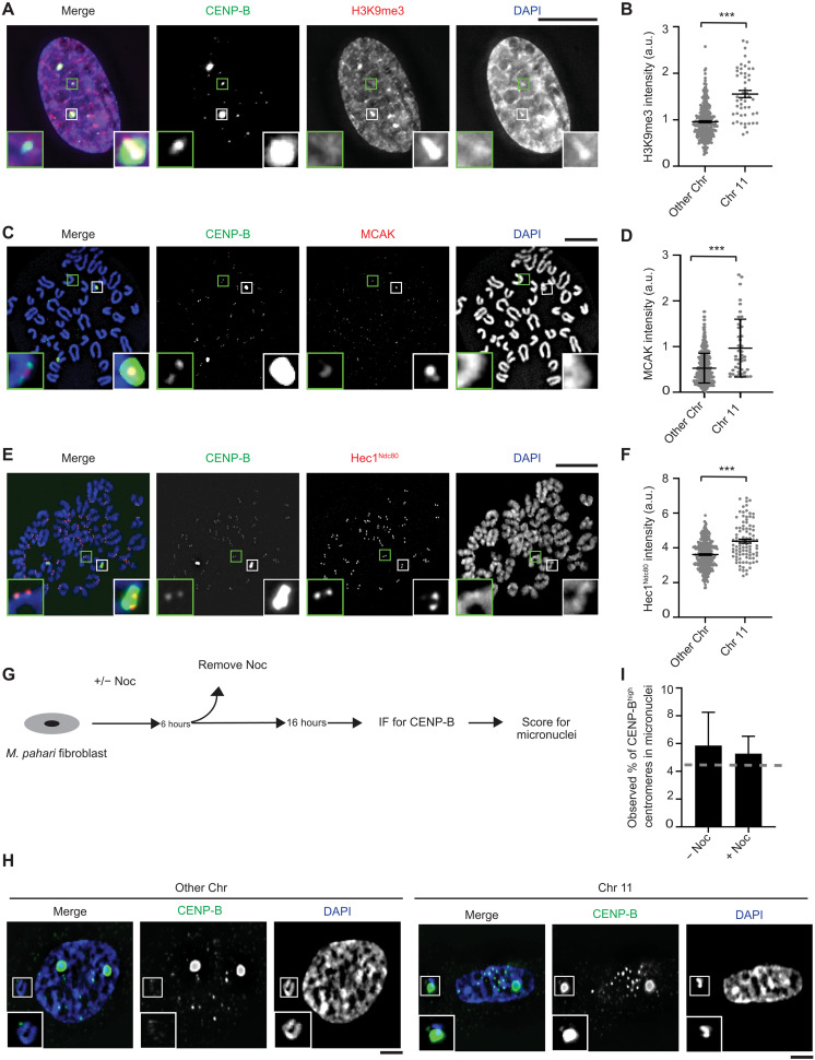
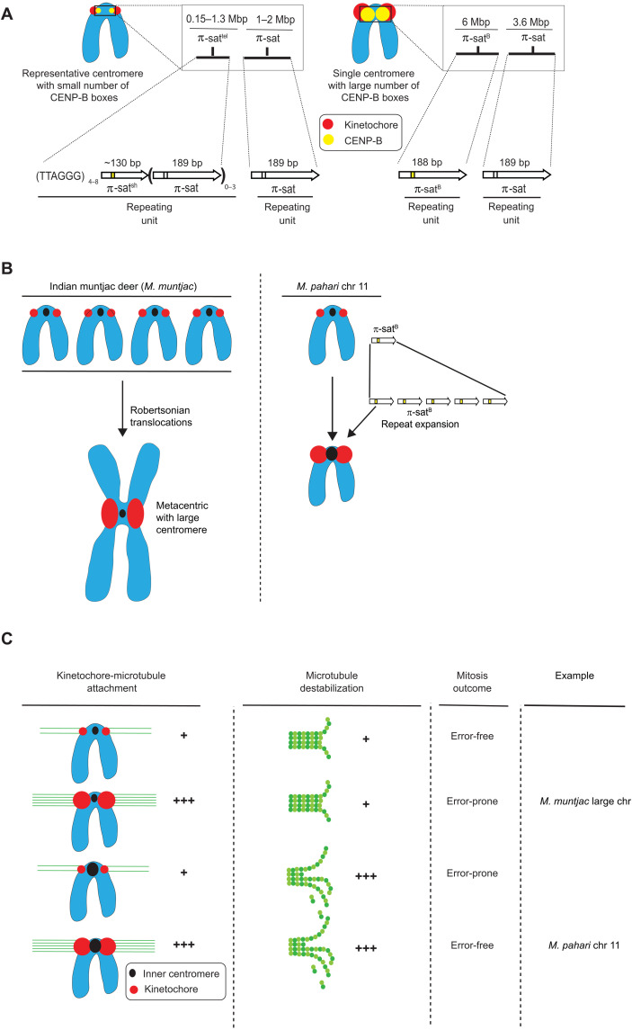
Mammalian centromeres direct faithful genetic inheritance and are typically characterized by regions of highly repetitive and rapidly evolving DNA. We focused on a mouse species, Mus pahari, that we found has evolved to house centromere-specifying centromere protein-A (CENP-A) nucleosomes at the nexus of a satellite repeat that we identified and termed π-satellite (π-sat), a small number of recruitment sites for CENP-B, and short stretches of perfect telomere repeats. One M. pahari chromosome, however, houses a radically divergent centromere harboring ~6 mega-base pairs of a homogenized π-sat-related repeat, π-satB, that contains >20,000 functional CENP-B boxes. There, CENP-B abundance promotes accumulation of microtubule-binding components of the kinetochore and a microtubule-destabilizing kinesin of the inner centromere. We propose that the balance of pro- and anti-microtubule binding by the new centromere is what permits it to segregate during cell division with high fidelity alongside the older ones whose sequence creates a markedly different molecular composition.


Figures

Fig. 1.
Fig. 1.. CENP-B occupancy on centromere DNA does not affect CENP-A nucleosome phasing but does vary widely between and within mouse species.
(A) Midpoint position of CENP-A ChIP H3K9Me3 or input reads (size 100 to 160 bp) from WT M. musculus along the trimer minor satellite consensus sequence. Vertical lines indicate the 17-bp CENP-B box. The major CENP-A nucleosome position (identified in the CENP-A ChIP samples) is indicated by a horizontal black line above the respective midpoint values and schematized (inset) for CENP-A ChIP with a triangle representing the dyad position. The same nucleosome position is indicated in the H3K9Me3 and input samples. Numbers to the left of the positions indicate the percentage of reads (means ± SEM; n = 3 independent experiments) where the midpoint spans the 10 bp at the 3′ end of the CENP-B box (yellow, labeled B). Horizontal gray lines indicate other major nucleosome positions in the H3K9Me3 and input samples. (B) Midpoint position of CENP-A ChIP H3K9Me3 or input reads (size 100 to 160 bp) from CENP-B KO M. musculus along the trimer minor satellite consensus sequence. (C) Centromere satellites from M. musculus, M. spretus, and M. pahari. (D) CENP-B is highly conserved in mouse species, with 100% identical sequences in both the DNA binding domain and the epitope targeted by the CENP-B antibody used in our study. (E) Immunofluorescence of CENP-A and CENP-B from lung fibroblast cells derived from M. musculus [with their nuclei identified by strong 4′,6-diamidino-2-phenylindole (DAPI)–staining pericentromeres] or M. pahari. Scale bar, 10 μm.
Fig. 2.
Fig. 2.. Identification of the most abundant form of centromere repeats in M. pahari: π-sat.
(A) Two approaches to identify M. pahari centromeric repeats. (B) Satellite sequences derived from TAREAN analysis on input sequencing data. Satellite probability was calculated as described in Materials and Methods. (C) Representative image of M. pahari centromeric DNA labeled with FISH probe using consensus sequence derived from k-mer approach. Insets: 7.9× magnification. Scale bar, 10 μm. (D) Schematized representation of the three satellites identified by TAREAN analysis. Differences between each satellite compared to π-sat are marked with black lines. The location corresponding to the CENP-B box position is indicated with dotted lines, and the functional CENP-B box from π-satB is indicated with yellow background and a blue “B.” (E) Alignment of the π-sat consensus sequence to minor sat consensus sequence. A dimer of π-sat was aligned to a trimer of the minor satellite, and the first monomer of π-sat is shown. The end of the first monomer of the minor satellite is marked with an asterisk. (F) Histograms show distribution of reads from input, CENP-A ChIP, or H3K9Me3 ChIP aligning to π-sat. (G) Representative example of a π-sat containing ONT long read that was divided into monomers. The percent identity of each monomer to π-sat is plotted.
Fig. 3.
Fig. 3.. π-satB is highly homogeneous, restricted to a single pair of chromosomes, and present in long, contiguous blocks that lack generic π-sat.
(A) Approach to identify CENP-B box containing satellite. (B) Alignment of π-sat and π-satB. (C) Histograms show distribution of reads from input, CENP-A ChIP, or H3K9Me3 ChIP aligning to π-satB. (D) Representative image of M. pahari centromeric DNA labeled with FISH probe using π-satB consensus sequence. Insets: 2.5× magnification. Scale bar, 10 μm. (E) Logo representation of the CENP-B box consensus of π-sat and π-satB. (F) Plots of the percent identity of satellites along a portion of representative ONT reads with (right) and without (left) CENP-B boxes to the π-sat and π-satB consensus sequences.
Fig. 4.
Fig. 4.. Genomic assembly reveals the identity and nature of the centromere harboring π-satB.
(A) The composition of the centromere of chromosome 11. The assembly consists of, in order, 8 kb of telomeric repeats, 6 Mbp of π-satB, 3.6 Mb of π-sat, and 400 kb of π-sattel, followed by other repetitive elements. The total number of CENP-B boxes (21,617) on this centromere is denoted. The fraction of π-sat repeats containing a functional CENP-B box (NTTCGNNNNANNCGGGN) and the frequency of telomeric repeats (TTAGGG) are shown. CENP-A ChIP-seq reads were aligned to the chromosome 11 centromere assembly. A pairwise sequence identity heatmap indicates that the centromere consists of 6 Mbp of highly homogeneous π-satB. (B) Schematized representation of the three types of repeats that make up repeating units of π-sattel. The repeating unit of π-sattel consists of a variable number of telomere repeats, a single unit of π-satsh, and from zero to three repeats of π-sat. Functional CENP-B boxes are typically found on π-satsh. An example of a single unit of π-sattel is shown. While the overall makeup of π-sattel contains the listed components, the number of telomere repeats and the units of π-sat can vary in different centromeres and even within a single centromere.
Fig. 5.
Fig. 5.. Restriction digest analysis confirms chromosome 11 assembly.
Schematic of predicted restriction digest sites of chromosome 11 with Bst XI and Hpa I. Pulsed-field gel Southern blot of Mus pahari DNA confirms the structure and organization of the chromosome 11 centromeric higher-order repeat (HOR) array. For each gel, left corresponds to ethidium bromide (EtBr) staining and right corresponds to 32P-labeled chromosome 11 π-satB–specific probe. The left gel was run at conditions to separate DNA from 0.6 to 5 Mb, and the right gel was run at conditions to separate DNA from 5 to 1000 kb.
Fig. 6.
Fig. 6.. Evolutionarily older Mus pahari centromeres harbor CENP-A nucleosomes near CENP-B boxes and π-sattel.
(A to D) The composition of a representative M. pahari centromere. Each of the assembly consists of, in order, an array of telomeric repeats, an array of π-sattel, and an array of π-sat followed by various repetitive elements. The fraction of π-sat repeats containing a functional CENP-B box (NTTCGNNNNANNCGGGN) and the frequency of telomeric repeats (TTAGGG) are shown. CENP-A ChIP-seq reads were aligned to the assembly revealing that CENP-A is primarily present on π-sattel. A pairwise sequence identity heatmap indicates the degree of homogeneity in centromeric DNA. (E) The types of repeating units found at M. pahari centromeres.
Fig. 7.
Fig. 7.. Chromosome 11 harbors levels of both pro- and anti-microtubule–binding proteins that are higher than the other Mus pahari centromeres.
(A) Immunofluorescence of H3K9Me3 from lung fibroblast cells derived from M. pahari. Insets: 4.0× magnification. Scale bar, 10 μm. (B) Quantification corresponding to (A). The mean ratio (± SEM) is shown. n = 314 for the centromeres with low abundance of CENP-B and n = 50 for the centromeres with high abundance of CENP-B, pooled from two independent experiments (***P < 0.0001). a.u., arbitrary units. (C) Immunofluorescence of MCAK from lung fibroblast cells derived from M. pahari. Insets: 6.5× magnification. Scale bar, 10 μm. (D) Quantification corresponding to (C). The mean ratio (± SEM) is shown. n = 389 for the centromeres with low abundance of CENP-B and n = 45 for the centromeres with high abundance of CENP-B, pooled from two independent experiments (***P < 0.0001). (E) Immunofluorescence of Hec1Ndc80 from lung fibroblast cells derived from M. pahari. Insets: 5.1× magnification. Scale bar, 10 μm. (F) Quantification corresponding to (E). The mean ratio (± SEM) is shown. n = 324 for the centromeres with low abundance of CENP-B and n = 94 for the centromeres with high abundance of CENP-B, pooled from three independent experiments (***P < 0.0001). (G) Schematic for measuring micronuclei containing chromosome 11 or other chromosomes. (H) Immunofluorescence of micronuclei with low and high abundance of CENP-B centromeres from lung fibroblast cells derived from M. pahari. Insets: 1.8× magnification. Scale bars, 10 μm. (I) Quantification corresponding to (H). Welch’s t test showed no significant difference between the actual micronuclei frequency and the expected frequency if there is no bias. A gray line represents the expected frequency given no bias, n = 133 (−Noc) and n = 419 (+Noc), pooled from four independent experiments, (P = 0.8374).
Fig. 8.
Fig. 8.. Divergent centromere DNA, molecular composition, and implications for mitotic chromosome segregation in M. pahari.
(A) Cartoon drawing summarizing the different types of M. pahari centromeres. Most M. pahari centromeres contain a low density of functional CENP-B boxes. Furthermore, these centromeres have two kinds of π-sat. First, the CENP-A–containing region is a stretch of repeating units of π-sat that is short (~130 bp) or long (189 bp) and interspersed with telomeric repeats. This is adjacent to a longer stretch of repeating units of 189-bp π-sat. The second type of M. pahari centromere has a high density of CENP-B boxes and is only found on chromosome 11. This centromere consists of 6 Mbp of homogeneous π-satB. The higher homogeneity of this centromeric DNA suggests that it is evolutionarily more recent relative to the other M. pahari centromeres. (B) A summary of the distinct mechanisms by which Indian munjtac and chromosome 11 from M. pahari centromeres likely became large repetitive arrays observed in the present day. (C) Model to understand different possible outcomes of centromere innovations during mitosis. The typical centromere has relatively low numbers of kinetochore attachments and relatively low amounts of microtubule destabilizer. These two factors balance each other, allowing normal segregation during mitosis. If either pro- or anti-microtubule binding factors are increased in the absence of the other, there will be an imbalance resulting in incorrect segregation during mitosis. Chromosome 11 has higher levels of microtubule destabilizer and more microtubule attachments, but because both factors are increased together, the chromosomes can still undergo error-free mitosis. Large Indian muntjac centromeres, on the other hand, have too strong pro-microtubule binding forces, compromising chromosome segregation (50).

Update of

References

    1. K. Kixmoeller, P. K. Allu, B. E. Black, The centromere comes into focus: From CENP-A nucleosomes to kinetochore connections with the spindle. Open Biol. 10, 200051 (2020). - PMC - PubMed
    1. N. Altemose, G. A. Logsdon, A. V. Bzikadze, P. Sidhwani, S. A. Langley, G. V. Caldas, S. J. Hoyt, L. Uralsky, F. D. Ryabov, C. J. Shew, M. E. G. Sauria, M. Borchers, A. Gershman, A. Mikheenko, V. A. Shepelev, T. Dvorkina, O. Kunyavskaya, M. R. Vollger, A. Rhie, A. M. McCartney, M. Asri, R. Lorig-Roach, K. Shafin, J. K. Lucas, S. Aganezov, D. Olson, L. G. de Lima, T. Potapova, G. A. Hartley, M. Haukness, P. Kerpedjiev, F. Gusev, K. Tigyi, S. Brooks, A. Young, S. Nurk, S. Koren, S. R. Salama, B. Paten, E. I. Rogaev, A. Streets, G. H. Karpen, A. F. Dernburg, B. A. Sullivan, A. F. Straight, T. J. Wheeler, J. L. Gerton, E. E. Eichler, A. M. Phillippy, W. Timp, Complete genomic and epigenetic maps of human centromeres. Science 376, eabl4178 (2022). - PMC - PubMed
    1. G. A. Logsdon, M. R. Vollger, P. Hsieh, Y. Mao, M. A. Liskovykh, S. Koren, S. Nurk, L. Mercuri, P. C. Dishuck, A. Rhie, L. G. de Lima, T. Dvorkina, D. Porubsky, W. T. Harvey, A. Mikheenko, A. V. Bzikadze, M. Kremitzki, T. A. Graves-Lindsay, C. Jain, K. Hoekzema, S. C. Murali, K. M. Munson, C. Baker, M. Sorensen, A. M. Lewis, U. Surti, J. L. Gerton, V. Larionov, M. Ventura, K. H. Miga, A. M. Phillippy, E. E. Eichler, The structure, function and evolution of a complete human chromosome 8. Nature 593, 101–107 (2021). - PMC - PubMed
    1. K. H. Miga, S. Koren, A. Rhie, M. R. Vollger, A. Gershman, A. Bzikadze, S. Brooks, E. Howe, D. Porubsky, G. A. Logsdon, V. A. Schneider, T. Potapova, J. Wood, W. Chow, J. Armstrong, J. Fredrickson, E. Pak, K. Tigyi, M. Kremitzki, C. Markovic, V. Maduro, A. Dutra, G. G. Bouffard, A. M. Chang, N. F. Hansen, A. B. Wilfert, F. Thibaud-Nissen, A. D. Schmitt, J.-M. Belton, S. Selvaraj, M. Y. Dennis, D. C. Soto, R. Sahasrabudhe, G. Kaya, J. Quick, N. J. Loman, N. Holmes, M. Loose, U. Surti, R. A. Risques, T. A. Graves Lindsay, R. Fulton, I. Hall, B. Paten, K. Howe, W. Timp, A. Young, J. C. Mullikin, P. A. Pevzner, J. L. Gerton, B. A. Sullivan, E. E. Eichler, A. M. Phillippy, Telomere-to-telomere assembly of a complete human X chromosome. Nature 585, 79–84 (2020). - PMC - PubMed
    1. C.-H. Chang, A. Chavan, J. Palladino, X. Wei, N. M. C. Martins, B. Santinello, C.-C. Chen, J. Erceg, B. J. Beliveau, C.-T. Wu, A. M. Larracuente, B. G. Mellone, Islands of retroelements are major components of Drosophila centromeres. PLoS Biol. 17, e3000241 (2019). - PMC - PubMed