. 2024 Jan 18;31(1):139-149.e14.
doi: 10.1016/j.chembiol.2023.10.019. Epub 2023 Nov 14.

Anticancer benzoxaboroles block pre-mRNA processing by directly inhibiting CPSF3

Affiliations

Anticancer benzoxaboroles block pre-mRNA processing by directly inhibiting CPSF3

Ye Tao et al. Cell Chem Biol. .

Abstract

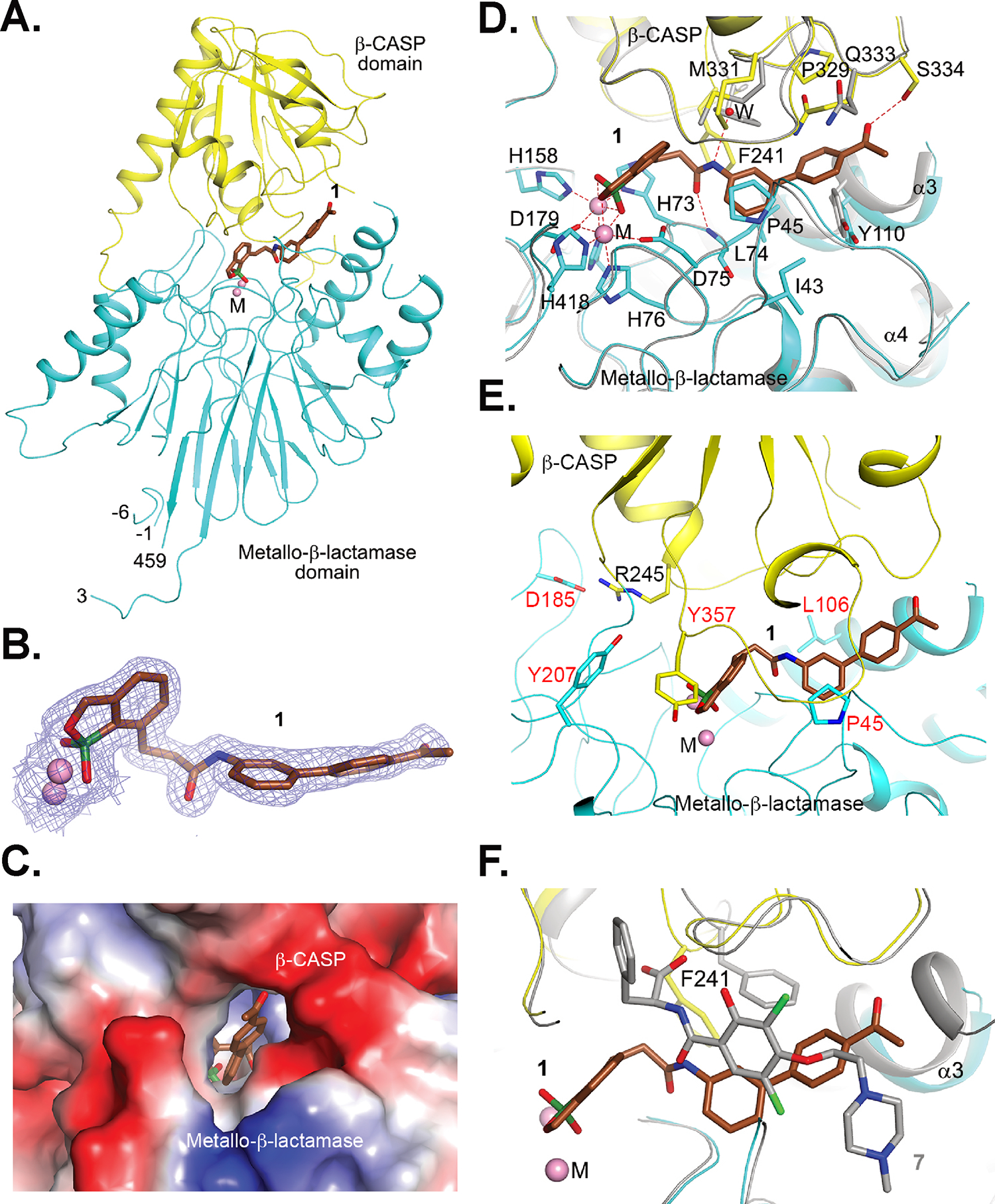
A novel class of benzoxaboroles was reported to induce cancer cell death but the mechanism was unknown. Using a forward genetics platform, we discovered mutations in cleavage and polyadenylation specific factor 3 (CPSF3) that reduce benzoxaborole binding and confer resistance. CPSF3 is the endonuclease responsible for pre-mRNA 3'-end processing, which is also important for RNA polymerase II transcription termination. Benzoxaboroles inhibit this endonuclease activity of CPSF3 in vitro and also curb transcriptional termination in cells, which results in the downregulation of numerous constitutively expressed genes. Furthermore, we used X-ray crystallography to demonstrate that benzoxaboroles bind to the active site of CPSF3 in a manner distinct from the other known inhibitors of CPSF3. The benzoxaborole compound impeded the growth of cancer cell lines derived from different lineages. Our results suggest benzoxaboroles may represent a promising lead as CPSF3 inhibitors for clinical development.


Conflict of interest statement

Declaration of interests The authors declare no competing interests.

Figures

Figure 1.
Figure 1.. Mutations in the active site of CPSF3 confer resistance to benzoxaborole compounds.
(A) Chemical structure of benzoxaborole compound, 1. (B) Dose response curve of 1 against HCT116 cells. Points and error bars represent mean ± SEM from three biological replicates. (C) Scatter plot of resazurin cell viability assay in engineered HCT116 cells treated with the indicated dose of 1 for two weeks, in the presence or absence of 500 μM IAA. Each dot represents one well containing viable cells in a 96-well plate. (D) Dose response curve of 1 against eleven HCT116 clones generated through a forward genetics screen (IC50 ranged from 5.55 μM to greater than 31.6 μM, the highest dose tested) and their parental population (IC50 = 170 nM). Points and error bars represent mean ± SEM from three biological replicates. (E) Table listing genes with recurrent missense mutations discovered through whole-exome sequencing and missense mutations found in each of the eleven resistant clones. (F) Dose response curve of 1 against eight genome-edited HeLa clones sequence verified to have incorporated the indicated CPSF3 mutations (IC50 ranged from 1.96 μM to greater than 31.6 μM, the highest dose tested) and wild-type HeLa cells (IC50 = 1.27 μM). Points and error bars represent mean ± SEM from two biological replicates.
Figure 2.
Figure 2.. Benzoxaborole compounds directly bind CPSF3 and inhibit mRNA cleavage in vitro.
(A) Chemical structure of the photocrosslinking benzoxaborole probe 3. (B) Photoaffinity labeling experiment of CPSF3 with photocrosslinking benzoxaborole probe 3. HeLa cells were treated with 1 μM 3 for 2 hours, then irradiated with UV. Clarified lysate was subjected to a click reaction with biotin azide. Labeled proteins were enriched with streptavidin beads, eluted, then resolved by SDS-PAGE. CPSF3 was detected by Western blot. (C) Photoaffinity labeling experiment of CPSF3 performed as described in (b) using either wild-type or mutant HeLa cells, treated with indicated doses of 3. (D) Dose response curve of the indicated compounds against HeLa cells. Points and error bars represent mean ± SEM from three biological replicates. (E) Photoaffinity labeling experiment of CPSF3 performed as in (b) but with the addition of co-treatment with the indicated competitors. (F) Gel electrophoresis of chemically synthesized histone pre-mRNA subject to mouse nuclear extract and indicated doses of 1. (G) Quantification of percentage of cut pre-mRNA observed in (f). (H) Correlation plot between quantified percentage of cut pre-mRNA and cellular IC50 of four benzoxaborole analogs. (I) Quantification of percentage of cut pre-mRNA observed in Figure S2(d) comparing 1 and 7.
Figure 3.
Figure 3.. Crystal structures of human CPSF3 in complex with inhibitors.
(A) Overall structure of human CPSF3 in complex with 1. The metallo-β-lactamase and β-CASP domains of CPSF3 are shown in cyan and yellow, respectively. 1 is shown as a stick model, with carbon atoms in brown and boron in dark green. The two metal ions in the CPSF3 active site are shown as pink spheres (labeled M). They are predominantly iron when CPSF3 is expressed in bacteria and insect cells, but more zinc is incorporated when it is expressed in human cells. A few residues from the N-terminal His-tag are observed (residues −6 to −1). (B) The 2Fo–Fc electron density for 1 at 1.7 Å resolution, contoured at 1σ. (C) Electrostatic surface of CPSF3, showing an opening that allows the compound to access the cavity in the active site region. (D) Detailed interactions between 1 and CPSF3. Side chains involved in the interactions are shown as stick models. Hydrogen-bonding and chelating interactions are indicated with the dashed lines in red. A water molecule is shown as a red sphere. The structure of free CPSF38 is shown in overlay (gray), and side chains with large conformational differences are shown as sticks. (E) Sites of resistance mutations to compound 1. Side chains of resistance mutation residues are shown as sticks and labeled in red. Asp185 is involved in an ion pair with Arg245. (F) Overlay of the binding mode of 1 with that of 7, the JTE-607 acid analog (in gray).
Figure 4.
Figure 4.. Benzoxaborole inhibition of CPSF3 causes mRNA read through.
(A) Volcano plot describing RNA-seq based quantification of read through expression in 1-treated versus DMSO-treated wild-type HeLa cells. Significance cutoffs were defined as changes greater than 50% from DMSO (log2(fold change) > 0.585 or < 0.585) and adjusted p-value/false discovery rate (FDR) < 0.05. (B) Volcano plot describing RNA-seq based quantification of read through expression in 1-treated versus DMSO-treated HeLa CPSF3Y207H mutant cells. (C) RNA-seq traces at the RILP locus and its downstream region from the experiments in (a) and (b). Each trace represents averages of three biological replicates from each experimental group. (D) RNA-seq traces at the CNKSR1 locus and its downstream region from the experiments in (a) and (b). Each trace represents averages of three biological replicates from each experimental group. (E) Volcano plot describing RNA-seq based quantification of genome-wide gene expression changes in 1-treated versus DMSO-treated wild-type HeLa cells. Significance cutoffs were defined as changes greater than 50% from DMSO (log2(fold change) > 0.585 or < 0.585 and FDR < 0.05. Genes with low baseline expression (average fragments per kilo base per million mapped reads (FPKM) < 0.1 in DMSO-treated cells) are highlighted in dark blue, dark grey, and dark red. (F) Volcano plot describing RNA-seq based quantification of genome-wide gene expression changes in 1-treated versus DMSO-treated HeLa CPSF3Y207H mutant cells.
Figure 5.
Figure 5.. Benzoxaboroles did not display lineage selectivity.
Average area under the dose response curve (AUC) for each lineage obtained from the PRISM assay on 902 cancer cell lines treated with 1. The AUC provides a measure for the cytotoxicity of 1 against the cell lines.

Similar articles

Cited by

References

    1. Nguyen TP, Fang M, Kim J, Wang B, Lin E, Khivansara V, Barrows N, Rivera-Cancel G, Goralski M, Cervantes CL, et al. (2023). Inducible mismatch repair streamlines forward genetic approaches to target identification of cytotoxic small molecules. Cell Chem Biol. 10.1016/j.chembiol.2023.07.017. - DOI - PMC - PubMed
    1. Rock FL, Mao W, Yaremchuk A, Tukalo M, Crépin T, Zhou H, Zhang YK, Hernandez V, Akama T, Baker SJ, et al. (2007). An antifungal agent inhibits an aminoacyl-tRNA synthetase by trapping tRNA in the editing site. Science (New York, N.Y.) 316, 1759–1761. 10.1126/science.1142189. - DOI - PubMed
    1. Akama T, Baker SJ, Zhang YK, Hernandez V, Zhou H, Sanders V, Freund Y, Kimura R, Maples KR, and Plattner JJ (2009). Discovery and structure-activity study of a novel benzoxaborole anti-inflammatory agent (AN2728) for the potential topical treatment of psoriasis and atopic dermatitis. Bioorganic & medicinal chemistry letters 19, 2129–2132. 10.1016/j.bmcl.2009.03.007. - DOI - PubMed
    1. Ding D, Zhao Y, Meng Q, Xie D, Nare B, Chen D, Bacchi CJ, Yarlett N, Zhang YK, Hernandez V, et al. (2010). Discovery of novel benzoxaborole-based potent antitrypanosomal agents. ACS medicinal chemistry letters 1, 165–169. 10.1021/ml100013s. - DOI - PMC - PubMed
    1. Zhang YK, Plattner JJ, Freund YR, Easom EE, Zhou Y, Gut J, Rosenthal PJ, Waterson D, Gamo FJ, Angulo-Barturen I, et al. (2011). Synthesis and structure-activity relationships of novel benzoxaboroles as a new class of antimalarial agents. Bioorganic & medicinal chemistry letters 21, 644–651. 10.1016/j.bmcl.2010.12.034. - DOI - PubMed

Publication types

MeSH terms

LinkOut - more resources