. 2023 Nov 15;14(11):743.
doi: 10.1038/s41419-023-06268-z.

BRISC is required for optimal activation of NF-κB in Kupffer cells induced by LPS and contributes to acute liver injury

Affiliations

BRISC is required for optimal activation of NF-κB in Kupffer cells induced by LPS and contributes to acute liver injury

Wen Zhang et al. Cell Death Dis. .

Abstract

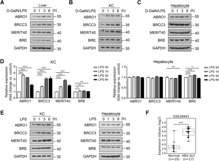
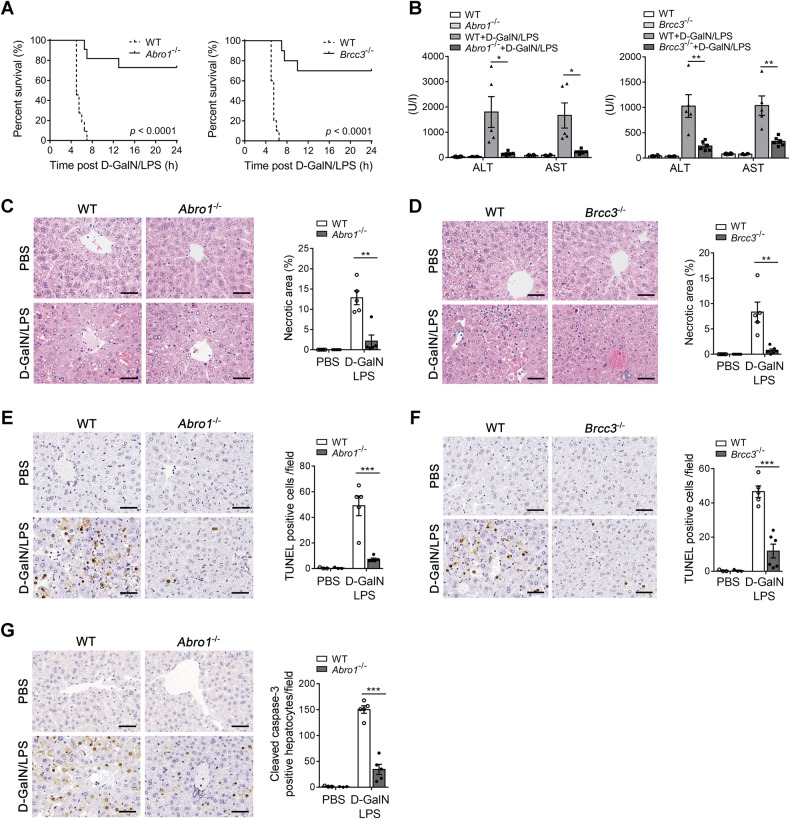
BRISC (BRCC3 isopeptidase complex) is a deubiquitinating enzyme that has been linked with inflammatory processes, but its role in liver diseases and the underlying mechanism are unknown. Here, we investigated the pathophysiological role of BRISC in acute liver failure using a mice model induced by D-galactosamine (D-GalN) plus lipopolysaccharide (LPS). We found that the expression of BRISC components was dramatically increased in kupffer cells (KCs) upon LPS treatment in vitro or by the injection of LPS in D-GalN-sensitized mice. D-GalN plus LPS-induced liver damage and mortality in global BRISC-null mice were markedly attenuated, which was accompanied by impaired hepatocyte death and hepatic inflammation response. Constantly, treatment with thiolutin, a potent BRISC inhibitor, remarkably alleviated D-GalN/LPS-induced liver injury in mice. By using bone marrow-reconstituted chimeric mice and cell-specific BRISC-deficient mice, we demonstrated that KCs are the key effector cells responsible for protection against D-GalN/LPS-induced liver injury in BRISC-deficient mice. Mechanistically, we found that hepatic and circulating levels of TNF-α, IL-6, MCP-1, and IL-1β, as well as TNF-α- and MCP-1-producing KCs, in BRISC-deleted mice were dramatically decreased as early as 1 h after D-GalN/LPS challenge, which occurred prior to the elevation of the liver injury markers. Moreover, LPS-induced proinflammatory cytokines production in KCs was significantly diminished by BRISC deficiency in vitro, which was accompanied by potently attenuated NF-κB activation. Restoration of NF-κB activation by two small molecular activators of NF-κB p65 effectively reversed the suppression of cytokines production in ABRO1-deficient KCs by LPS. In conclusion, BRISC is required for optimal activation of NF-κB-mediated proinflammatory cytokines production in LPS-treated KCs and contributes to acute liver injury. This study opens the possibility to develop new strategies for the inhibition of KCs-driven inflammation in liver diseases.


Conflict of interest statement

The authors declare no competing interests.

Figures

Fig. 1
Fig. 1. BRISC expression is induced in KCs by LPS and increased in the liver tissue of patients with ALF.
WT mice were treated with a sublethal dose of D-GalN/LPS. Immunoblot analysis of BRISC components expression in A liver, B KCs, and C hepatocytes at the indicated times. KCs and hepatocytes were isolated and treated with LPS for various times. D Real-time PCR analysis and E immunoblot analysis of BRISC components expression at the indicated times. F BRCC3 expression level in the liver samples of patients with hepatitis B virus-associated ALF from published transcriptome dataset (GSE38941). Data are presented as mean ± SEM; Expression scores are shown as box plots, with the horizontal lines representing the median; the bottoms and tops of the boxes represent the 25th and 75th percentiles, respectively, and the vertical bars represent the range of data; *P < 0.05, **P < 0.01, ***P < 0.001; two-tailed unpaired t-test.
Fig. 2
Fig. 2. Deletion of BRISC potently protects mice from D-GalN/LPS-induced fatal hepatitis.
A Survival rate of WT and Abro1−/− mice or Brcc3−/− mice after intraperitoneally injected with a lethal dose of D-GalN (700 mg/kg) plus LPS (15 μg/kg) (N = 10–11). Log-rank test. WT and Abro1−/− mice or WT and Brcc3−/− mice were treated with a sublethal dose of D-GalN (700 mg/kg) plus LPS (10 μg/kg) or PBS for 6 h (N = 3–6). B Serum levels of ALT and AST of WT and Abro1−/− mice or Brcc3−/− mice. Representative hematoxylin and eosin (H&E) staining of liver sections from C WT and Abro1−/− mice or D WT and Brcc3−/− mice. Necrotic area was shown as a percentage of the total field area. Representative images of TUNEL-stained liver sections of E WT and Abro1−/− mice or F WT and Brcc3−/− mice. TUNEL-positive cells per field were counted. G Representative liver sections of WT and Abro1−/− mice that were stained with cleaved caspase-3. Cleaved caspase-3 positive hepatocytes in each field were counted. Scale bar, 50 μm. Data are presented as mean ± SEM; *P < 0.05, **P < 0.01, ***P < 0.001; two-tailed unpaired t-test.
Fig. 3
Fig. 3. D-GalN/LPS-induced hepatic inflammation is attenuated in BRISC-deficient mice.
WT and Abro1−/− mice were treated with D-GalN/LPS for the indicated times (N = 3–6). A Immunohistochemistry (IHC) staining analysis of F4/80+ cells and Ly6G+ cells. Representative IHC pictures were shown and positive cells per high-power field (×400) were counted. Cytometric bead array (CBA) analysis of B the hepatic levels of MCP-1, MIP-1α, and MIP-1β, C the serum levels of TNF-α, IL-6, MCP-1, and IL-1β, and D the hepatic levels of TNF-α, IL-6, and IL-1β. E Relative mRNA levels of hepatic TNF-α, IL-6, MCP-1, and IL-1β. Scale bar, 50 μm. Data are presented as mean ± SEM; *P < 0.05, **P < 0.01, ***P < 0.001; two-tailed unpaired t-test.
Fig. 4
Fig. 4. BRISC deficiency-mediated hepatoprotective effect is dependent on hematopoietic cells.
BM chimeras were generated by BM transplantation with depletion of KCs prior to irradiation. AD BM cells were transplanted from WT mice (CD45.2) to WT mice (CD45.2) or from Abro1−/− mice (CD45.2) to Abro1−/− mice (CD45.2) (N = 5). EH BM cells from WT mice (CD45.2) or Abro1−/− mice (CD45.2) were transplanted into WT mice (CD45.1) (N = 5–6). IL BM cells from WT mice (CD45.1) were transplanted into WT mice (CD45.2) or Abro1−/− mice (CD45.2) (N = 5–7). The mice were treated with D-GalN/LPS 10 weeks after transplantation and the liver injury was examined at 6 h after D-GalN/LPS injection. A, E, I Serum levels of ALT and AST. B, F, J Representative H&E staining and percentage of necrotic area of liver sections. CBA analysis of the C, G, K serum and D, H, L hepatic levels of TNF-α and IL-6. WT and Abro1−/− mice were treated with D-GalN/TNF-α or PBS for 6 h (N = 3–6). Liver injury was evaluated by M serum ALT level and N H&E staining 6 h after D-GalN/LPS injection. Necrotic area was shown as a percentage of the total field area. Scale bar, 50 μm. Data are presented as mean ± SEM; *P < 0.05, **P < 0.01, ***P < 0.001; two-tailed unpaired t-test.
Fig. 5
Fig. 5. Kupffer cells contribute to the protection of BRISC-deficient mice from LPS-induced liver injury.
A Survival curve for Abro1flox/flox and Abro1-MKO mice challenged with a lethal dose of D-GalN/LPS (N = 11). Log-rank test. Abro1flox/flox and Abro1-MKO mice were treated with a sublethal dose of D-GalN/LPS for 6 h (N = 5–12). Liver injury was evaluated by B serum ALT level and C H&E staining. D Hepatocyte apoptosis was evaluated by TUNEL staining. E Hepatic inflammatory cells infiltration was measured by F4/80+ and Ly6G+ immunohistochemistry staining. F Survival curves for Abro1flox/flox and Abro1-KCKO mice challenged with a lethal dose of D-GalN/LPS (N = 12). Log-rank test. Abro1flox/flox and Abro1-KCKO mice were treated with a sublethal dose of D-GalN/LPS for 6 h (N = 4–13). G Serum ALT and H H&E staining 6 h after D-GalN/LPS injection. Necrotic area was shown as a percentage of the total field area. I Hepatocyte apoptosis was evaluated by TUNEL staining. J Hepatic inflammatory cells infiltration was measured by F4/80+ and Ly6G+ immunohistochemistry staining. Representative images were shown and the number of positive cells in each high-power field was counted. Scale bar, 50 μm. Data are presented as mean ± SEM; *P < 0.05, **P < 0.01, ***P < 0.001; two-tailed unpaired t-test.
Fig. 6
Fig. 6. BRISC deficiency selectively suppresses LPS-induced proinflammatory cytokines production in KCs in vitro.
CBA analysis of TNF-α and IL-6 from A WT and Abro1−/− or B WT and Brcc3−/− KCs stimulated with various doses of LPS for 3 h. C CBA analysis of TNF-α, IL-6, and IL-1β from WT, Abro1−/−, and Brcc3−/− KCs stimulated with 100 ng/ml LPS for the indicated times. D Relative mRNA levels of TNF-α, IL-6, and IL-1β in WT and Abro1−/− KCs treated with 100 ng/ml LPS for the indicated times. E Relative mRNA levels of TNF-α and IL-6 in WT and Abro1−/− BMDMs, PMs, and NEUTs stimulated with 100 ng/ml LPS for the indicated times. F CBA analysis of TNF-α and IL-6 from WT and Abro1−/− BMDMs, PMs, and neutrophils stimulated with 100 ng/ml LPS for 12 h. Data are presented as mean ± SEM; *P < 0.05, **P < 0.01; ***P < 0.001; two-way ANOVA with Bonferroni’s multiple comparisons test (AE) or two-tailed unpaired t-test (F).
Fig. 7
Fig. 7. BRISC positively regulates NF-κB activation in LPS-stimulated KCs.
A WT and Abro1−/− KCs were stimulated with 100 ng/ml LPS for various times. Immunoblot analysis of the indicated target proteins. B WT and Brcc3−/− KCs were stimulated with 100 ng/ml LPS for various times, followed by immunoblot analysis of the indicated target proteins. C WT and Abro1−/− PMs, neutrophils, and BMDMs were stimulated with 100 ng/ml LPS for the indicated time points, followed by immunoblot analysis of the indicated target proteins. D WT and Abro1−/− KCs transduced with lentivirus expressing NF-κB-luciferase reporter gene were treated with 100 ng/ml LPS for 3 h. The luminescence levels were measured and normalized to control values. Nuclear and cytoplasmic proteins of WT and Abro1−/− KCs were extracted after stimulation with LPS for 30 min. E Immunoblot analysis of p65 expression in the cytoplasm and nucleus. F ELISA of the DNA binding activity of nuclear NF-κB p65. WT and Abro1−/− KCs were pre-treated with NF-κΒ activator 1 G or NF-κΒ activator 2 H for 6 h and then treated with 100 ng/ml LPS for 3 h. CBA analysis of TNF-α and IL-6. Data are presented as means ± SEM; **P < 0.01; ***P < 0.001; two-tailed unpaired t-test.
Fig. 8
Fig. 8. Pharmacological targeting of BRISC attenuates D-GalN/LPS-induced liver injury.
A Survival curves for WT and Abro1−/− mice challenged with a lethal dose of D-GalN/LPS or D-GalN/TNF-α (N = 10). Log-rank test. WT mice received two intraperitoneal injections of THL (2.5 mg/kg) 1 h before and 1 h after PBS or a sublethal dose of D-GalN/LPS administration (N = 3–5). B Serum levels of ALT and AST, C representative H&E staining and percentage of necrotic area of liver sections 6 h after D-GalN/LPS injection. D CBA analysis of the serum levels of TNF-α and MCP-1. E WT KCs pre-treated with 50 nM THL or vehicle control for 2 h were left unstimulated or stimulated with 1 μg/ml LPS for 6 h. CBA analysis of TNF-α and IL-6. Scale bar, 50 μm. Data are presented as mean ± SEM; *P < 0.05, **P < 0.01; ***P < 0.001; two-tailed unpaired t-test.

References

    1. Stravitz RT, Lee WM. Acute liver failure. Lancet. 2019;394:869–81. doi: 10.1016/S0140-6736(19)31894-X. - DOI - PMC - PubMed
    1. Rajaram P, Subramanian R. Acute liver failure. Semin Resp Crit Care Med. 2018;39:513–22. doi: 10.1055/s-0038-1673372. - DOI - PubMed
    1. Triantafyllou E, Woollard KJ, McPhail MJW, Antoniades CG, Possamai LA. The role of monocytes and macrophages in acute and acute-on-chronic liver failure. Front Immunol. 2018;9:2948. doi: 10.3389/fimmu.2018.02948. - DOI - PMC - PubMed
    1. An L, Wirth U, Koch D, Schirren M, Drefs M, Koliogiannis D, et al. The role of gut-derived lipopolysaccharides and the intestinal barrier in fatty liver diseases. J Gastrointest Surg. 2022;26:671–83. doi: 10.1007/s11605-021-05188-7. - DOI - PMC - PubMed
    1. Hamesch K, Borkham-Kamphorst E, Strnad P, Weiskirchen R. Lipopolysaccharide-induced inflammatory liver injury in mice. Lab Anim. 2015;49:37–46. doi: 10.1177/0023677215570087. - DOI - PubMed

Publication types

MeSH terms