. 2023 Nov 15;14(1):7372.
doi: 10.1038/s41467-023-43269-0.

Mucosal-associated invariant T cells contribute to suppression of inflammatory myeloid cells in immune-mediated kidney disease

Affiliations

Mucosal-associated invariant T cells contribute to suppression of inflammatory myeloid cells in immune-mediated kidney disease

Ann-Christin Gnirck et al. Nat Commun. .

Abstract

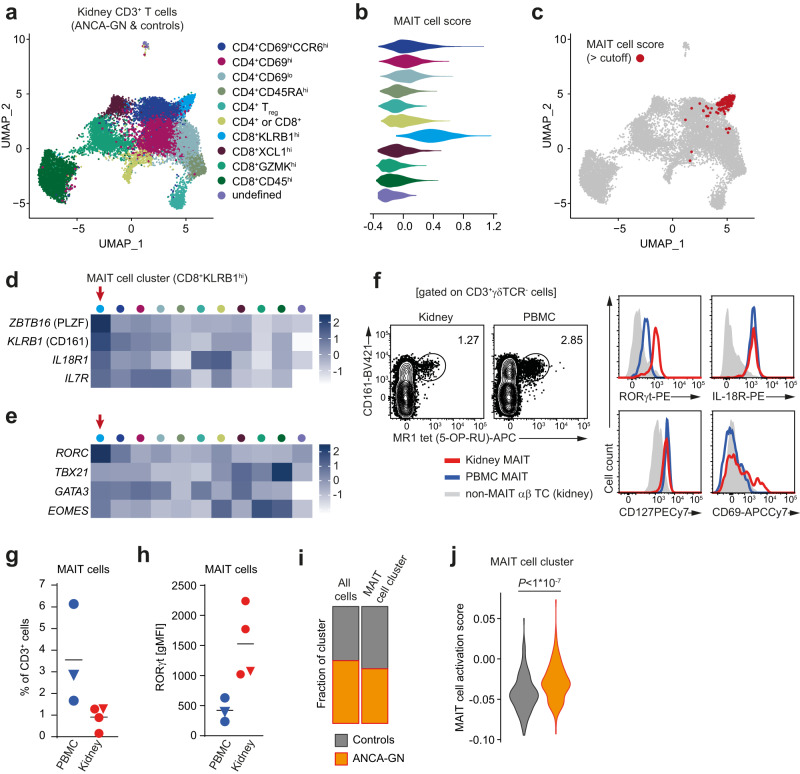
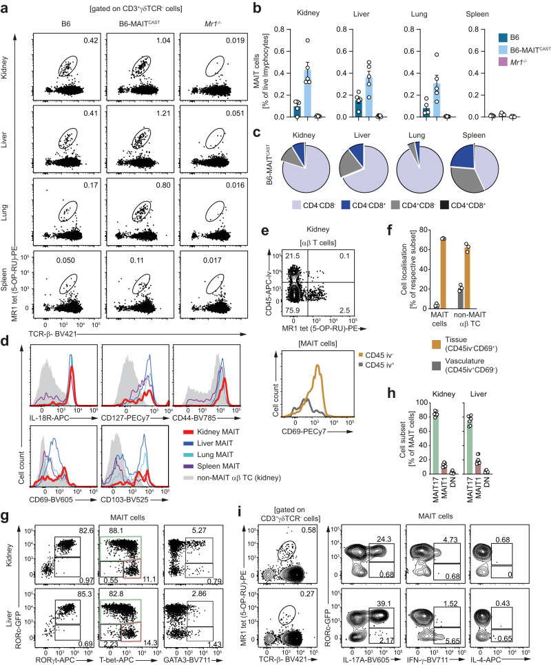
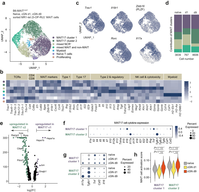
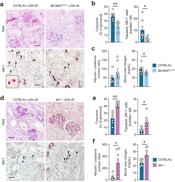
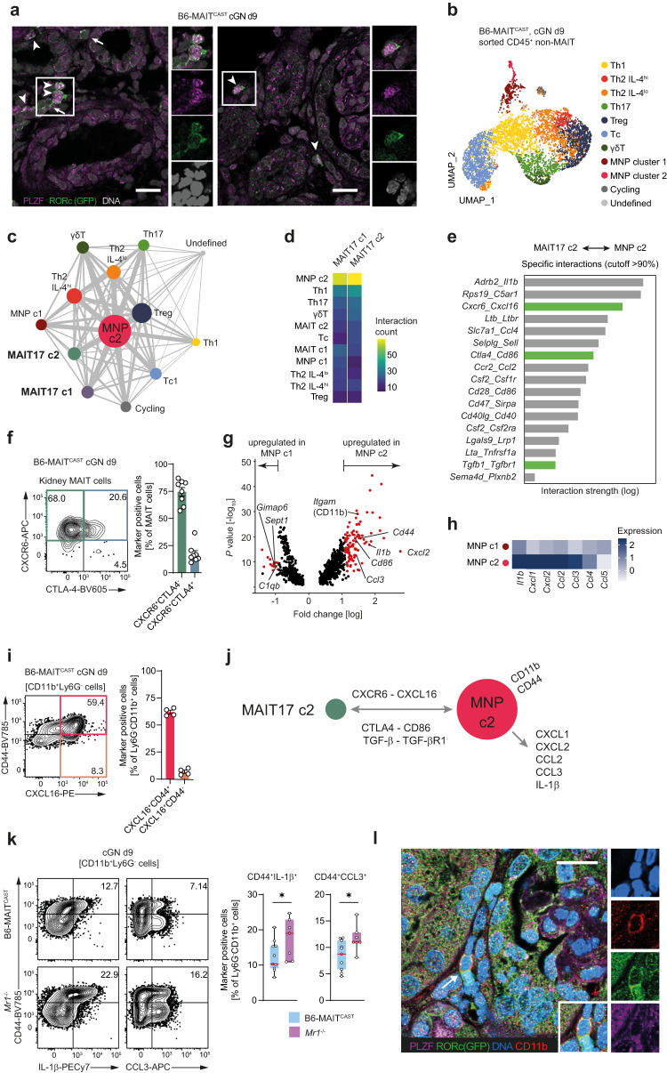
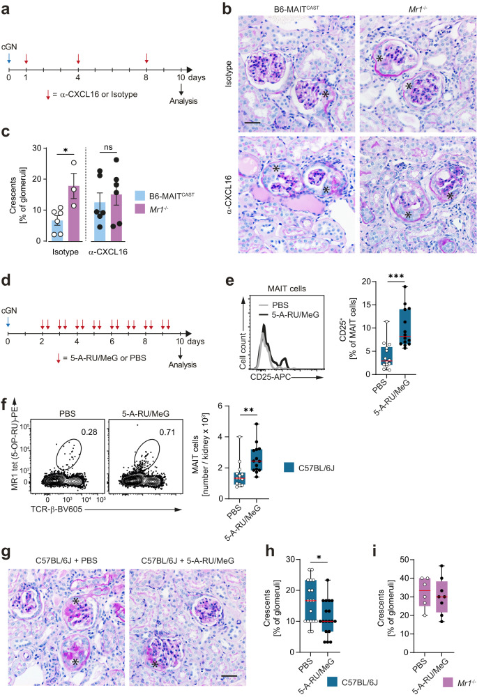
Mucosal-associated invariant T (MAIT) cells have been implicated in various inflammatory diseases of barrier organs, but so far, their role in kidney disease is unclear. Here we report that MAIT cells that recognize their prototypical ligand, the vitamin B2 intermediate 5-OP-RU presented by MR1, reside in human and mouse kidneys. Single cell RNAseq analysis reveals several intrarenal MAIT subsets, and one, carrying the genetic fingerprint of tissue-resident MAIT17 cells, is activated and expanded in a murine model of crescentic glomerulonephritis (cGN). An equivalent subset is also present in kidney biopsies of patients with anti-neutrophil cytoplasmatic antibody (ANCA)-associated cGN. MAIT cell-deficient MR1 mice show aggravated disease, whereas B6-MAITCAST mice, harboring higher MAIT cell numbers, are protected from cGN. The expanded MAIT17 cells express anti-inflammatory mediators known to suppress cGN, such as CTLA-4, PD-1, and TGF-β. Interactome analysis predicts CXCR6 - CXCL16-mediated cross-talk with renal mononuclear phagocytes, known to drive cGN progression. In line, we find that cGN is aggravated upon CXCL16 blockade. Finally, we present an optimized 5-OP-RU synthesis method which we apply to attenuating cGN in mice. In summary, we propose that CXCR6+ MAIT cells might play a protective role in cGN, implicating them as a potential target for anti-inflammatory therapies.


Conflict of interest statement

The authors declare no competing interests.

Figures

Fig. 1
Fig. 1. Mucosal-associated invariant T (MAIT) 17 cells reside in the human kidney and are activated in anti-neutrophil cytoplasmatic antibody (ANCA)-associated glomerulonephritis (GN).
a Unsupervised Uniform Manifold Approximation and Projection (UMAP) clustering (resolution 0.6) of a published data set of pooled single-cell RNA sequencing (scRNAseq) data from CD3+ T cells isolated from kidney biopsy specimens of ANCA-GN patients (n = 6) and unaffected kidney tissue of tumor nephrectomy patients (controls, n = 3). Eleven clusters were identified and named according to cluster defining genes. b Expression of an uncurated MAIT cell-defining gene signature generated from a MAIT cell cluster identified in the bronchoalveolar lavage fluid of Coronavirus Disease 2019 patients (MAIT cell score, see Supplementary Fig. 1) in the indicated clusters. Color coding as in (a). c Cells that show high expression of the MAIT cell score were marked in red and plotted on the original UMAP plot. Gene expression heat map of MAIT cell-defining genes (d) and T cell lineage-defining transcription factors (e) in the indicated clusters. Color coding as in (a). f Representative flow cytometric identification and characterization of MAIT cells in paired kidney and blood samples of a tumor nephrectomy patient. Gating strategy is specified in brackets and numbers indicate the percentage of cells in the gate. MAIT cell frequencies in the CD3+ T cell fraction (g) and RORγt expression in MAIT cells (h) from unaffected kidney tissue and blood of tumor nephrectomy patients, as well as from the blood of healthy human donors. Samples from the same patient are marked with a triangle symbol. Each symbol in (g) and (h) represents one biologically independent sample (n = 3 for PBMC, n = 4 for kidney) and horizontal lines represent the mean. i Contribution of cells from controls and ANCA-GN patients to the MAIT cell cluster (CD8+KLRB1hi) identified in scRNAseq analyses. j Expression of an uncurated gene signature defining in vitro activated human peripheral blood MAIT cells (MAIT cell activation score (Hinks et al.)) in the MAIT cell cluster of controls and ANCA-GN patients. The two-sided Student’s t test was used for comparison between the two groups. (UMAP uniform manifold approximation and projection for dimension reduction, ANCA-GN anti-neutrophil cytoplasmic antibody-associated glomerulonephritis, PBMC peripheral blood mononuclear cells, gMFI geometric mean fluorescent intensity). Source data are provided as a Source Data file.
Fig. 2
Fig. 2. Mucosal-associated invariant T (MAIT) cells display a tissue-resident MAIT17 phenotype in the murine kidney.
a Representative flow cytometry plots of leukocytes isolated from kidney, liver, lung, and spleen of naïve B6, B6-MAITCAST, and Mr1−/− mice. b Quantification of MAIT cell frequencies in the live lymphocyte gate in the respective organs and mouse strains (see (a)). c Distribution of CD4 and CD8 surface expression in the MAIT cell population of naïve B6-MAITCAST mice in kidney, liver, lung, and spleen. d Representative flow cytometric analysis of surface marker expression on MAIT cells in kidney, liver, lung, and spleen of naïve B6-MAITCAST mice. e Representative flow cytometry plots of αβ T cells from kidneys of naïve B6-MAITCAST mice euthanized shortly after intravenous (i.v.) injection of an fluorochrome coupled α-CD45 antibody (upper panel) and histogram of CD69 surface expression in the CD45 iv+ and CD45 iv MAIT cell population (lower panel). f Distribution of CD45 iv+ CD69 (intravascular) and CD45 iv CD69+ (tissue) populations in the MAIT cell and non-MAIT αβ T cell compartment in the kidneys of naïve B6-MAITCAST mice (n = 3 biologically independent animals per group). Representative flow cytometry plots of transcription factor expression (g) and quantification of MAIT cell subsets (RORc-GFP+T-bet+/− = MAIT17, RORc-GFP-T-bet+ = MAIT1, RORc-GFPT-bet = double negative (DN)) (h) in kidney and liver MAIT cells from naïve RORc-GFP B6-MAITCAST mice (n = 7 biologically independent animals per group). i Cytokine production (after restimulation with phorbol 12-myristate 13-acetate/ionomycin) in kidney and liver MAIT cells from naïve RORc-GFP B6-MAITCAST mice. Gating strategies are specified in brackets and numbers indicate percentage of cells in the gates. Bars represent mean ± SEM and symbols represent individual animals. All experiments were performed twice with at least three mice per group. Source data are provided as a Source Data file.
Fig. 3
Fig. 3. Kidney mucosal-associated invariant T (MAIT) 17 cells express pro- and anti-inflammatory mediators in experimental crescentic glomerulonephritis (cGN).
a Uniform Manifold Approximation and Projection (UMAP) dimensionality reduction (resolution 0.2) of pooled single-cell RNA sequencing data of MR1 tetramer (5-OP-RU)+ MAIT cells isolated by flow cytometric cell sorting from kidneys of naïve B6-MAITCAST mice and on day 1 and 9 after induction of cGN (naïve n = 18, cGN day 1 n = 11, cGN day 9 n = 13; n represents the number of biologically independent, male, 9–14 week-old mice that were pooled for the isolation). Seven clusters were identified and named according to cluster defining genes and expression of selected marker genes. b Heat map of normalized marker gene expression of the indicated clusters. c UMAP plots showing expression of MAIT cell-defining genes (upper panels) and MAIT17 cell markers in the identified clusters. d Contribution of different MAIT cell clusters to the dataset. Numbers below the bars indicate total number of MAIT cells analyzed for each time point. e Volcano plot of differential gene expression in MAIT17 clusters 1 and 2. f Gene expression of all interleukins expressed in the data set and other selected cytokines in MAIT17 cluster 1 and 2. g Expression of selected cytokine genes in MAIT17 clusters 1 and 2 at the different time points. h Expression of an uncurated gene signature defining in vivo activated murine MAIT cells (MAIT cell activation score (Hinks et al.)) in MAIT cell clusters 1 and 2 at the different time points. One-way ANOVA was used for statistical analysis followed by a post hoc analysis with Newman-Keuls test for multiple comparisons.
Fig. 4
Fig. 4. Mucosal-associated invariant T (MAIT) cells protect from renal tissue damage in experimental crescentic glomerulonephritis (cGN).
a Representative photomicrographs of kidney sections from C57BL/6J wild type and B6-MAITCAST mice on day 9 of cGN stained with periodic acid-Schiff (PAS) (upper panel) and immunohistochemistry for the neutrophil marker GR-1 (lower panel). Asterisks mark glomerular crescents, arrow heads mark neutrophils in the tubulointerstitial area. b Quantification of crescent formation and tubulointerstitial neutrophil infiltration (C57BL/6J wild type n = 7, B6-MAITCAST n = 7; n represents the number of biologically independent animals). c Kidney function parameters in C57BL/6J wild type and B6-MAITCAST mice (groups as in b). Data in (ac) are representative for three independent experiments. d Representative PAS and GR-1 stainings of kidney sections from C57BL/6J wild type and Mr1−/− mice on day 9 of cGN. Asterisks mark glomerular crescents, arrow heads mark neutrophils in the tubulointerstitial area. e Quantification of crescent formation and tubulointerstitial neutrophil infiltration (C57BL/6J wild type n = 7, Mr1−/− n = 8; n represents the number of biologically independent animals). f Kidney function parameters in C57BL/6J wild type and Mr1−/− mice (C57BL/6J wild type n = 7, Mr1−/− n = 8; n represents the number of biologically independent animals). Data in (df) are representative for two independent experiments. Scale bar represents 20 µm. Symbols represent individual animals. Bars represent mean ± SEM. (*p < 0.05, **p < 0.01 in two-sided Student’s t test). Source data are provided as a Source Data file.
Fig. 5
Fig. 5. Immunomodulatory mucosal-associated invariant T (MAIT) 17 cells interact with proinflammatory monocytes in experimental crescentic glomerulonephritis (cGN).
a Representative immunofluorescence staining for identification of PLZF+RORc+ MAIT17 cells in kidney sections of RORc-GFP B6-MAITCAST mice on day 9 after cGN induction (representative for n = 3 biologically independent animals). b Uniform Manifold Approximation and Projection (UMAP) dimensionality reduction (resolution 0.5) of single-cell RNA sequencing data of CD45+ non-MAIT cells isolated by flow cytometric cell sorting from kidneys of B6-MAITCAST mice on 9 after induction of cGN. The 11 clusters were named according to cluster defining genes and expression of selected marker genes. c Interaction network based on ligand–receptor interactions between all CD45+ cell clusters identified in kidneys of B6-MAITCAST mice with cGN (pooled non-MAIT cell and MAIT cell data sets). Dot size represents the number of total significant interactions with other clusters and line thickness represents number of interactions between individual clusters. d Heatmap showing the absolute number of significant interactions between the indicated cell clusters. e List of top ligand–receptor interactions between MAIT17 cluster 2 and mononuclear phagocyte (MNP) cluster 2 filtered for interaction strength (>1) and specificity (>90%). Interaction pathways that have been implicated in amelioration of cGN are marked in green. f Representative flow cytometry plot and quantification of CXCR6 and CTLA4 expression in kidney MAIT cells of B6-MAITCAST (cGN day 9, n = 8 biologically independent animals). g Volcano plot of differential gene expression between MNP clusters 1 and 2. Significantly upregulated genes are marked in red. h Normalized gene expression heatmap of proinflammatory cytokines and chemokines MNP clusters 1 and 2. i Representative flow cytometry plot and quantification of CXCL16 and CD44 expression (after restimulation with PMA/ionomycin) in kidney CD11b+ MNPs from B6-MAITCAST mice (cGN day 9, n = 4 biologically independent animals). j Hypothetic working model for interaction between MAIT17 cluster 2 and MNP cluster 2 based on the interactome analysis. k Representative flow cytometry plots and quantification of IL-1β and CCL3 production (after restimulation with phorbol 12-myristate 13-acetate/ionomycin) by CD11b+CD44hi MNPs in the kidney of B6-MAITCAST and Mr1−/− mice with cGN (day 9, B6-MAITCAST n = 11 biologically independent animals, Mr1−/− n = 7 biologically independent animals). l Representative immunofluorescence staining of MAIT17 cells located in close proximity to CD11b+ mononuclear phagocytes (arrow) in kidney sections of B6-MAITCAST mice (cGN day 9, representative for n = 3 biologically independent animals). Data in (f) and (k) are pooled from two independent experiments. Data in (a, l) are representative for two independent experiments with n = 3 biologically independent animals with similar results. Scale bar represents 15 µm. Circles in bar graphs represent individual animals and bars represent mean ± SEM. (*p < 0.05 in two-sided Student’s t test). Source data are provided as a Source Data file.
Fig. 6
Fig. 6. CXCL16 blockade abrogates protective effect of mucosal-associated invariant T (MAIT) cells and pharmacological MAIT cell activation ameliorates experimental crescentic glomerulonephritis (cGN).
a Schematic representation of experimental setup for α-CXCL16 treatment in cGN. b Representative photomicrographs of periodic acid-Schiff-stained kidney sections from isotype- and α-CXCL16-treated B6-MAITCAST and Mr1−/− mice on day 10 of cGN. c Quantification of crescent formation in the respective groups of mice (B6-MAITCAST + Isotype n = 6, Mr1−/− + Isotype n = 3, B6-MAITCAST + α-CXCL16 n = 6, Mr1−/− + α-CXCL16 n = 6, n represents biologically independent animals for each group). Data are pooled from two independent experiments. d Schematic representation of experimental setup for 5-A-RU/MeG treatment in cGN. e Representative histogram and quantification of CD25 expression in PBS- and 5-A-RU/MeG-treated C57BL/6J wild type mice with cGN. (day 10, PBS n = 14, 5-A-RU/MeG n = 14, n represents biologically independent animals for each group). f Representative flow cytometry plots and quantification of MAIT cells in kidneys of PBS- and 5-A-RU/MeG-treated C57BL/6J wild type mice with cGN (day 10, PBS n = 14, 5-A-RU/MeG n = 14, n represents biologically independent animals for each group). Data in (e and f) are pooled from three independent experiments with similar results. g Representative photomicrographs of periodic acid-Schiff-stained kidney sections from PBS- and 5-A-RU/MeG -treated C57BL/6J wild type on day 10 of cGN. Quantification of crescent formation in PBS- and 5-A-RU/MeG-treated C57BL/6J wild type mice (h) and Mr1−/− mice (i) with cGN (wild type day 10, PBS n = 18, 5-A-RU/MeG n = 18, Mr1−/− day 10, PBS n = 6, 5-A-RU/MeG n = 8; n represents biologically independent animals for each group). Data in (f) are pooled from four independent experiments and data in (i) are pooled from two independent experiments. Asterisks mark glomerular crescents. Scale bar represents 20 µm. Circles represent individual animals and bars represent mean ± SEM (*p < 0.05, **p < 0.01, ***p < 0.001 in two-sided Student’s t test). Source data are provided as a Source Data file.

References

    1. Floege J, Amann K. Primary glomerulonephritides. Lancet. 2016;387:2036–2048. doi: 10.1016/S0140-6736(16)00272-5. - DOI - PubMed
    1. Kurts C, Panzer U, Anders HJ, Rees AJ. The immune system and kidney disease: basic concepts and clinical implications. Nat. Rev. Immunol. 2013;13:738–753. doi: 10.1038/nri3523. - DOI - PubMed
    1. Krebs CF, et al. Autoimmune renal disease is exacerbated by S1P-receptor-1-dependent intestinal Th17 cell migration to the kidney. Immunity. 2016;45:1078–1092. doi: 10.1016/j.immuni.2016.10.020. - DOI - PMC - PubMed
    1. Tipping PG, Holdsworth SR. T cells in crescentic glomerulonephritis. J. Am. Soc. Nephrol. 2006;17:1253–1263. doi: 10.1681/ASN.2005091013. - DOI - PubMed
    1. Tilloy F, et al. An invariant T cell receptor alpha chain defines a novel TAP-independent major histocompatibility complex class Ib-restricted alpha/beta T cell subpopulation in mammals. J. Exp. Med. 1999;189:1907–1921. doi: 10.1084/jem.189.12.1907. - DOI - PMC - PubMed

Publication types

Substances