

Surface curvature-induced oriented assembly of sushi-like Janus therapeutic nanoplatform for combined chemodynamic therapy
- PMID: 37968644
- PMCID: PMC10647176
- DOI: 10.1186/s12951-023-02138-0
Surface curvature-induced oriented assembly of sushi-like Janus therapeutic nanoplatform for combined chemodynamic therapy
Abstract
Background: Chemodynamic therapy (CDT) based on Fenton/Fenton-like reaction has emerged as a promising cancer treatment strategy. Yet, the strong anti-oxidation property of tumor microenvironment (TME) caused by endogenous glutathione (GSH) still severely impedes the effectiveness of CDT. Traditional CDT nanoplatforms based on core@shell structure possess inherent interference of different subunits, thus hindering the overall therapeutic efficiency. Consequently, it is urgent to construct a novel structure with isolated functional units and GSH depletion capability to achieve desirable combined CDT therapeutic efficiency.
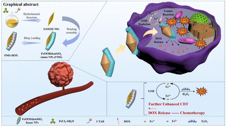
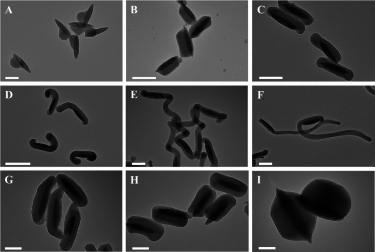
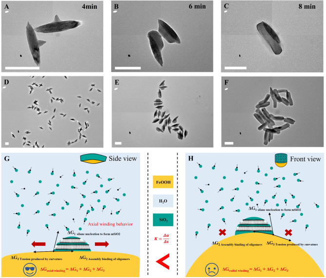
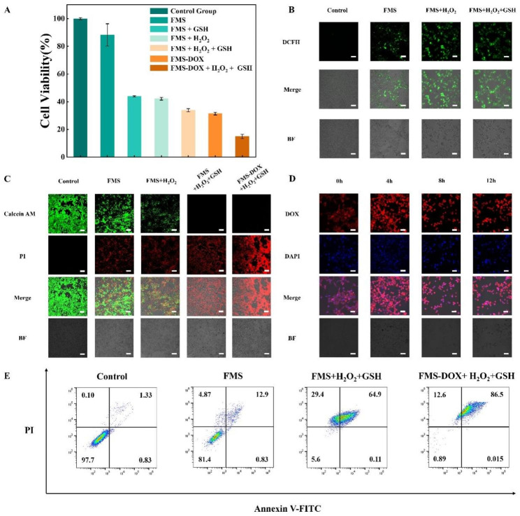
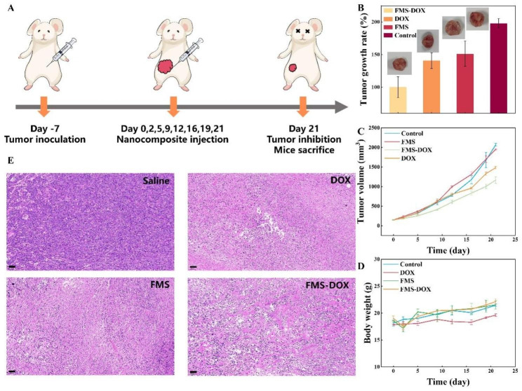
Results: Herein, a surface curvature-induced oriented assembly strategy is proposed to synthesize a sushi-like novel Janus therapeutic nanoplatform which is composed of two functional units, a FeOOH nanospindle serving as CDT subunit and a mSiO2 nanorod serving as drug-loading subunit. The FeOOH CDT subunit is half covered by mSiO2 nanorod along its long axis, forming sushi-like structure. The FeOOH nanospindle is about 400 nm in length and 50 nm in diameter, and the mSiO2 nanorod is about 550 nm in length and 100 nm in diameter. The length and diameter of mSiO2 subunit can be tuned in a wide range while maintaining the sushi-like Janus structure, which is attributed to a Gibbs-free-energy-dominating surface curvature-induced oriented assembly process. In this Janus therapeutic nanoplatform, Fe3+ of FeOOH is firstly reduced to Fe2+ by endogenous GSH, the as-generated Fe2+ then effectively catalyzes overexpressed H2O2 in TME into highly lethal ·OH to achieve efficient CDT. The doxorubicin (DOX) loaded in the mSiO2 subunit can be released to achieve combined chemotherapy. Taking advantage of Fe3+-related GSH depletion, Fe2+-related enhanced ·OH generation, and DOX-induced chemotherapy, the as-synthesized nanoplatform possesses excellent therapeutic efficiency, in vitro eliminating efficiency of tumor cells is as high as ~ 87%. In vivo experiments also show the efficient inhibition of tumor, verifying the synthesized sushi-like Janus nanoparticles as a promising therapeutic nanoplatform.
Conclusions: In general, our work provides a successful paradigm of constructing novel therapeutic nanoplatform to achieve efficient tumor inhibition.
Keywords: Asymmetric nanostructure; Chemodynamic therapy; Janus nanoparticles; Mesoporous; Nanocatalytic medicine.
© 2023. The Author(s).
Conflict of interest statement
The authors declare no competing interests.
Figures


Similar articles
-
A multivalent polyphenol-metal-nanoplatform for cascade amplified chemo-chemodynamic therapy.Acta Biomater. 2024 Jan 1;173:389-402. doi: 10.1016/j.actbio.2023.11.006. Epub 2023 Nov 14. Acta Biomater. 2024. PMID: 37967695
-
A tetrasulfide bond-bridged mesoporous organosilica-based nanoplatform for triple-enhanced chemodynamic therapy combined with chemotherapy and H2S therapy.J Mater Chem B. 2023 Nov 22;11(45):10822-10835. doi: 10.1039/d3tb02147e. J Mater Chem B. 2023. PMID: 37920970
-
Facile Synthesis of Fe3O4@Au/PPy-DOX Nanoplatform with Enhanced Glutathione Depletion and Controllable Drug Delivery for Enhanced Cancer Therapeutic Efficacy.Molecules. 2022 Jun 22;27(13):4003. doi: 10.3390/molecules27134003. Molecules. 2022. PMID: 35807249 Free PMC article.
-
Recent Advances on NIR-II Light-Enhanced Chemodynamic Therapy.Adv Healthc Mater. 2024 Apr;13(10):e2303451. doi: 10.1002/adhm.202303451. Epub 2023 Nov 27. Adv Healthc Mater. 2024. PMID: 37983596 Review.
-
Recent advances in multifunctional nanomaterials for photothermal-enhanced Fenton-based chemodynamic tumor therapy.Mater Today Bio. 2022 Jan 4;13:100197. doi: 10.1016/j.mtbio.2021.100197. eCollection 2022 Jan. Mater Today Bio. 2022. PMID: 35036895 Free PMC article. Review.
References
-
- Siegel RL, Miller KD, Jemal A, Cancer Statistics. 2016. CA: A Cancer Journal for Clinicians 2016, 66 (1), 7–30. - PubMed
MeSH terms
Substances
Grants and funding
- 20QA1401200, 22YF1402200/Shanghai Rising-Star Program
- 20QA1401200, 22YF1402200/Shanghai Rising-Star Program
- 22075049, 21875043, 22088101, 21701027, 21733003, 21905052, 51961145403/National Natural Science Foundation of China
- 2018YFA0209401, 2018YFE0201701/National Key Research and Development Program of China
- 17JC1400100/Key Basic Research Program of Science and Technology Commission of Shanghai Municipality
LinkOut - more resources
Full Text Sources
Medical
