

doi: 10.1097/eus.0000000000000037.
Epub 2023 Oct 23.
Pancreatic leiomyosarcoma: EUS findings of an uncommon pancreatic mass (with video)
Affiliations
- PMID: 37969164
- PMCID: PMC10631615
- DOI: 10.1097/eus.0000000000000037
Item in Clipboard
Pancreatic leiomyosarcoma: EUS findings of an uncommon pancreatic mass (with video)
Endosc Ultrasound.
2023 Sep-Oct.
No abstract available
Conflict of interest statement
The authors declare no conflict of interest.
Figures
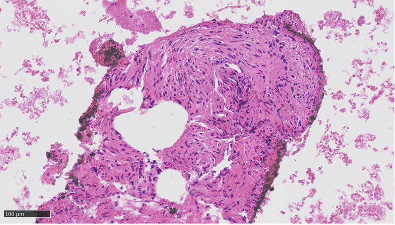
Cytological sample showed spindle cells proliferation with elongated, cigar-shaped and blunt-ended nuclei with severe atypia, variable pleomorphism, and fascicular grown pattern.
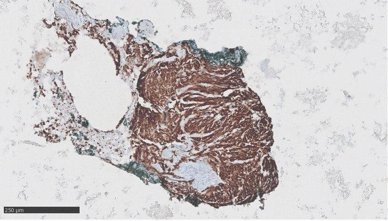
Immunohistochemical tests revealed these cells to be diffusely positive for desmin, focally positive for smooth muscle actin, and negative for CD117, DOG1, cytokeratin AE1–3, and S100. MIB1 antibody for Ki67 highlighted a proliferation rate of 10%.
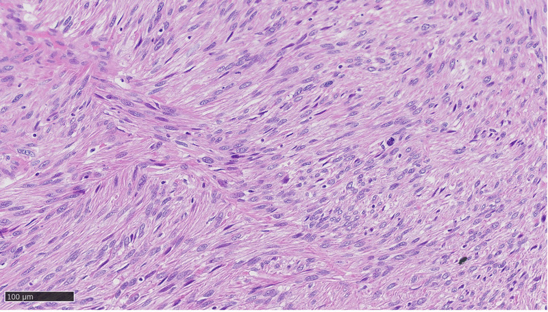
Histological examination of the neoplasm showed spindle-shaped cells with plump, blunt-ended nuclei, fields of nuclear pleomorphism, and distinctly eosinophilic fibrillary cytoplasm. Mitotic figures were common, including atypical ones. The cells merged with blood vessel's wall and were set in long intersecting fascicles. Hyalinized hypocellular areas and tumor necrosis were focally present.
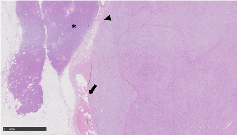
At low magnification, the tumor merges with the blood vessel's wall (arrow), and the tumor borders appear well circumscribed (arrowhead) without invasion of pancreatic parenchyma (*).
References
-
- Sweeney JT, Crabtree DK, Yassin R, Somogyi L. Metastatic uterine leiomyosarcoma involving the pancreas diagnosed by EUS with fine-needle aspiration. Gastrointest Endosc 2002;56(4):596–597. - PubMed
-
- Miyajima S Takeda S Goda K, et al. Metastatic primary pulmonary leiomyosarcoma to the pancreas diagnosed by endoscopic ultrasound-guided fine-needle aspiration. Clin J Gastroenterol 2021;14(6):1779–1784. - PubMed
