Review
. 2022 Feb 25;1(2):60-73.
doi: 10.1016/j.cjcpc.2022.02.003. eCollection 2022 Apr.

Approach to Wide Complex Tachycardia in Paediatric Patients

Affiliations
Review

Approach to Wide Complex Tachycardia in Paediatric Patients

Carolina A Escudero et al. CJC Pediatr Congenit Heart Dis. .

Abstract

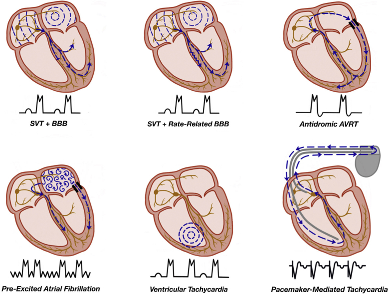
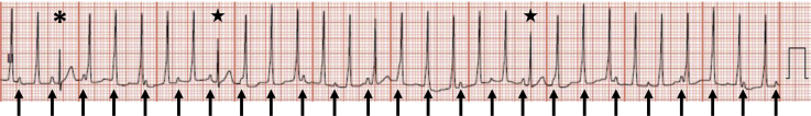
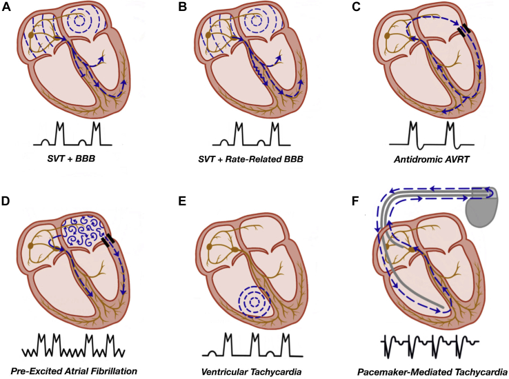
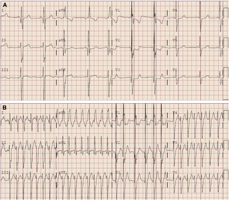
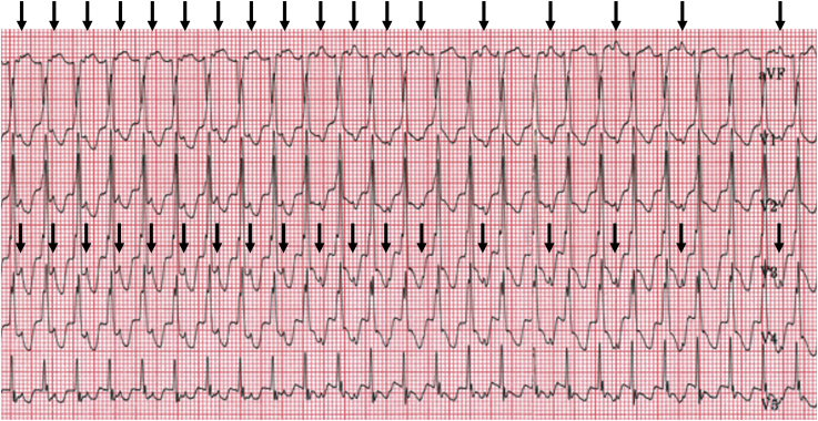
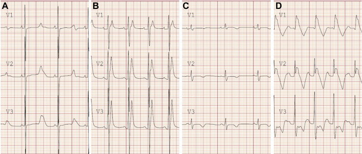
Wide complex tachycardia (WCT) is an infrequently encountered condition in paediatric patients and may be due to a variety of causes including supraventricular tachycardia with aberrant conduction, ventricular activation via an accessory pathway, ventricular pacing, or ventricular tachycardia. Immediate tachycardia termination is required in haemodynamically unstable patients. After stabilization or in those with haemodynamically tolerated WCT, a careful review of electrocardiographic tracings and diagnostic manoeuvres are essential to help elucidate the cause. Subacute and chronic management for WCT will depend on the underlying cause as well as features of the patient and the tachycardia presentation. This article will review the epidemiology, potential causes, and management of WCT in children. A detailed review of the pathophysiology, differential diagnosis, and diagnostic and treatment options is provided to enable the reader to develop a practical approach to managing this condition in young patients.

La tachycardie à complexes QRS larges est rare en pédiatrie et peut avoir diverses causes, notamment une tachycardie supraventriculaire avec trouble de la conduction, l’activation ventriculaire par une voie accessoire, une stimulation ventriculaire ou une tachycardie ventriculaire. La suppression immédiate de la tachycardie est primordiale lorsque l’état hémodynamique du patient est instable. Une fois l’état du patient stabilisé, ou en cas de tachycardie à complexes QRS larges tolérée sur le plan hémodynamique, l’examen minutieux des tracés électrocardiographiques et des manœuvres diagnostiques est crucial pour en élucider la cause. La prise en charge des cas subaigus et chroniques de tachycardie à complexes QRS larges dépend de sa cause sous-jacente ainsi que des caractéristiques du patient et du tableau clinique de la tachycardie. Cet article porte sur l’épidémiologie, les causes possibles et la prise en charge de la tachycardie à complexes QRS larges chez les enfants. Un examen approfondi de la physiopathologie, du diagnostic différentiel et des options diagnostiques et thérapeutiques est présenté pour permettre au lecteur d’élaborer une approche pratique pour la prise en charge de cette affection chez leurs jeunes patients.


Figures

None
Graphical abstract
Figure 1
Figure 1
Accelerated idioventricular rhythm with ventriculoatrial dissociation in an infant. The QRS duration of the ventricular tachycardia beats is 80 milliseconds. The arrows indicate sinus P waves, which are dissociated from the QRS complexes. The asterisk indicates a sinus capture beat. The stars demonstrate fusion beats.
Figure 2
Figure 2
Mechanisms of wide complex tachycardia. (A) SVT with an underlying bundle branch block (BBB). (B) SVT with a rate-related bundle branch block. (C) Antidromic tachycardia as seen in a patient with ventricular pre-excitation, with the antegrade limb of the circuit being the accessory pathway and the retrograde limb the AV node. (D) Pre-excited atrial fibrillation with antegrade conduction via the accessory pathway and the AV node, leading to an irregularly irregular wide complex tachycardia. (E) Ventricular tachycardia with VA dissociation due to the absence of retrograde AV nodal conduction. (F) Pacemaker-mediated tachycardia with a ventricular paced beat followed by retrograde conduction via the AV node. The retrograde atrial activity is sensed by the pacemaker with subsequent ventricular pacing to complete the reentrant circuit. AV, atrioventricular; AVRT, atrioventricular reciprocating tachycardia; SVT, supraventricular tachycardia; VA, ventriculoatrial.
Figure 3
Figure 3
Wide complex tachycardia in a patient with a baseline conduction abnormality. (A) Baseline electrocardiogram (ECG) showing sinus rhythm and a right bundle branch block in a child with congenital heart disease. (B) The same patient’s ECG during an atrial tachycardia showing a QRS complex similar to baseline. The rhythm is irregular due to variable atrioventricular conduction of the atrial tachycardia.
Figure 4
Figure 4
Supraventricular tachycardia (SVT) with rate-related aberrancy in a child with tachycardia. A wide complex tachycardia with rate-related left bundle branch block (rhythm strip from lead V1) transitioning to a narrow complex tachycardia; diagnostic of SVT. Reproduced with permission.
Figure 5
Figure 5
Pre-excited atrial fibrillation. The characteristic features of an irregularly irregular wide complex tachycardia, variable QRS duration, and consistent QRS axis are shown. The degree of ventricular pre-excitation is a reflection of fusion between atrial wavefronts conducting via the atrioventricular node and the accessory pathway. Reproduced with permission.
Figure 6
Figure 6
Right ventricular outflow tract ventricular tachycardia. This tachycardia typically has an inferior axis and left bundle branch block morphology. The arrows indicate P waves, which are best seen in the right precordial leads, and therefore demonstrate ventriculoatrial dissociation.
Figure 7
Figure 7
Left ventricular posterior fascicular ventricular tachycardia. The electrocardiogram demonstrates a superior QRS axis and right bundle branch block morphology. Note the characteristic sharp initial deflection of the QRS (indicative of activation of the fascicular tissue) and relatively narrow appearance of this wide complex tachycardia.
Figure 8
Figure 8
Bidirectional ventricular tachycardia with alternating axes, as demonstrated by the asterisks and stars, followed by degeneration into ventricular fibrillation in a patient with catecholaminergic polymorphic ventricular tachycardia.
Figure 9
Figure 9
Initiation of torsades de pointes with characteristic “twisting” appearance around the isoelectric line. This patient had idiopathic ventricular fibrillation thought to be due to intermediate-coupled premature ventricular contractions as described by Li et al. Reproduced with permission.
Figure 10
Figure 10
Coarse ventricular fibrillation characterized by a disorganized, low-amplitude, and polymorphic appearance.
Figure 11
Figure 11
Ventricular tachycardia with concordance. The QRS complexes of the ventricular ectopic beats (indicated by asterisks) are monophasic with the same polarity in the precordial leads (positive concordance), suggesting a ventricular origin.
Figure 12
Figure 12
Pacemaker-mediated tachycardia. Ventricular paced beats conduct retrograde to the atrium, with subsequent ventricular tracking of the retrograde atrial activation, continuing the tachycardia circuit. The arrows indicate the retrograde atrial activation. The black triangles indicate the pacing spikes.
Figure 13
Figure 13
Ventriculoatrial (VA) dissociation with adenosine. The arrows indicate the P waves. There is initially 1:1 VA conduction during the wide complex tachycardia. With adenosine administration, there is gradual VA prolongation followed by VA dissociation with continuation of the tachycardia. This finding is consistent with a diagnosis of ventricular tachycardia. The differential diagnosis for this pattern is junctional ectopic tachycardia in a patient with an underlying bundle branch block, as could be seen after surgery for congenital heart disease.
Figure 14
Figure 14
Electrocardiographic findings in leads V1-V3 in 3 primary channelopathies and arrhythmogenic right ventricular cardiomyopathy (ARVC). (A) Long QT syndrome with prolonged QT interval and abnormal T-wave morphology; (B) short QT syndrome with severely shortened QT interval and peaked T waves; (C) ARVC with low voltage QRS complexes and T-wave inversion in the right precordial leads; (D) Brugada syndrome with coved (type I) ST-segment elevation with T-wave inversion in the right precordial leads.

References

    1. Salerno J.C., Seslar S.P. Supraventricular tachycardia. Arch Pediatr Adolesc Med. 2009;163:268–274. - PubMed
    1. Roggen A., Pavlovic M., Pfammatter J.P. Frequency of spontaneous ventricular tachycardia in a pediatric population. Am J Cardiol. 2008;101:852–854. - PubMed
    1. Ceresnak S.R., Liberman L., Avasarala K., et al. Are wide complex tachycardia algorithms applicable in children and patients with congenital heart disease? J Electrocardiol. 2010;43:694–854. - PubMed
    1. Topjian A.A., Raymond T.T., Atkins D., et al. Part 4: pediatric basic and advanced life support: 2020 American Heart Association guidelines for cardiopulmonary resuscitation and emergency cardiovascular care. Circulation. 2020;142(suppl 2):469–523. - PubMed
    1. Crosson J.E., Callans D.J., Bradley D.J., et al. PACES/HRS expert consensus statement on the evaluation and management of ventricular arrhythmias in the child with a structurally normal heart. Heart Rhythm. 2014;11:55–78. - PubMed

LinkOut - more resources