

Rare Case of an Adult With Double-Chambered Left Ventricle
- PMID: 37969559
- PMCID: PMC10642131
- DOI: 10.1016/j.cjcpc.2021.10.001
Rare Case of an Adult With Double-Chambered Left Ventricle
Abstract
The rare case of an adult with a double-chambered left ventricle was revealed using multimodality imaging using echocardiography and cardiac magnetic resonance imaging in a 38-year-old asymptomatic male patient. The congenital malformation was dominated by a second, coarsely trabeculated muscular shelf dividing the left ventricle into 2 chambers without signs for left ventricular inflow or outflow tract obstruction. The partition wall did not show any signs for intramyocardial fibrosis in late gadolinium enhancement cardiovascular magnetic resonance imaging. Flow measurements excluded a relevant intracardial shunt across the additive perimembranous ventricular septal defect. There were no signs for global right and left ventricular dysfunction with left and right ventricular volumes and ejection fraction within normal limits. A conservative approach was recommended. In summary, we are able to present the case of an adult with a double-chambered left ventricle with a second muscular "septum" partially dividing the left ventricular cavity without causing a relevant impact on cardiac function or clinical signs for heart failure.
Le cas rare d’un adulte présentant un ventricule gauche à double chambre a été révélé par une imagerie multimodale utilisant l’échocardiographie et l’imagerie par résonnance magnétique cardiaque chez un homme asymptomatique de 38 ans. La malformation congénitale était dominée par une deuxième bande musculaire grossièrement trabéculaire divisant le ventricule gauche en deux chambres sans signes d’obstruction des chambres d’admission et d’éjection du ventricule gauche. La cloison de partition ne montrait aucun signe de fibrose intramyocardique à l’imagerie par résonnance magnétique cardiovasculaire avec rehaussement tardif au gadolinium. Les mesures du débit ont exclu un shunt intracardiaque significatif à travers le défaut septal ventriculaire transmembranaire supplémentaire. Il n’y avait pas de signe de dysfonction ventriculaire droite et gauche globale, les volumes ventriculaires gauche et droit et la fraction d’éjection étant dans les limites normales. Une approche conservatrice a été recommandée. En résumé, nous pouvons présenter le cas d’un adulte porteur d’un ventricule à double chambre avec une deuxième « cloison » musculaire divisant partiellement la cavité ventriculaire gauche sans causer d’effet notable sur la fonction cardiaque ou de signes cliniques d’insuffisance cardiaque.
© 2021 The Author(s).
Figures
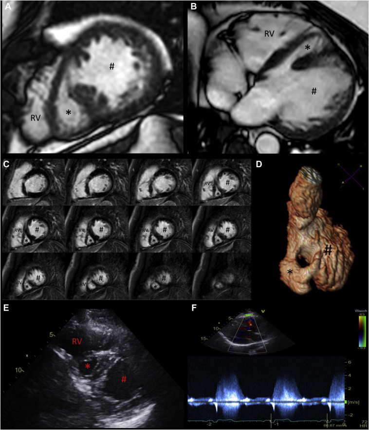
 ) showing the right ventricle (RV) as well as left ventricle divided by an accessory muscle bundle in a main principle (#) and second smaller (∗) chamber. (C) CMR late gadolinium enhancement short-axis stack. The left ventricle is divided in a principle and a small accessory chamber by a prominent muscle bundle with a basal and apical connection between the 2 chambers. No thrombus was detected. (D) Contrast-enhanced 3D reconstruction of the left ventricular cavity illustrating the accessory left ventricular chamber. (E) Echocardiographic short-axis view of the RV, left ventricle principle chamber (#) and left ventricle accessory chamber (∗). (F) Doppler pulse measurement of the restrictive ventricular septal defect that presented independently of the accessory left ventricular accessory muscle bundle.
References
-
- Anderson R.H., Gufler H. Commentary: what makes the morphologically left ventricle double chambered? J Thorac Cardiovasc Surg. 2020;159:e195–e196. - PubMed
-
- Kumar G.R., Vaideswar P., Agrawal N., et al. Double chambered ventricles: a retrospective clinicopathological study. Indian J Thoracic Cardiovasc Surg. 2007;23:135–140.
LinkOut - more resources
Full Text Sources
