

A novel network architecture combining central-peripheral deviation with image-based convolutional neural networks for diffusion tensor imaging studies
- PMID: 37969894
- PMCID: PMC10637193
- DOI: 10.1080/02664763.2022.2108386
A novel network architecture combining central-peripheral deviation with image-based convolutional neural networks for diffusion tensor imaging studies
Abstract
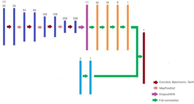
Brain imaging research is a very challenging topic due to complex structure and lack of explicitly identifiable features in the image. With the advancement of magnetic resonance imaging (MRI) technologies, such as diffusion tensor imaging (DTI), developing classification methods to improve clinical diagnosis is crucial. This paper proposes a classification method for DTI data based on a novel neural network strategy that combines a convolutional neural network (CNN) with a multilayer neural network using central-peripheral deviation (CPD), which reflects diffusion dynamics in the white matter by spatially evaluating the deviation of diffusion coefficients between the inner and outer parts of the brain. In our method, a multilayer perceptron (MLP) using CPD is combined with the final layers for classification after reducing the dimensions of images in the convolutional layers of the neural network architecture. In terms of training loss and the classification error, the proposed classification method improves the existing image classification with CNN. For real data analysis, we demonstrate how to process raw DTI image data sets obtained from a traumatic brain injury study (MagNeTS) and a brain atlas construction study (ICBM), and apply the proposed approach to the data, successfully improving classification performance with two age groups.
Keywords: Concentric circle pooling; convolutional neural network; diffusion tensor image; image classification; multi-layer perceptron.
© 2022 Informa UK Limited, trading as Taylor & Francis Group.
Conflict of interest statement
No potential conflict of interest was reported by the author(s).
Figures


References
-
- MicroDicom viewer, 2021.
-
- Balu N., Chu B., Hatsukami T.S., Yuan C., and Yarnykh V.L., Comparison between 2D and 3D high-resolution black-blood techniques for carotid artery wall imaging in clinically significant atherosclerosis. Journal of Magnetic Resonance Imaging: JMRI 27 (2008), pp. 918–924. Available at https://pubmed.ncbi.nlm.nih.gov/18383253 https://www.ncbi.nlm.nih.gov/pmc/articles/PMC2830087/. - PMC - PubMed
-
- Bisong E., Building Machine Learning and Deep Learning Models on Google Cloud Platform: A Comprehensive Guide for Beginners, Apress, Ottawa, ON, 2019.
-
- Bottou L., Online learning and stochastic approximations. On-line Learning in Neural Networks 17 (1998), pp. 142.
Grants and funding
LinkOut - more resources
Full Text Sources