. 2023:3:55.
doi: 10.1038/s43586-023-00247-6. Epub 2023 Jul 20.

In vitro selection of aptamers and their applications

Affiliations

In vitro selection of aptamers and their applications

M C DeRosa et al. Nat Rev Methods Primers. 2023.

Abstract

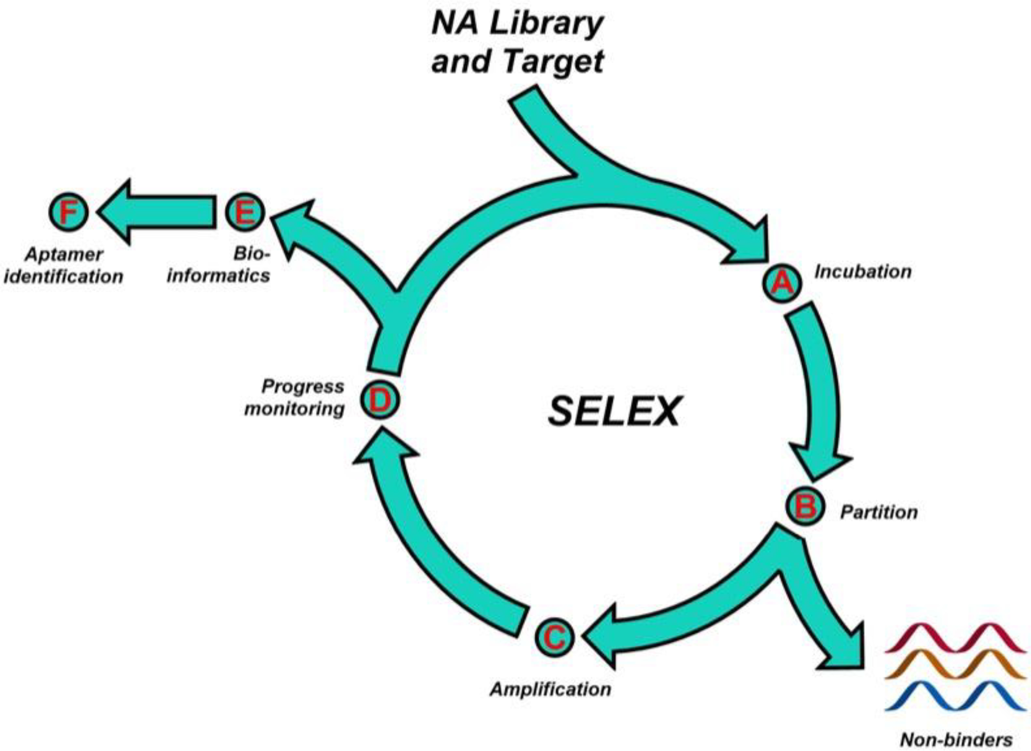
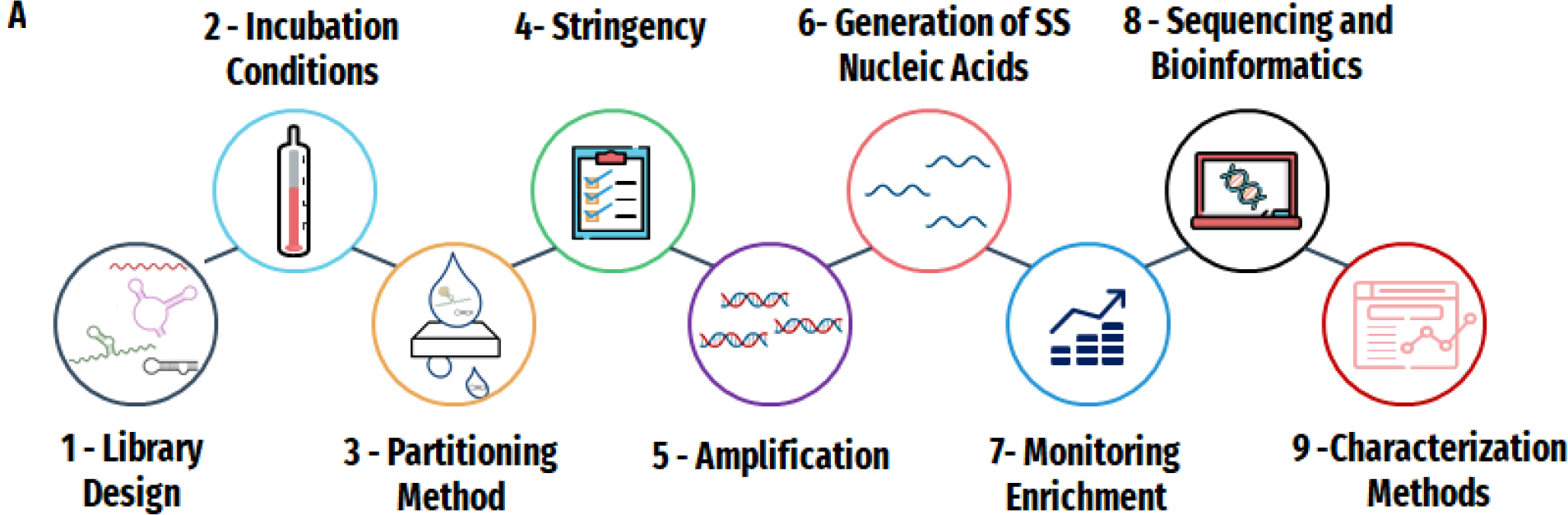
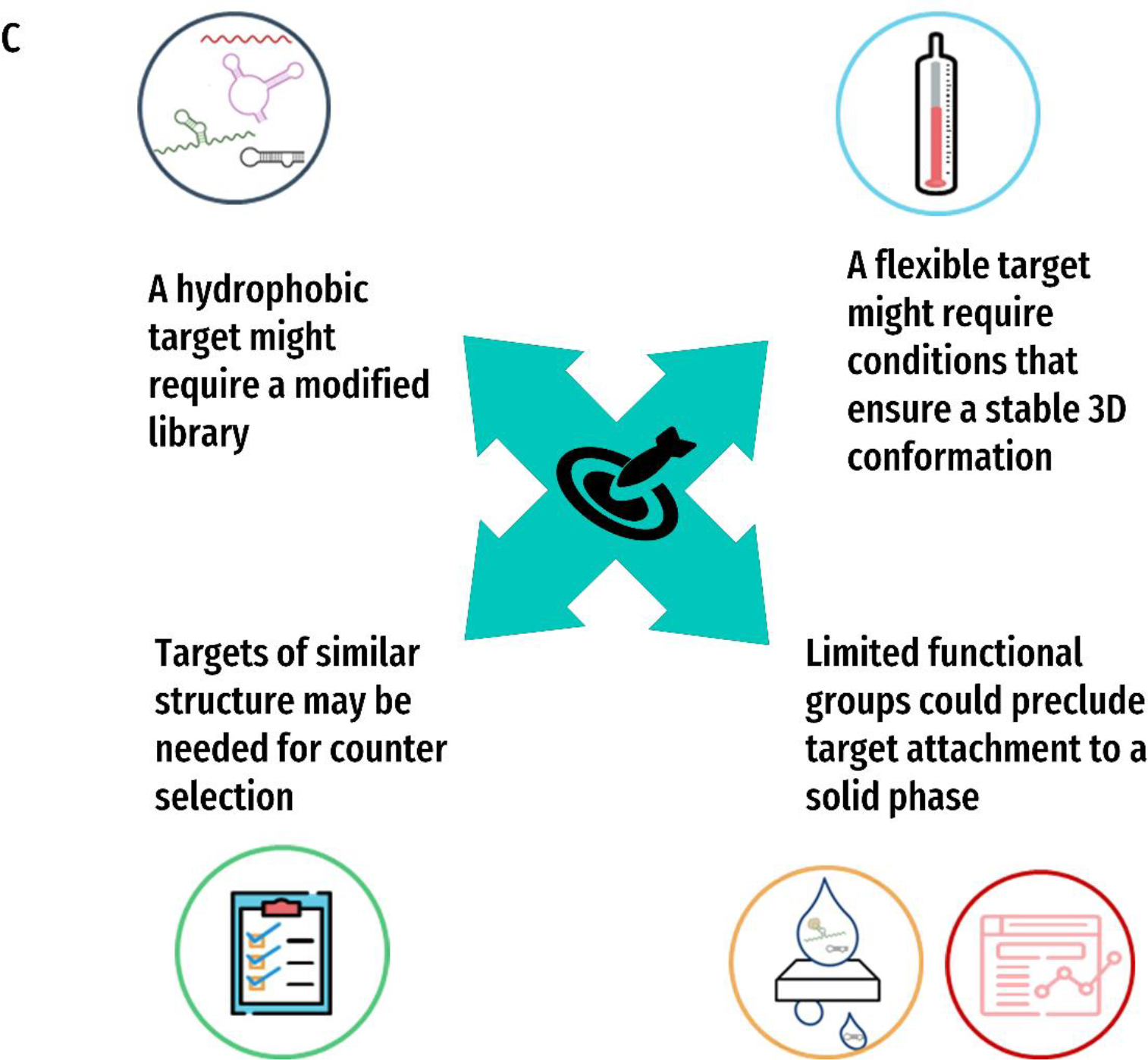
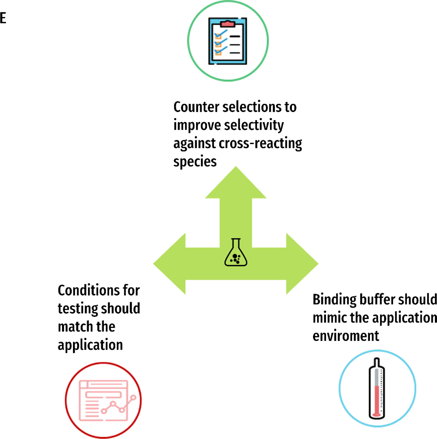
The introduction of the in-vitro evolution method known as SELEX (Systematic Evolution of Ligands by Exponential enrichment) more than 30 years ago led to the conception of versatile synthetic receptors known as aptamers. Offering many benefits such as low cost, high stability and flexibility, aptamers have sparked innovation in molecular diagnostics, enabled advances in synthetic biology and have facilitated new therapeutic approaches. The SELEX method itself is inherently adaptable and offers near limitless possibilities in yielding functional nucleic acid ligands. This Primer serves to provide guidance on experimental design and highlight new growth areas for this impactful technology.


Conflict of interest statement

The authors declare no competing interests.

Figures

Figure 1.
Figure 1.. General outline of the SELEX process and aptamer identification.
A SELEX library is chemically synthesized using a DNA synthesizer, and PCR optimization prior to SELEX ensures efficient amplification. The SELEX library is incubated with the target, and binders are separated from non-binders using an appropriate partitioning method. The SELEX library with potential binders is then amplified and utilized in the next round of SELEX. The evolved libraries are monitored for enrichment and sequenced to identify hit aptamer candidates.
Figure 2.
Figure 2.. Decision-making for SELEX design.
A) The nine main aspects of a selection experiment: 1) Library Design 2) Incubation Conditions 3) Partitioning Method 4) Stringency 5) Amplification 6) Generation of Single Stranded Nucleic acids 7) Monitoring Enrichment 8) Sequencing and Bioinformatics and 9) Characterization Methods. B) The four key factors that will inform those decisions: 1) the target of interest 2) the eventual application of the aptamer 3) the medium and 4) the resources available. C)-F) Examples of ways these queries may influence the different aspects of the SELEX experiment for each of the four key factors C) Target D) Application E) Medium and F) Resources
Figure 3.
Figure 3.. Representative analysis of the in vitro selection process.
A. Ideal Polymerase Chain Reaction (PCR) amplification analyzed by gel electrophoresis. Lane 1 ladder, lane 2 positive control, lane 3 negative control, lane 4 amplification of the library following a round of selection. B. One method for monitoring the in vitro selection process is by comparing the quantity of library that interacts with the target compared to controls. Blue bars represent percent of library from each positive selection round (the target of interest); orange bars represent the library recovered from a parallel negative control selection round (absence of target). C. Landscape of sequence enrichment throughout rounds ofin vitro selection. In early rounds, the library is not enriched and therefore sequencing yields a diverse sequence pool with very few copies of the same sequence and are therefore mostly unique sequences. In later rounds, after enrichment has occurred, more sequences will emerge from the data that have numerous copies, thus resulting in fewer unique sequences.
Figure 4.
Figure 4.. Representative characterization techniques of aptamer candidates
A) Aptamer binding measured using fluorescence polarization. The aptamer is modified with a fluorophore and is excited by polarized light. In solution, the aptamer rotates thus resulting in a depolarized emission of light. As increasing concentrations of the target are added and bind to the aptamer, the larger aptamer-target complex moves slower thus the emitted fluorescence remains more polarized. The change in polarization with increasing concentrations of target binding to the aptamer compared to a negative control sequence can be plotted as a binding isotherm to solve for the dissociation constant. B) Aptamer binding kinetics measured using a real-time surface plasmon resonance assay. The aptamer is immobilized to a surface and the target is flowed over a surface plasmon biosensor chip. The interaction at the surface is detected in real time resulting in on and off binding curves that can be fit to obtain kinetic data. C). Secondary structure prediction of an aptamer. By synthesizing truncated sequences, a simple binding assay can determine the importance of different regions of the full-length aptamer necessary for binding. Here, aptamer designs are synthesized with each colored region removed and then tested for relative binding compared to the full-length sequence. D). Aptamer stability and half-life measured using denaturing gel electrophoresis. A labelled aptamer can be incubated with serum or nucleases and loaded into a gel at various time points to determine the time required to fully degrade the aptamer of interest.
Figure 5:
Figure 5:
Aptamers are powerful tools for molecular imaging. A) Schematic of staining live cells with fluorescently labeled aptamer. B) Aptamers that bind to epidermal growth factor receptor (EGFR), and two other proteins were compared to control cells to demonstrate the ability of aptamers for molecular imaging.. C) Schematic illustration of the Simoa assay,. Briefly, capture antibodies are immobilized on microbeads, which are then incubated with the target protein and biotinylated SOMAmer. Finally a streptavidin modified beta–galactosidase enzyme is added. The beads are distributed in microwells, and the presence of the target is revealed in the presence ofresorufin β-D-galactopyranoside substrate. Digital detection (black panel with lit up wells) is then translated into a concentration based reading (calibration curve shown). D) Schematic illustration of EGFR labelling by SOMAmer. The SOMAmer binds to the target protein, and a fluorescently labelled imager strand binds to a docking region of the SOMAmer. The SOMAmer DNA-PAINT based method allows for sensitive fluorescent detection of target protein expressed in cells. Panel A and B were reproduced under Creative Commons Attribution License from Gomes de Castro et al., 2017. Part C reproduced with permission from Wu et al., 2016. Part D Reproduced with permission from Strauss et al., 2018.
Figure 6:
Figure 6:
Aptamers that have been selected to bind to light up fluorophores When bound to a fluorogen (DFHBI, DFHO, and TO1-biotin), aptamers can substantially enhance their emitted fluorescence. The spinach (a), corn (b), and mango (c) aptamers are shown Adapted from Neubacher and Hennig (2018).

References

    1. Tuerk C & Gold L Systematic evolution of ligands by exponential enrichment: RNA ligands to bacteriophage T4 DNA polymerase. Science 249, 505–510 (1990).

      a. One of the original three selection/aptamer papers, this work that describes in vitro selection of RNA aptamers that binds T4 DNA polymerase that coined the term ‘SELEX’, is required reading by anyone interested in the aptamer field.

    1. Ellington AD & Szostak JW In vitro selection of RNA molecules that bind specific ligands. Nature 346, 818–822, doi:10.1038/346818a0 (1990).

      a. One of the original three selection/aptamer papers, this work that describes in vitro selection of RNA aptamers that binds organic dyes and coins the term ‘aptamer’, is required reading by anyone interested in the aptamer field.

    1. Mills DR, Peterson R & Spiegelman S An extracellular Darwinian experiment with a self-duplicating nucleic acid molecule. Proceedings of the National Academy of Sciences 58, 217–224 (1967). - PMC - PubMed
    1. Robertson DL & Joyce GF Selection in vitro of an RNA enzyme that specifically cleaves single-stranded DNA. Nature 344, 467–468, doi:10.1038/344467a0 (1990).

      a. One of the original three selection/aptamer papers, this work that describes in vitro selection of an RNA enzyme that can cleave single-stranded DNA, is required reading by anyone interested in the functional nucleic acid field.

    1. Jarmoskaite I, AlSadhan I, Vaidyanathan PP & Herschlag D How to measure and evaluate binding affinities. eLife 9, e57264, doi:10.7554/eLife.57264 (2020). - DOI - PMC - PubMed

LinkOut - more resources