

Radiologic and clinicopathologic features of eosinophilic solid and cystic renal cell carcinoma: report of two cases and review of literature
- PMID: 37970333
- PMCID: PMC10641369
Radiologic and clinicopathologic features of eosinophilic solid and cystic renal cell carcinoma: report of two cases and review of literature
Abstract
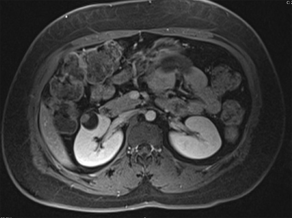
Eosinophilic Solid and Cystic Renal Cell Carcinoma (ESC RCC) is a rare entity described in the latest WHO Classification of Urinary and Male Genital Tumours (2022 edition). It is a neoplasm that occurs most often in a sporadic setting, with no association with tuberous sclerosis complex (TSC). It typically presents as a well demarcated, non-encapsulated lesion, with solid and cystic architecture, composed of cells with voluminous eosinophilic cytoplasm and cytoplasmic stippling. Tumor cells are at least focally immunohistochemically (IHC) reactive for CK20. CD10 and Cathepsin K are positive in most cases. Consistent somatic mutually exclusive mutations in the TSC1 and TSC2 genes are detected in ESC RCC. We describe two ESC RCC cases diagnosed at our institution. Both cases occurred in female patients, ages of 33 and 64, respectively. Both patients had no evidence of TSC and both lesions were found incidentally, by imaging studies, at an early stage. Macroscopic and microscopic findings in both neoplasms were classic. One case was analyzed by molecular testing and TSC2 gene mutation was detected. Both cases had focal positivity of CD10 and Cathepsin K by IHC. Both tumors were stage pT1a at diagnosis and the patients remained free of disease after resection. It has been proposed that TSC1/2 can be a molecular marker for ESC RCC and be used to expand the morphologic spectrum of ESC RCC. As a novel rare subtype of renal cell carcinoma, with very limited data on molecular evaluation, it is useful to document these newly diagnosed ESC RCC cases.
Keywords: Eosinophilic Solid and Cystic Renal Cell Carcinoma; TSC1; TSC2; immunohistochemistry; molecular study; morphology; radiography.
IJCEP Copyright © 2023.
Conflict of interest statement
None.
Figures


References
-
- Schreiner A, Daneshmand S, Bayne A, Countryman G, Corless CL, Troxell ML. Distinctive morphology of renal cell carcinomas in tuberous sclerosis. Int J Surg Pathol. 2010;18:409–418. - PubMed
-
- Guo J, Tretiakova MS, Troxell ML, Osunkoya AO, Fadare O, Sangoi AR, Shen SS, Lopez-Beltran A, Mehra R, Heider A, Higgins JP, Harik LR, Leroy X, Gill AJ, Trpkov K, Campbell SC, Przybycin C, Magi-Galluzzi C, McKenney JK. Tuberous sclerosis-associated renal cell carcinoma: a clinicopathologic study of 57 separate carcinomas in 18 patients. Am J Surg Pathol. 2014;38:1457–1467. - PubMed
-
- Trpkov K, Hes O, Bonert M, Lopez JI, Bonsib SM, Nesi G, Comperat E, Sibony M, Berney DM, Martinek P, Bulimbasic S, Suster S, Sangoi A, Yilmaz A, Higgins JP, Zhou M, Gill AJ, Przybycin CG, Magi-Galluzzi C, McKenney JK. Eosinophilic, solid, and cystic renal cell carcinoma: clinicopathologic study of 16 unique, sporadic neoplasms occurring in women. Am J Surg Pathol. 2016;40:60–71. - PubMed
-
- Trpkov K, Williamson SR, Gill AJ, Adeniran AJ, Agaimy A, Alaghehbandan R, Amin MB, Argani P, Chen YB, Cheng L, Epstein JI, Cheville JC, Comperat E, da Cunha IW, Gordetsky JB, Gupta S, He H, Hirsch MS, Humphrey PA, Kapur P, Kojima F, Lopez JI, Maclean F, Magi-Galluzzi C, McKenney JK, Mehra R, Menon S, Netto GJ, Przybycin CG, Rao P, Rao Q, Reuter VE, Saleeb RM, Shah RB, Smith SC, Tickoo S, Tretiakova MS, True L, Verkarre V, Wobker SE, Zhou M, Hes O. Novel, emerging and provisional renal entities: the Genitourinary Pathology Society (GUPS) update on renal neoplasia. Mod Pathol. 2021;34:1167–1184. - PubMed
Publication types
LinkOut - more resources
Full Text Sources
Research Materials