

Epiphyseal cartilage vascular architecture at the distal humeral osteochondritis dissecans predilection site in juvenile pigs
- PMID: 37971288
- PMCID: PMC10978299
- DOI: 10.1002/jor.25732
Epiphyseal cartilage vascular architecture at the distal humeral osteochondritis dissecans predilection site in juvenile pigs
Abstract
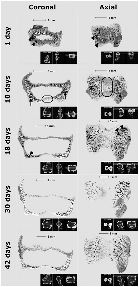
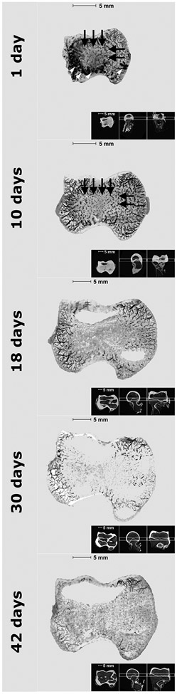
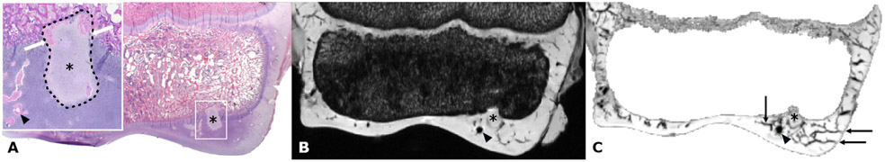
Failure of endochondral ossification due to interruption of the vascular supply to the epiphyseal cartilage is a critical step in the development of osteochondritis dissecans (OCD). Herein we describe the vascular architecture of the distal humeral epiphyseal cartilage in pigs and identify characteristic features that have been associated with sites predisposed to OCD development across species. Distal humeral specimens were harvested from pigs (n = 5, ages = 1, 10, 18, 30, and, 42 days old) and imaged at 9.4T magnetic resonance imaging (MRI) using a 3D gradient recalled echo sequence. The MRI data were processed using a quantitative susceptibility mapping (QSM) pipeline to visualize the vascular architecture. Specimens were also evaluated histologically to identify the presence of ischemic epiphyseal cartilage necrosis (osteochondrosis [OC]-latens) and associated failure of endochondral ossification (OC-manifesta). The QSM data enabled visualization of two distinct vascular beds arising from the perichondrium at the lateral and medial aspects of the distal humeral epiphysis. Elongated vessels originating from these beds coursed axially to supply the lateral and medial thirds of epiphyseal cartilage. At 18 days of age and older, a shift from perichondrial to transosseous blood supply was noted axially, which appeared more pronounced on the lateral side. This shift coincided with histologic identification of OC-latens (30- and 42-day-old specimens) and OC-manifesta (18- and 42-day-old specimens) lesions in the corresponding regions. The vascular anatomy and its evolution at the distal humeral epiphysis closely resembles that previously reported at predilection sites of knee OCD, suggesting a shared pathophysiology between the knee and elbow joints.
Keywords: MRI; OCD; elbow; epiphyseal vasculature.
© 2023 The Authors. Journal of Orthopaedic Research® published by Wiley Periodicals LLC on behalf of Orthopaedic Research Society.
Figures

Similar articles
-
Novel Application of Magnetic Resonance Imaging Demonstrates Characteristic Differences in Vasculature at Predilection Sites of Osteochondritis Dissecans.Am J Sports Med. 2015 Oct;43(10):2522-7. doi: 10.1177/0363546515596410. Epub 2015 Aug 18. Am J Sports Med. 2015. PMID: 26286878 Free PMC article.
-
Identification of Areas of Epiphyseal Cartilage Necrosis at Predilection Sites of Juvenile Osteochondritis Dissecans in Pediatric Cadavers.J Bone Joint Surg Am. 2018 Dec 19;100(24):2132-2139. doi: 10.2106/JBJS.18.00464. J Bone Joint Surg Am. 2018. PMID: 30562294 Free PMC article.
-
Transection of vessels in epiphyseal cartilage canals leads to osteochondrosis and osteochondrosis dissecans in the femoro-patellar joint of foals; a potential model of juvenile osteochondritis dissecans.Osteoarthritis Cartilage. 2013 May;21(5):730-8. doi: 10.1016/j.joca.2013.02.005. Epub 2013 Feb 18. Osteoarthritis Cartilage. 2013. PMID: 23428601
-
Pathogenesis of epiphyseal osteochondrosis.Vet J. 2013 Jul;197(1):3-12. doi: 10.1016/j.tvjl.2013.03.035. Epub 2013 May 4. Vet J. 2013. PMID: 23647656 Review.
-
An Update on the Pathogenesis of Osteochondrosis.Vet Pathol. 2015 Sep;52(5):785-802. doi: 10.1177/0300985815588778. Epub 2015 Jun 16. Vet Pathol. 2015. PMID: 26080832 Review.
Cited by
-
Relationship between the extent of vascular injury and the evolution of surgically induced osteochondrosis lesions in a piglet model.PLoS One. 2024 Aug 8;19(8):e0308641. doi: 10.1371/journal.pone.0308641. eCollection 2024. PLoS One. 2024. PMID: 39116161 Free PMC article.
References
-
- Chambers HG, Shea KG, Anderson AF, Jojo Brunelle TJ, Carey JL, Ganley TJ, Paterno M, Weiss JM, Sanders JO, Watters WC et al. 2012. American academy of orthopaedic surgeons clinical practice guideline on: The diagnosis and treatment of osteochondritis dissecans. J Bone Joint Surg Am. 94(14):1322–1324. - PubMed
-
- Olstad K, Kongsro J, Grindflek E, Dolvik NI. 2014. Ossification defects detected in ct scans represent early osteochondrosis in the distal femur of piglets. J Orthop Res. 32(8):1014–1023. - PubMed
-
- Olstad K, Ekman S, Carlson CS. 2015. An update on the pathogenesis of osteochondrosis. Vet Pathol. 52(5):785–802. - PubMed
Publication types
MeSH terms
Grants and funding
LinkOut - more resources
Full Text Sources
Medical
