. 2023 Dec 11;51(22):12508-12521.
doi: 10.1093/nar/gkad1075.

Structural and biochemical insights into NEIL2's preference for abasic sites

Affiliations

Structural and biochemical insights into NEIL2's preference for abasic sites

Brian E Eckenroth et al. Nucleic Acids Res. .

Abstract

Cellular DNA is subject to damage from a multitude of sources and repair or bypass of sites of damage utilize an array of context or cell cycle dependent systems. The recognition and removal of oxidatively damaged bases is the task of DNA glycosylases from the base excision repair pathway utilizing two structural families that excise base lesions in a wide range of DNA contexts including duplex, single-stranded and bubble structures arising during transcription. The mammalian NEIL2 glycosylase of the Fpg/Nei family excises lesions from each of these DNA contexts favoring the latter two with a preference for oxidized cytosine products and abasic sites. We have determined the first liganded crystal structure of mammalian NEIL2 in complex with an abasic site analog containing DNA duplex at 2.08 Å resolution. Comparison to the unliganded structure revealed a large interdomain conformational shift upon binding the DNA substrate accompanied by local conformational changes in the C-terminal domain zinc finger and N-terminal domain void-filling loop necessary to position the enzyme on the DNA. The detailed biochemical analysis of NEIL2 with an array of oxidized base lesions indicates a significant preference for its lyase activity likely to be paramount when interpreting the biological consequences of variants.


Figures

Graphical Abstract
Graphical Abstract
Figure 1.
Figure 1.
Crystal structure of NEIL2 from Monodelphis domestica bound to DNA. (A) MdoNEIL2 is colored by domain with tan for the N-terminal and brown for the C-terminal domains. Anomalous difference Fourier maps contoured at 3 σ for selenomethionine (orange), gold (maroon), barium (cyan), hexamine cobalt (green) and sulfur/phosphate (blue) used for structure determination. In the boxes are methionines 36 and 39 at the crystallographic two-fold axis, showing peak duplication indicating the dual conformation. The distance measurement of the two rotamers for the equivalent residues in the previously solved unliganded structure PDBID 6VJI are shown for (13). (B) Structure based sequence alignment using Promals3D (63) between HsaNEIL2 and MdoNEIL2. Greyed region is insert D1 and boxed region is insert D2. Rows for the alignment from top to bottom are conservation, HsaNEIL2, MdoNEIL2, PDBID:6vji, consensus sequence, consensus secondary structure.
Figure 2.
Figure 2.
Overall structure of NEIL2 in complex with AP-site containing DNA. Crystal structure of Monodelphis domestica NEIL2 (MdoNEIL2) with dsDNA containing an AP-site analog. (A) NEIL2 is colored blue to red from the N- to C-terminus, the damaged-containing DNA strand in black and complementary strand in grey. The NEIL2 large insert D1 removed for crystallization is indicated by a dashed line (cyan) and insert D2 is indicated (green). (B) The pale blue model is a second conformation of the N-terminal domain in the crystal.
Figure 3.
Figure 3.
(A) Global and local conformational changes are observed in NEIL2 upon binding damaged DNA. Shown are the unliganded MdoNEIL2 (13) (magenta) and DNA-bound (cyan) after least squares superposition of the C-terminal domains with DNA omitted for clarity. Boxed areas indicate local conformational changes between unliganded and DNA-bound. (B) Local conformations after least squares superposition of the N-terminal domains.
Figure 4.
Figure 4.
N-terminal domain interactions between NEIL2 and DNA. (A) NEIL2 has a single loop containing Leu141-Phe142 for filling the void generated by the everted lesion. It also contains a short loop containing Asn182 in coordinating position for bases immediately 3′ to the damage site. Both N-terminal domain conformations converge at the AP-site, as do active site residues. Shown in cyan and dark blue are the two N-terminal domain conformations. The complementary strand would be in the foreground and has been omitted for clarity. (B) The unique insert 2 in NEIL2 coordinates the flipped out orphaned base of the dsDNA complementary strand. The data indicate that this insert is flexible.
Figure 5.
Figure 5.
Interaction between the C-terminal domain and the damaged strand (A) or complementary strand (B). The C-terminal domain and zinc-finger arm bind the backbone of the damaged strand. All residues are conserved between MdoNEIL2 and the human enzyme, except Phe311, which is a conservative change to a bulky hydrophobic leucine residue. The phosphate backbone of the complementary strand (−2 and −3 positions) is bound by His285 and His286 sidechains along with the amide nitrogen of Leu284 and His286. Outside of the orphaned base contact with the base of insert 2 shown in Figure 4B, this is the only significant contact with the complementary strand. Shown is the 2.08 Å resolution MIR (multiple isomorphous replacement) phased map after density modification contoured at 1 σ.
Figure 6.
Figure 6.
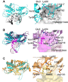
Comparison of NEIL1, 2 and 3 in complex with AP-site containing DNA. NEIL1 and NEIL2 orient dsDNA differently but maintain a similar lesion position in the active site while NEIL3 is bound to a ssDNA hairpin with partial dsDNA in the active site. (A) Shown is NEIL2 (cyan), the AP-site containing strand (black) and complementary strand (grey). (B) Shown is NEIL1 (purple) (PDBID 5ITT) (64), its AP-site containing strand (maroon) and complementary strand (pink). AP-sites for both are highlighted (black or magenta spheres). Zoom panels show NEIL1 orthologs have two void-filling loops, including one with a residue (R118) to stabilize the intrahelical orphaned base whereas NEIL2 has the shorter Asn182 containing loop. (C) Comparison to NEIL3 (orange) in complex with AP-site trapped ssDNA hairpin (PDBID 7Z5A (42)). Asp133 is equivalent to Asn182 in NEIL2 and was proposed to contribute to ssDNA requirement of NEIL3 via charge repulsion. NEIL3 also has a shortened form of this loop similar to NEIL2.
Figure 7.
Figure 7.
Base excision versus AP lyase activity in ssDNA and dsDNA of WT full-length HsaNEIL2 and WT full-length MdoNEIL2 under STO conditions. (A) STO production curves for WT HsaNEIL2 (top) and WT MdoNEIL2 (bottom) processing AP, Gh, 5-OHU and Ug substrates in single strand (left) and duplex (right) DNA. The plots show that amount product formed over the course of 10 min, and the data was fit to single exponential (see Materials and methods). Duplex lesion substrates were placed across from pre-replicative base pairs (dG for 5-OHU and Ug, and dC for Gh), and AP was placed across from dC; data were fitted to a single-exponential equation (see methods), and represent averages of at least three trials from separate enzyme aliquots with three sub-replicates for each aliquot. (B) Relative processing of substrates was gauged by plotting the % product formed at 10 min by WT HsaNEIL2 (left) and WT MdoNEIL2 (right) with AP, Gh, 5-OHU and Ug substrates in single strand and duplex DNA. Bar graphs were created using GraphPad prism 9. (C) Represents structure activity relationships (SAR) for NEIL2 defining substrates versus non-substrates. (D) Shown is the active site of MdoNEIL2 bound to the AP- site (dark grey) with a cytosine modeled in two orientations. The sphere indicates the C5 position. Also shown are the complementary strand (light grey), N-terminal domain (cyan), C-terminal domain (blue). The magenta cytosine is modeled into the catalytic pocket via the compressed minor groove whereas the green cytosine is modeled flipped out towards the major groove.
Figure 8.
Figure 8.
Impact of insert 2 deletion on NEIL2 dsDNA activity under STO conditions. (A) AP lyase activity with WT full-length HsaNEIL2 (circles), WT full-length MdoNEIL2 (upward triangle), MdoNEIL2ΔD1 (square), MdoNEIL2ΔD2 (rhombus), MdoNEIL2ΔD1 Asn182Asp (hexagon) in single strand (left) and duplex across from dC (right). Curves represent the amount of product formed over the course of 10 minutes at various time points under STO conditions; data were fitted to a single exponential equation (see methods), and represent averages of trials of three separate enzyme aliquots, with three sub-replicates for each aliquot. Note, reactions were analyzed within the first 10 minutes to prevent interference due to loss of enzyme activity during the reaction. (B) The total amount of product produced at 10 min for AP lyase activity in single-strand (left) and duplex DNA across from dC (right). Bar graphs represent the average and standard deviation.

Similar articles

Cited by

References

    1. Chatterjee N., Walker G.C.. Mechanisms of DNA damage, repair, and mutagenesis. Environ. Mol. Mutagen. 2017; 58:235–263. - PMC - PubMed
    1. Shen L., Song C.X., He C., Zhang Y.. Mechanism and function of oxidative reversal of DNA and RNA methylation. Annu. Rev. Biochem. 2014; 83:585–614. - PMC - PubMed
    1. Drohat A.C., Coey C.T.. Role of base excision “repair” enzymes in erasing epigenetic marks from DNA. Chem. Rev. 2016; 116:12711–12729. - PMC - PubMed
    1. Sarker A.H., Cooper P.K., Hazra T.K.. DNA glycosylase NEIL2 functions in multiple cellular processes. Prog. Biophys. Mol. Biol. 2021; 164:72–80. - PMC - PubMed
    1. Jacobs A.L., Schar P.. DNA glycosylases: in DNA repair and beyond. Chromosoma. 2012; 121:1–20. - PMC - PubMed