. 2024 Jan 1;10(1):32-41.
doi: 10.1001/jamaoncol.2023.4853.

Nivolumab for Patients With High-Risk Oral Leukoplakia: A Nonrandomized Controlled Trial

Affiliations

Nivolumab for Patients With High-Risk Oral Leukoplakia: A Nonrandomized Controlled Trial

Glenn J Hanna et al. JAMA Oncol. .

Abstract

Importance: Proliferative verrucous leukoplakia (PVL) is an aggressive oral precancerous disease characterized by a high risk of transformation to invasive oral squamous cell carcinoma (OSCC), and no therapies have been shown to affect its natural history. A recent study of the PVL immune landscape revealed a cytotoxic T-cell-rich microenvironment, providing strong rationale to investigate immune checkpoint therapy.

Objective: To determine the safety and clinical activity of anti-programmed cell death 1 protein (PD-1) therapy to treat high-risk PVL.

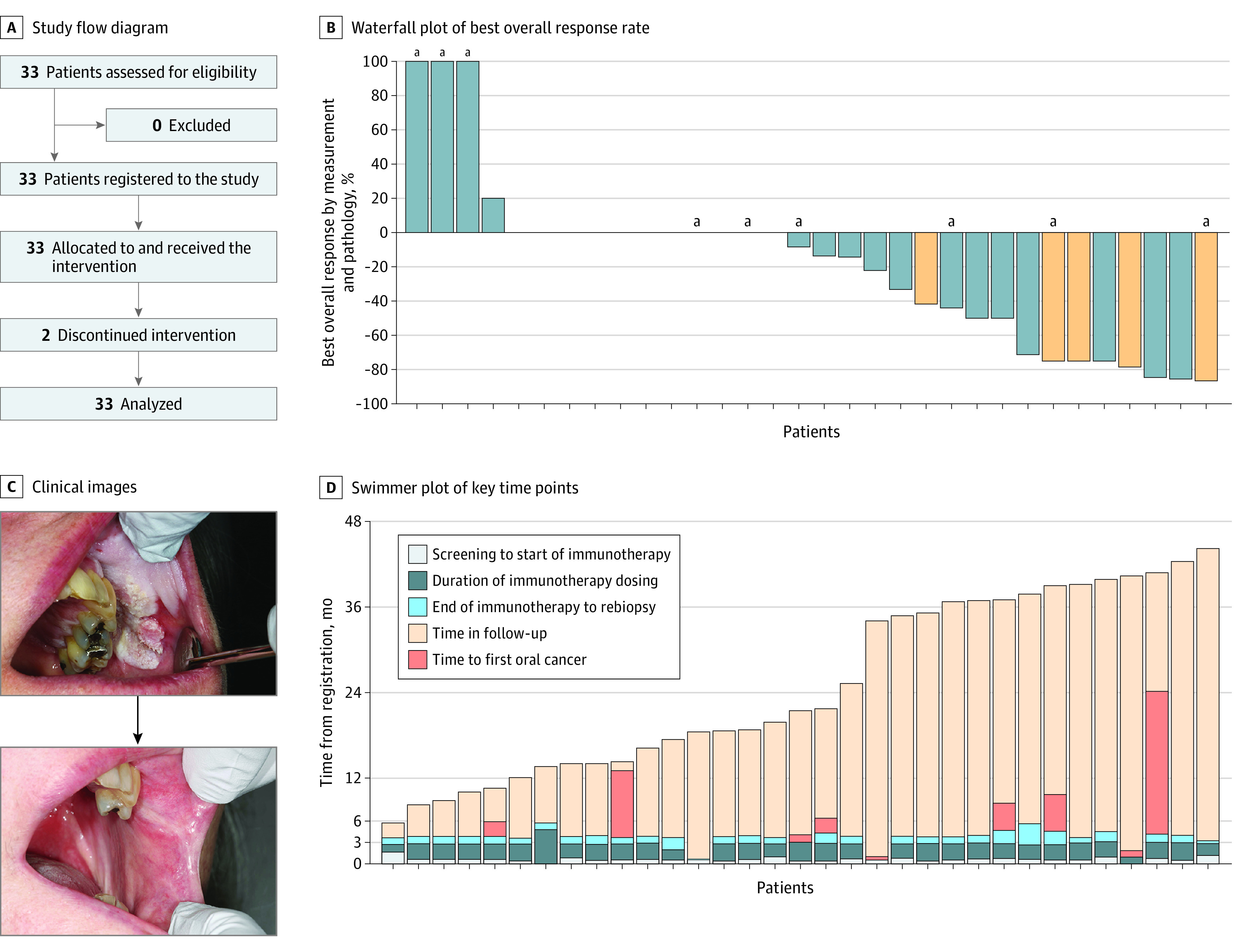
Design, setting, and participants: This nonrandomized, open-label, phase 2 clinical trial was conducted from January 2019 to December 2021 at a single academic medical center; median (range) follow-up was 21.1 (5.4-43.6) months. Participants were a population-based sample of patients with PVL (multifocal, contiguous, or a single lesion ≥4 cm with any degree of dysplasia).

Intervention: Patients underwent pretreatment biopsy (1-3 sites) and then received 4 doses of nivolumab (480 mg intravenously) every 28 days, followed by rebiopsy and intraoral photographs at each visit.

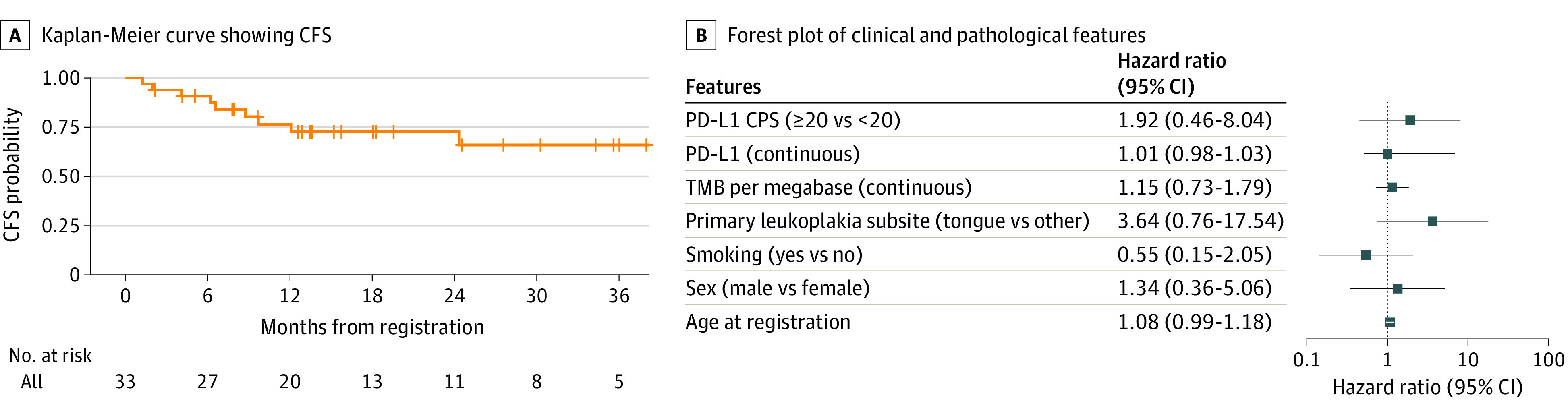
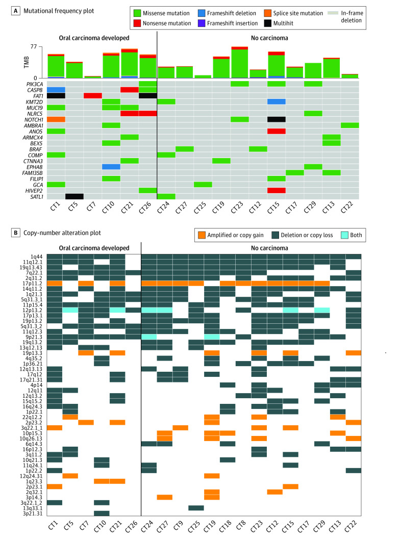
Main outcomes and measures: The primary end point was the change in composite score (size and degree of dysplasia) from before to after treatment (major response [MR]: >80% decrease in score; partial response: 40%-80% decrease). Secondary analyses included immune-related adverse events, cancer-free survival (CFS), PD-1 ligand 1 (PD-L1) expression, 9p21.3 deletion, and other exploratory immunologic and genomic associations of response.

Results: A total of 33 patients were enrolled (median [range] age, 63 [32-80] years; 18 [55%] were female), including 8 (24%) with previously resected early-stage OSCC. Twelve patients (36%) (95% CI, 20.4%-54.8%) had a response by composite score (3 MRs [9%]), 4 had progressive disease (>10% composite score increase, or cancer). Nine patients (27%) developed OSCC during the trial, with a 2-year CFS of 73% (95% CI, 53%-86%). Two patients (6%) discontinued because of toxic effects; 7 (21%) experienced grade 3 to 4 immune-related adverse events. PD-L1 combined positive scores were not associated with response or CFS. Of 20 whole-exome sequenced patients, all 6 patients who had progression to OSCC after nivolumab treatment exhibited 9p21.3 somatic copy-number loss on pretreatment biopsy, while only 4 of the 14 patients (29%) who did not develop OSCC had 9p21.3 loss.

Conclusions and relevance: This immune checkpoint therapy precancer nonrandomized clinical trial met its prespecified response end point, suggesting potential clinical activity for nivolumab in high-risk PVL. Findings identified immunogenomic associations to inform future trials in this precancerous disease with unmet medical need that has been difficult to study.

Trial registration: ClinicalTrials.gov Identifier: NCT03692325.


Conflict of interest statement

Conflict of Interest Disclosures: Dr Hanna reported grants from Bristol Myers Squibb (BMS) during the conduct of the study; grants from Bicara, Exicure, Gateway for Cancer Research, GSK, ImmunityBio, Kite Pharma, KSQ Therapeutics, Regeneron, Repertoire, Sanofi Genzyme, and Merck; and personal fees from Replimune and Surface Oncology outside the submitted work. Dr Villa reported grants from BMS during the conduct of the study; and serving as a consultant for Merck, Lipella Pharmaceuticals, and AFYX Therapeutics outside the submitted work. Dr Nandi reported a provisional patent under EFS_ID: 46746025, pending, regarding this work. Dr Rettig reported nonfinancial support from Naveris outside the submitted work. Dr Jo reported that her spouse works for Merck and Co as an Executive Director of Clinical Research and receives salary and benefits (including stock options). Dr Uppaluri reported personal fees from Merck, Inc (advisory board) outside the submitted work. Dr Haddad reported personal fees from Coherus, Scholar Rock, Bluedot Bio, EMD Serono, Merck, BMS, Pfizer, GSK, Boehringer Ingelheim, Eisai, Bayer, AstraZeneca, Kura, National Comprehensive Cancer Network, Nanobiotix, ISA, Bayer, and Mirati outside the submitted work. Dr Cohen reported personal fees from MSD outside the submitted work. Dr Alexandrov reported personal fees from io9 LLC and Genome Insight outside the submitted work; in addition, Dr Alexandrov had a patent for 63/412,835, pending; and Dr Alexandrov is also an inventor of a US Patent 10,776,718 for source identification by non-negative matrix factorization; Dr Alexandrov also declares US provisional applications with serial numbers: 63/366,392; 63/367,846; 63/289,601; 63/269,033; 63/483,237; and 63/492,348. Dr William reported personal fees from BMS, Roche, AstraZeneca, Merck, Pfizer, Novartis, Eli Lilly, Takeda, Janssen, Sanofi, and Seagen, and other from io9 (scientific advisory board) during the conduct of the study. Dr Lippman reported a patent for PCT/US 63/412,835 SD2023-089, pending. Dr Woo reported grants from BMS during the conduct of the study. No other disclosures were reported.

Figures

Figure 1.
Figure 1.. Clinical and Pathologic Response
A, Study flow diagram. B, Waterfall plot showing best overall response rate to up to 4 doses of immunotherapy (nivolumab) in patients with high-risk oral proliferative verrucous leukoplakia. Response was determined by the relative change in composite score determined pre- to posttherapy using bidimensional lesion measurements and biopsy degree of dysplasia (mild, moderate, severe). A decrease by greater than 80% equated to a major response, a decrease by 40% to 80% was a partial response, and an increase by 10% or more or new carcinoma in situ or oral squamous cell carcinoma development was deemed progressive disease. Orange bars indicate patients with pretreatment epithelial dysplasia tissue demonstrating a programmed cell death 1 ligand 1 combined positive score of 20 or greater. C, Photographs of a responder (top, pretreatment; bottom, posttreatment) showing a 4 × 3 cm verrucous lesion contiguous with a left maxillary gingival sulcus lesion defined by proliferative and hyperkeratotic change with inferior erythematous friability at a heaped-up border, which resolved postimmunotherapy. Of note, teeth No. 25 to 27 were extracted prior to the posttreatment photo being taken. D, Swimmer plot showing key time points throughout the trial duration and follow-up period. Each bar represents an individual participant. All patients were alive at last follow-up. aDeveloped oral cancer.
Figure 2.
Figure 2.. Survival Outcomes
A, Kaplan-Meier curve showing cancer-free survival (CFS) reported in months from the time of trial registration to the first of oral squamous cell carcinoma, death, or censored at last follow-up. B, Forest plot showing the hazard ratio and 95% CIs (log 10) of the association of clinical and pathologic variables with CFS (hazard ratio >1 indicates higher risk of CFS event). Univariate Cox proportional hazard model. CPS indicates combined positive score; PD-L1, programmed cell death 1 ligand 1; TMB, tumor mutational burden.
Figure 3.
Figure 3.. Genomic Associations of Response and Survival
A, Mutational frequency plot with each column representing an individual dysplastic sample (sample IDs noted, CT#). Genes are arranged top to bottom by mutational frequency. Tumor mutational burden (TMB) for dysplastic samples is plotted in the bar graph above the mutational plot. Only the top 20 most frequently altered genes are displayed. B, Copy-number alteration plot demonstrating allelic imbalance (amplifications, deletions, or both [polyploidy]) among pretreatment oral dysplastic epithelial samples prior to treatment with 4 doses of nivolumab arranged by those patients who developed oral carcinoma (left) vs not (at last known follow-up).

Comment in

References

    1. Mello FW, Miguel AFP, Dutra KL, et al. . Prevalence of oral potentially malignant disorders: a systematic review and meta-analysis. J Oral Pathol Med. 2018;47(7):633-640. doi:10.1111/jop.12726 - DOI - PubMed
    1. Chaturvedi AK, Udaltsova N, Engels EA, et al. . Oral leukoplakia and risk of progression to oral cancer: a population-based cohort study. J Natl Cancer Inst. 2020;112(10):1047-1054. doi:10.1093/jnci/djz238 - DOI - PMC - PubMed
    1. Speight PM, Khurram SA, Kujan O. Oral potentially malignant disorders: risk of progression to malignancy. Oral Surg Oral Med Oral Pathol Oral Radiol. 2018;125(6):612-627. doi:10.1016/j.oooo.2017.12.011 - DOI - PubMed
    1. Torrejon-Moya A, Jané-Salas E, López-López J. Clinical manifestations of oral proliferative verrucous leukoplakia: a systematic review. J Oral Pathol Med. 2020;49(5):404-408. doi:10.1111/jop.12999 - DOI - PubMed
    1. Villa A, Menon RS, Kerr AR, et al. . Proliferative leukoplakia: proposed new clinical diagnostic criteria. Oral Dis. 2018;24(5):749-760. doi:10.1111/odi.12830 - DOI - PubMed

Associated data