. 2024 Jan 9;9(1):e173440.
doi: 10.1172/jci.insight.173440.

Efficient pulmonary lymphatic drainage is necessary for inflammation resolution in ARDS

Affiliations

Efficient pulmonary lymphatic drainage is necessary for inflammation resolution in ARDS

Pu-Hong Zhang et al. JCI Insight. .

Abstract

The lymphatic vasculature is the natural pathway for the resolution of inflammation, yet the role of pulmonary lymphatic drainage function in sepsis-induced acute respiratory distress syndrome (ARDS) remains poorly characterized. In this study, indocyanine green-near infrared lymphatic living imaging was performed to examine pulmonary lymphatic drainage function in septic mouse models. We found that the pulmonary lymphatic drainage was impaired owing to the damaged lymphatic structure in sepsis-induced ARDS. Moreover, prior lymphatic defects by blocking vascular endothelial growth factor receptor-3 (VEGFR-3) worsened sepsis-induced lymphatic dysfunction and inflammation. Posttreatment with vascular endothelial growth factor-C (Cys156Ser) (VEGF-C156S), a ligand of VEGFR-3, ameliorated lymphatic drainage by rejuvenating lymphatics to reduce the pulmonary edema and promote draining of pulmonary macrophages and neutrophils to pretracheal lymph nodes. Meanwhile, VEGF-C156S posttreatment reversed sepsis-inhibited CC chemokine ligand 21 (CCL21), which colocalizes with pulmonary lymphatic vessels. Furthermore, the advantages of VEGF-C156S on the drainage of inflammatory cells and edema fluid were abolished by blocking VEGFR-3 or CCL21. These results suggest that efficient pulmonary lymphatic drainage is necessary for inflammation resolution in ARDS. Our findings offer a therapeutic approach to sepsis-induced ARDS by promoting lymphatic drainage function.

Keywords: Pulmonology; Respiration; Vascular Biology.


Conflict of interest statement

Conflict of interest: The authors have declared that no conflict of interest exists.

Figures

Figure 1
Figure 1. The pulmonary lymphatic drainage function is impaired in an LPS-induced sepsis model.
(A) ICG was intratracheally poured into unilateral lung, and ICG drained from the pulmonary alveoli into the pretracheal lymph nodes (pLNs). (B) Procedure and timeline: At 6 hours after the intraperitoneal injection of LPS (10 mg/kg) or the CLP, the sepsis model was induced. ICG (1 mg/mL, 10 μL) was intratracheally poured into unilateral lung. Fluorescence intensities of ICG were determined at 0 hours, 24th hour, and 48th hour using an IVIS associated with the clearance rate to reflect the effect of tissue fluid clearance by lymphatic flow. The lung tissue samples were collected at the 24th hour. (C and D) ICG was intratracheally poured into unilateral lung and quantified and presented as relative radiance (photons/s per cm2 /steradian) at 0 hours, 24th hour, and 48th hour using IVIS in LPS-induced sepsis model. (E and F) The ICG clearance rate in 24th hour and 48th hour (control n = 6, LPS n = 7; representative data from 3 independent experiments). (G and H) Fluorescence intensities of ICG were determined in pLNs at 30 minutes after the pour of ICG by IVIS and confocal microscopy in LPS-induced sepsis model at the 24th hour (control n = 6, LPS n = 6; representative data from 3 independent experiments). All n values refer to the number of mice used, and the error bars depict mean ± SD. P values were calculated by 2-tailed paired or unpaired Student’s t test.
Figure 2
Figure 2. The pulmonary lymphatic drainage function is impaired in a CLP-induced sepsis model.
(A and B) ICG was intratracheally poured into unilateral lung and quantified and presented at 0 hours, 24th hour, and 48th hour using IVIS in CLP-induced sepsis model. (C and D) The ICG clearance rate at 24th hour and 48th hour (sham n = 6, CLP n = 6; representative data from 3 independent experiments). (E and F) Fluorescence intensities of ICG were determined in pLNs at 30 minutes after the pour of ICG by IVIS and confocal microscopy in CLP-induced sepsis model at 24th hour (sham n = 6, CLP n = 6; representative data from 3 independent experiments). All n values refer to the number of mice used, and the error bars depict mean ± SD. P values were calculated by 2-tailed paired or unpaired Student’s t test.
Figure 3
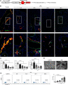
Figure 3. The pulmonary lymphatic vessels are damaged in an LPS-induced sepsis model.
(A) Schematic representation of a conditional, lymph-specific fluorescence mouse model (Prox1-CreERT2+ Rosa26-tdTomato+). Prox1-CreERT2 mice were crossed with Rosa26-tdTomato mice and treated with tamoxifen for 4 consecutive days before initiation of sepsis studies. (B) The pulmonary lymphatic vessels were labeled with an immunofluorescence stain of VEGFR-3 (green) and Prox1-tdTomato (red) signals from Prox1-CreERT2+ R26-tdTomato+ mice after formation of sepsis. Scale bars, 100 μm (top), 50 μm (bottom). (C) Quantification of the percentage area coverage, the relative fluorescence intensity, and the diameter of lymphatic vessels (n = 9 per group; representative data from 3 independent experiments). (D) Scanning electron microscopy of the pulmonary lymphatic endothelium. Scale bars, 1 μm. (E) Representative flow cytometry images and the quantification of dead lymphatic endothelial cells (LECs) from lung suspension. The frame showed the percentage of annexin V+ LECs. (n = 9 per group; representative data from 3 independent experiments.) All n values refer to the number of mice used, and the error bars depict mean ± SD. P values were calculated by a 1-way ANOVA with Tukey’s multiple-comparison test.
Figure 4
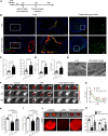
Figure 4. Posttreatment with VEGF-C156S ameliorated pulmonary lymphatic drainage function by rejuvenating lymphatics in LPS-induced sepsis.
(A) Procedure and timeline: Recombinant VEGF-C156S protein was administrated to an LPS-induced sepsis model 6 hours afterward. Then, the lung tissue and the pLNs were obtained at the 24th hour. (B) Lymphatic vessels were labeled with an immunofluorescence stain of VEGFR-3 (green) and Prox1-tdTomato signals (red). Scale bars for lung, 100 μm (left), 50 μm (right). Scale bar for pLNs, 50 μm. (C) Quantification of the percentage area coverage and the relative fluorescence intensity of lymphatic vessels in lungs (LPS = 9, LPS + VEGF-C156S = 9; representative data from 3 independent experiments). (D) Quantification of the percentage area coverage and the relative fluorescence intensity of lymphatic vessels in pLNs (LPS = 9, LPS + VEGF-C156S = 9; representative data from 3 independent experiments). (E) Scanning electron microscopy of the pulmonary lymphatic endothelium. Scale bar, 1 μm. (F and G) ICG was intratracheally poured into unilateral lung and quantified and presented as relative radiance at 0 hours, 24th hour, and 48th hour using IVIS. (H and I) The ICG clearance rate at the 24th hour and 48th hour (control n = 6, LPS n = 6, LPS + VEGF-C156S n = 6; representative data from 3 independent experiments). (J and K) At the 24th hour, ICG was intratracheally poured into unilateral lung. Fluorescence intensities of ICG were determined in pLNs at 30 minutes after the pour by IVIS and confocal microscopy (control n = 6, LPS n = 6, LPS + VEGF-C156S n = 6; representative data from 3 independent experiments). Scale bar, 200 μm. All n values refer to the number of mice used, and the error bars depict mean ± SD. P values were calculated by a 1-way ANOVA with Tukey’s multiple-comparison test.
Figure 5
Figure 5. VEGF-C156S posttreatment promoted pulmonary inflammatory cells draining to pLNs in LPS-induced sepsis.
Recombinant VEGF-C156S protein was administrated to LPS-induced sepsis model. Then, the lung tissue and the pLNs were obtained at the 24th hour. (A and B) Representative immunofluorescence images and quantification of F4/80+ cells (red, macrophages) and LY6G+ cells (green, neutrophil) in lung sections and pLN sections (LPS = 9, LPS + VEGF-C156S = 9; representative data from 3 independent experiments). Scale bars, 50 µm. (C and D) Representative flow cytometry images and quantification of F4/80+/CD45.2+ cells (macrophages) and LY6G+ cells (neutrophils) in lung tissue and pLNs. The frame showed the percentage of F4/80+/CD45.2+ cells or LY6G+ cells. (LPS = 9, LPS + VEGF-C156S = 9; representative data from 3 independent experiments). All n values refer to the number of mice used, and the error bars depict mean ± SD. P values were calculated by 2-tailed unpaired Student’s t test.
Figure 6
Figure 6. VEGF-C156S posttreatment promotes pulmonary inflammation resolution in LPS-induced sepsis.
(A) The concentrations of inflammatory factors in lung tissue homogenate or the serum, such as IL-1β, TNF-α, MPO, and IL-6, were measured by ELISA (n = 9~10; representative data from 3 independent experiments). (B) Wet-to-dry ratios for lungs (LPS = 9, LPS + VEGF-C156S = 9; representative data from 3 independent experiments). (C) Representative images of lung H&E-stained sections and the acute lung injury scores (LPS = 9, LPS + VEGF-C156S = 9; representative data from 3 independent experiments). Box plots show the interquartile range (box), median (line), and minimum and maximum (whiskers). (D) The survival curve. Mice received a single intraperitoneal injection with a lethal dose of LPS (40 mg/kg of body weight), followed by a single tail vein injection of VEGF-C156S (0.1 μg/g of body weight, 6 hours apart; LPS n = 10, LPS + VEGF-C156S n = 10). All n values refer to the number of mice used, and the error bars depict mean ± SD. P values were calculated by 2-tailed unpaired Student’s t test. The lung injury scores were shown as median (quartile) [M (P25, P75)] and analyzed by using the Kruskal-Wallis test. The survival rate in each subgroup was estimated by Kaplan-Meier survival curves and compared by the pairwise log-rank test.
Figure 7
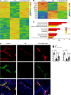
Figure 7. VEGF-C156S ameliorates LPS-inhibited CCL21 in pulmonary lymphatic vessels during sepsis.
(A) Heatmap of DEGs after VEGF-C156S administration in LPS-induced sepsis model (up, 168; down, 199; power > 0.4). (B and C) Gene sets involved in inflammatory response, lymphatic remodeling, body fluid drainage, as well as immunological response as shown by the representative upregulated pathways after VEGF-C156S administration. (D) Prox1-tdTomato (red) and CCL21 (green) were colocalized in lung sections. Quantification of the percentage area coverage and the relative intensity of CCL21 staining (n = 6 per group; representative data from 3 independent experiments). All n values refer to the number of mice used, and the error bars depict mean ± SD. P values were calculated by a 1-way ANOVA with Tukey’s multiple-comparison test.
Figure 8
Figure 8. Enhancement of lymphatic drainage by VEGF-C156S is dependent on the VEGFR-3/CCL21 pathway.
(A) Monitoring and treatment scheme. MAZ51 was intraperitoneally injected at 10 mg/kg of body weight for 30 days (5 days per week) for blocking VEGFR-3. CCL21 was blocked on days 2, 4, and 6 by CCL21-blocking antibody (αCCL21). VEGF-C156S was administrated to LPS-induced sepsis model following the administration of anti-CCL21 (αCCL21)/IgG (Iso) antibodies or MAZ51/Vector. (BE) The lymphatic drainage function was determined by IVIS. The remaining ICG was quantified and presented as relative radiance at 0 hours, 24th hour, and 48th hour. The ICG clearance rate at the 24th hour and 48th hour (Vector Iso n = 6, MAZ51 n = 6; αCCL21 n = 6; representative data from 3 independent experiments). (F and G) Fluorescence intensities of ICG were determined in the pLNs at 30 minutes after the pour of ICG by IVIS and confocal microscopy at the 24th hour (Vector Iso n = 6, MAZ51 n = 6; αCCL21 n = 6; representative data from 3 independent experiments). All n values refer to the number of mice used, and the error bars depict mean ± SD. P values were calculated by a 1-way ANOVA with Tukey’s multiple-comparison test.
Figure 9
Figure 9. Enhancement of inflammation resolution by VEGF-C156S was dependent on VEGFR-3/CCL21 pathway.
(A and B) Representative flow cytometry images and quantification of F4/80+/CD45.2+ cells and LY6G+ cells in lung and pLNs. (Vector Iso n = 9, MAZ51 n = 9; αCCL21 n = 9; representative data from 3 independent experiments). (C) Representative images of lung H&E-stained sections and the acute lung injury scores (Vector Iso n = 9, MAZ51 n = 9; αCCL21 n = 9; representative data from 3 independent experiments). The lung injury scores were shown as median (quartile) [M (P25, P75)] and analyzed by using the Kruskal-Wallis test. (D) Wet-to-dry ratios for lungs (Vector Iso n = 9, MAZ51 n = 9; αCCL21 n = 9; representative data from 3 independent experiments). Box plots show the interquartile range (box), median (line), and minimum and maximum (whiskers). All n values refer to the number of mice used, and the error bars depict mean ± SD. P values were calculated by a 1-way ANOVA with Tukey’s multiple-comparison test. (E) Survival of mouse with vehicle or VEGF-C156S treatment sepsis mice following the administration of anti-CCL21 (αCCL21)/IgG (Iso) antibodies, or MAZ51/Vector, were detected. Survival in each subgroup was estimated by Kaplan-Meier survival curves and compared by the pairwise log-rank test (n = 15 per group).

References

    1. Guillen-Guio B, et al. Sepsis-associated acute respiratory distress syndrome in individuals of European ancestry: a genome-wide association study. Lancet Respir Med. 2020;8(3):258–266. doi: 10.1016/S2213-2600(19)30368-6. - DOI - PMC - PubMed
    1. Thompson BT, et al. Acute respiratory distress syndrome. N Engl J Med. 2017;377(6):562–572. doi: 10.1056/NEJMra1608077. - DOI - PubMed
    1. Griffiths M, Evans T. Inhaled nitric oxide therapy in adults. N Engl J Med. 2005;353(25):2683–2695. doi: 10.1056/NEJMra051884. - DOI - PubMed
    1. Steinberg K, et al. Efficacy and safety of corticosteroids for persistent acute respiratory distress syndrome. N Engl J Med. 2006;354(16):1671–1684. doi: 10.1056/NEJMoa051693. - DOI - PubMed
    1. Levy B, Serhan C. Resolution of acute inflammation in the lung. Annu Rev Physiol. 2014;76:467–492. doi: 10.1146/annurev-physiol-021113-170408. - DOI - PMC - PubMed

Publication types

MeSH terms

Substances