High Throughput Tomography (HiTT) on EMBL beamline P14 on PETRA III
- PMID: 37971957
- PMCID: PMC10833423
- DOI: 10.1107/S160057752300944X
High Throughput Tomography (HiTT) on EMBL beamline P14 on PETRA III
Abstract
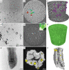
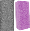
Here, high-throughput tomography (HiTT), a fast and versatile phase-contrast imaging platform for life-science samples on the EMBL beamline P14 at DESY in Hamburg, Germany, is presented. A high-photon-flux undulator beamline is used to perform tomographic phase-contrast acquisition in about two minutes which is linked to an automated data processing pipeline that delivers a 3D reconstructed data set less than a minute and a half after the completion of the X-ray scan. Combining this workflow with a sophisticated robotic sample changer enables the streamlined collection and reconstruction of X-ray imaging data from potentially hundreds of samples during a beam-time shift. HiTT permits optimal data collection for many different samples and makes possible the imaging of large sample cohorts thus allowing population studies to be attempted. The successful application of HiTT on various soft tissue samples in both liquid (hydrated and also dehydrated) and paraffin-embedded preparations is demonstrated. Furthermore, the feasibility of HiTT to be used as a targeting tool for volume electron microscopy, as well as using HiTT to study plant morphology, is demonstrated. It is also shown how the high-throughput nature of the work has allowed large numbers of `identical' samples to be imaged to enable statistically relevant sample volumes to be studied.
Keywords: X-ray tomography; beamlines; biology; phase-contrast imaging.
open access.
Figures


Similar articles
-
BioSAXS Sample Changer: a robotic sample changer for rapid and reliable high-throughput X-ray solution scattering experiments.Acta Crystallogr D Biol Crystallogr. 2015 Jan 1;71(Pt 1):67-75. doi: 10.1107/S1399004714026959. Epub 2015 Jan 1. Acta Crystallogr D Biol Crystallogr. 2015. PMID: 25615861 Free PMC article.
-
Automation of the EMBL Hamburg protein crystallography beamline BW7B.J Synchrotron Radiat. 2004 Sep 1;11(Pt 5):372-7. doi: 10.1107/S090904950401516X. Epub 2004 Aug 17. J Synchrotron Radiat. 2004. PMID: 15310952
-
P13, the EMBL macromolecular crystallography beamline at the low-emittance PETRA III ring for high- and low-energy phasing with variable beam focusing.J Synchrotron Radiat. 2017 Jan 1;24(Pt 1):323-332. doi: 10.1107/S1600577516016465. Epub 2017 Jan 1. J Synchrotron Radiat. 2017. PMID: 28009574 Free PMC article.
-
Designing a synchrotron micro-focusing beamline for macromolecular crystallography.Postepy Biochem. 2016;62(3):395-400. Postepy Biochem. 2016. PMID: 28132495 Review. English.
-
[SPring-8 structural biology beamline].Yakugaku Zasshi. 2010 May;130(5):649-55. doi: 10.1248/yakushi.130.649. Yakugaku Zasshi. 2010. PMID: 20460859 Review. Japanese.
Cited by
-
Present and future structural biology activities at DESY and the European XFEL.J Synchrotron Radiat. 2025 Mar 1;32(Pt 2):474-485. doi: 10.1107/S1600577525000669. Epub 2025 Feb 18. J Synchrotron Radiat. 2025. PMID: 39964790 Free PMC article.
-
Applications of synchrotron light in seed research: an array of x-ray and infrared imaging methodologies.Front Plant Sci. 2025 Feb 17;15:1395952. doi: 10.3389/fpls.2024.1395952. eCollection 2024. Front Plant Sci. 2025. PMID: 40034948 Free PMC article.
-
Current and future perspectives for structural biology at the Grenoble EPN campus: a comprehensive overview.J Synchrotron Radiat. 2025 May 1;32(Pt 3):577-594. doi: 10.1107/S1600577525002012. Epub 2025 Apr 14. J Synchrotron Radiat. 2025. PMID: 40226912 Free PMC article. Review.
-
Integrated 3D imaging of FFPE lung tissue combining microCT, light and electron microscopy allows for contextualized ultrastructural and histological analysis.Sci Rep. 2025 May 28;15(1):18656. doi: 10.1038/s41598-025-02770-w. Sci Rep. 2025. PMID: 40437062 Free PMC article.
-
Studies of Fractal Microstructure in Nanocarbon Polymer Composites.Polymers (Basel). 2024 May 10;16(10):1354. doi: 10.3390/polym16101354. Polymers (Basel). 2024. PMID: 38794548 Free PMC article.
References
-
- Albers, J., Pacilé, S., Markus, M. A., Wiart, M., Vande Velde, G., Tromba, G. & Dullin, C. (2018). Mol. Imaging Biol. 20, 732–741. - PubMed
-
- Bartkiewicz, P. & Duval, P. (2007). Meas. Sci. Technol. 18, 2379–2386.
-
- Cipriani, F., Felisaz, F., Lavault, B., Brockhauser, S., Ravelli, R., Launer, L., Leonard, G. & Renier, M. (2007). AIP Conf. Proc. 879, 1928–1931.
-
- Cloetens, P., Ludwig, W., Baruchel, J., Guigay, J.-P., Pernot-Rejmánková, P., Salomé-Pateyron, M., Schlenker, M., Buffière, J.-Y., Maire, E. & Peix, G. (1999). J. Phys. D Appl. Phys. 32, A145–A151.
MeSH terms
Grants and funding
LinkOut - more resources
Full Text Sources
Research Materials

