. 2023 Nov 21;120(47):e2305215120.
doi: 10.1073/pnas.2305215120. Epub 2023 Nov 16.

A peptidomimetic modulator of the CaV2.2 N-type calcium channel for chronic pain

Affiliations

A peptidomimetic modulator of the CaV2.2 N-type calcium channel for chronic pain

Kimberly Gomez et al. Proc Natl Acad Sci U S A. .

Abstract

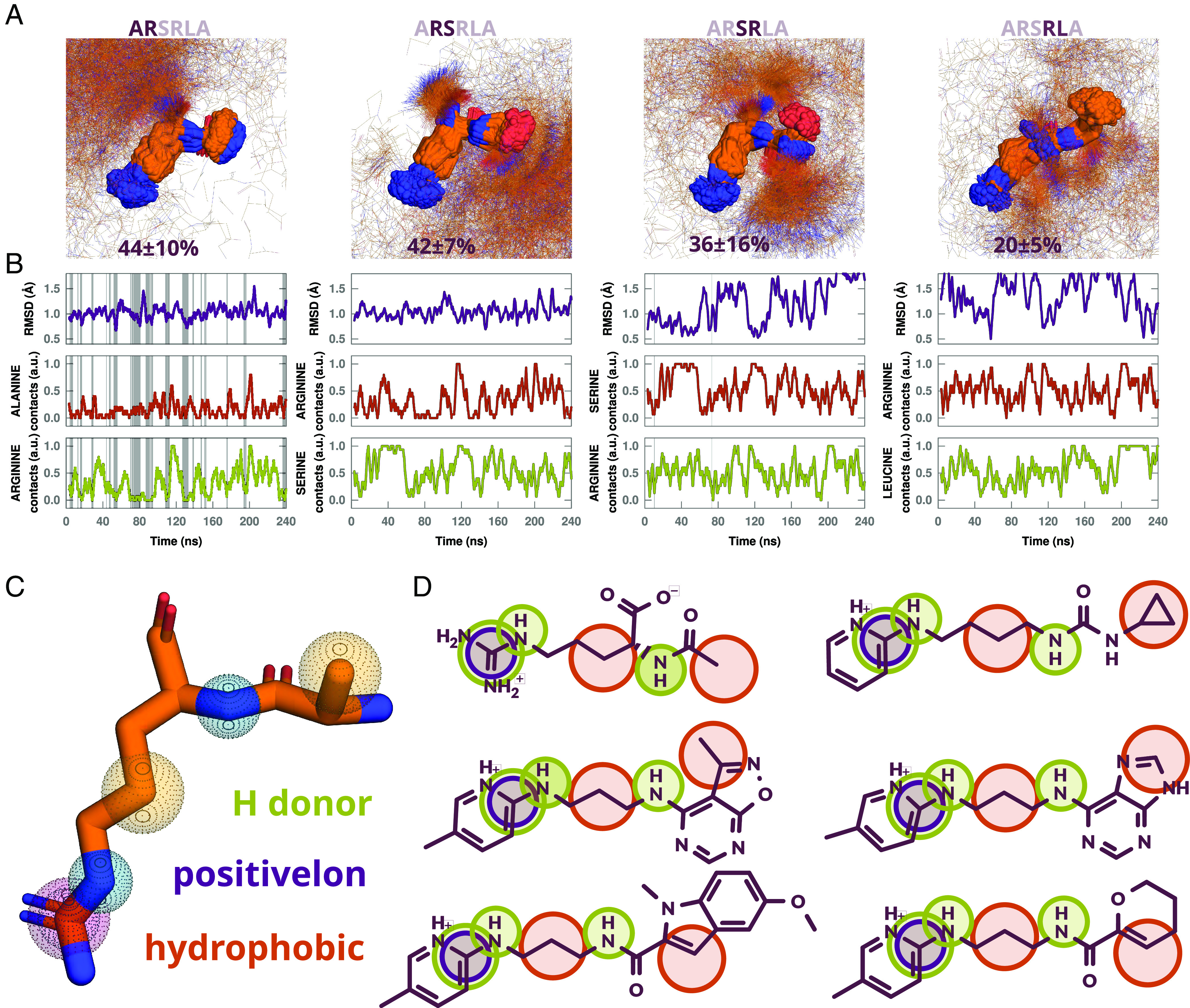
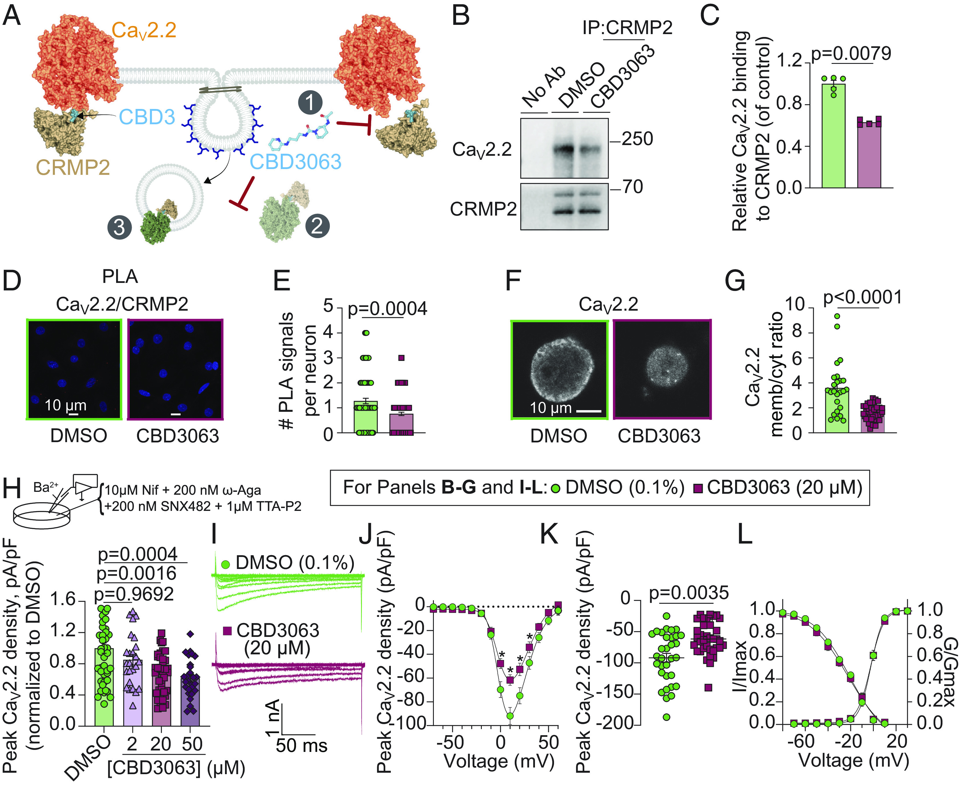
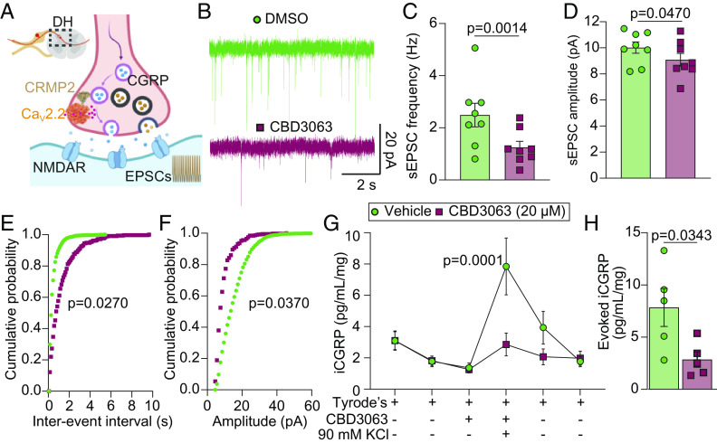
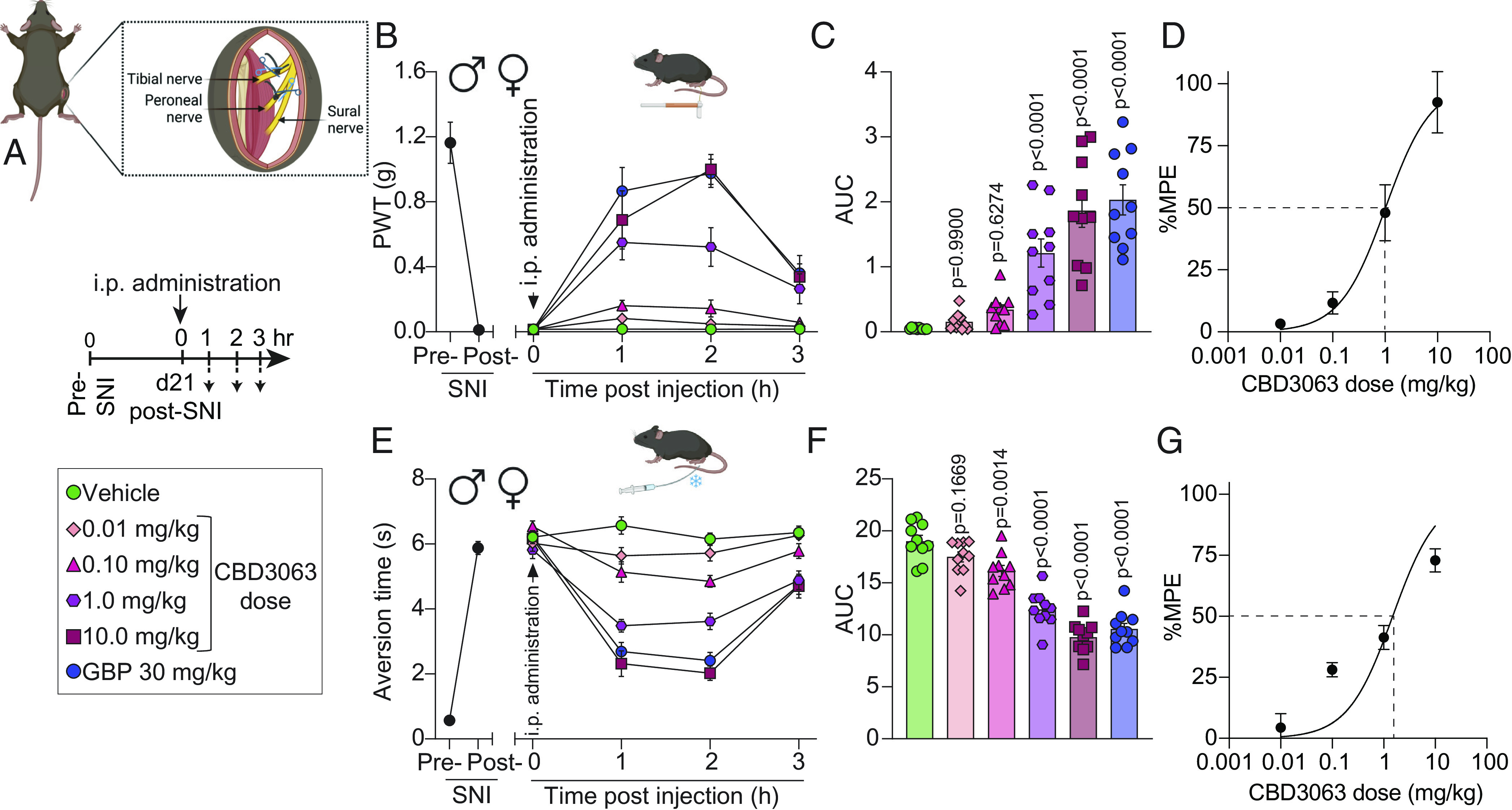
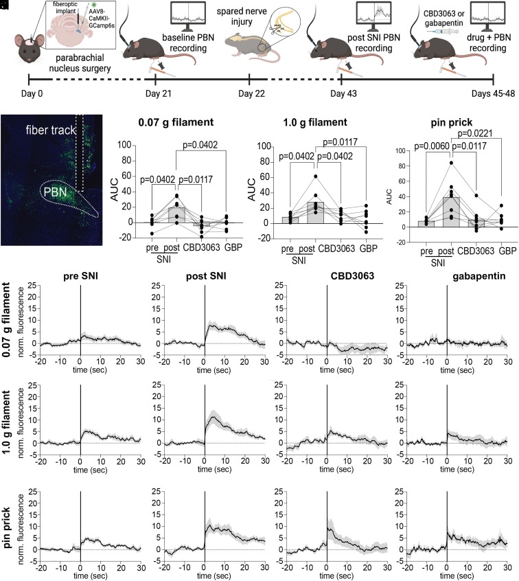
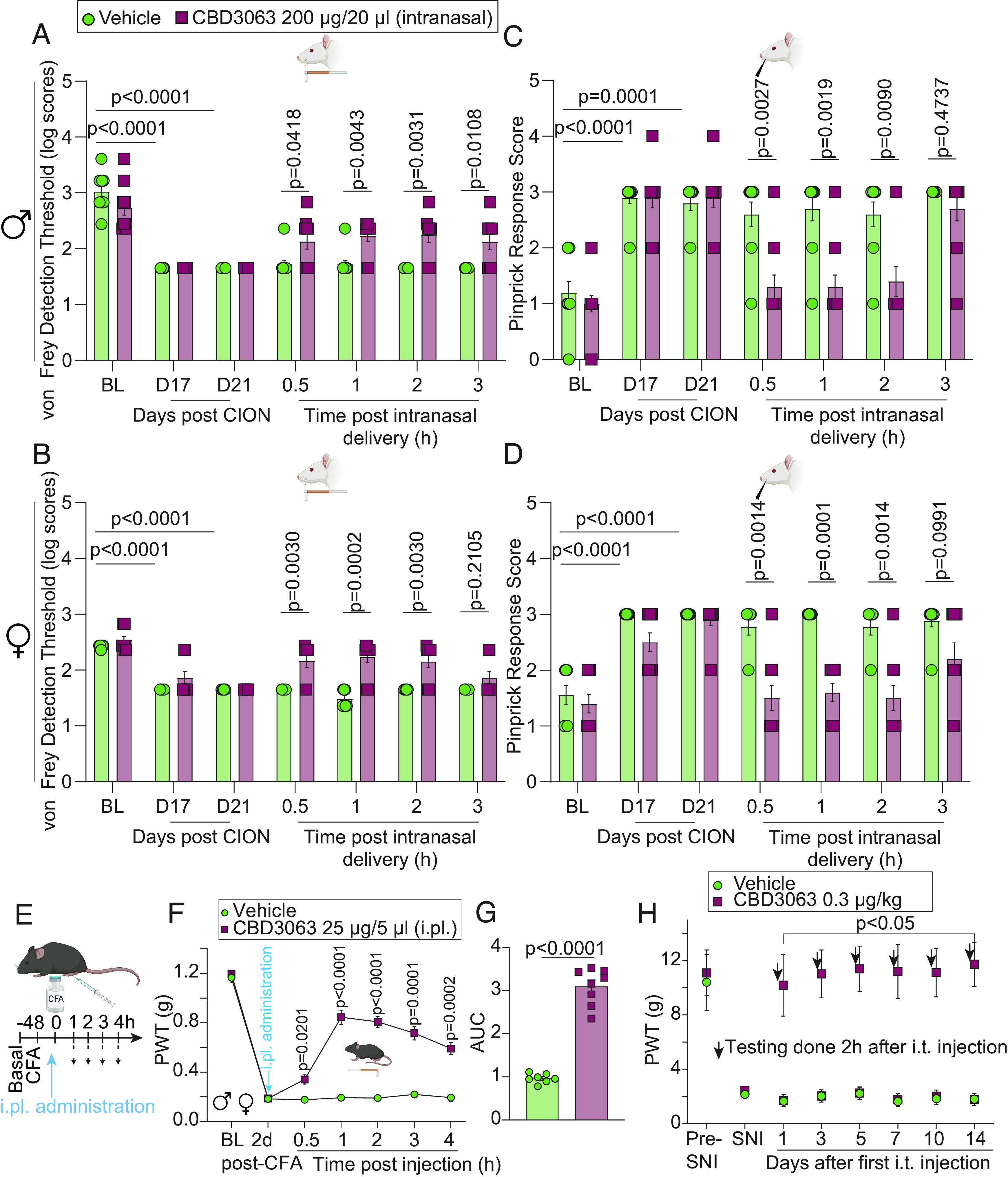
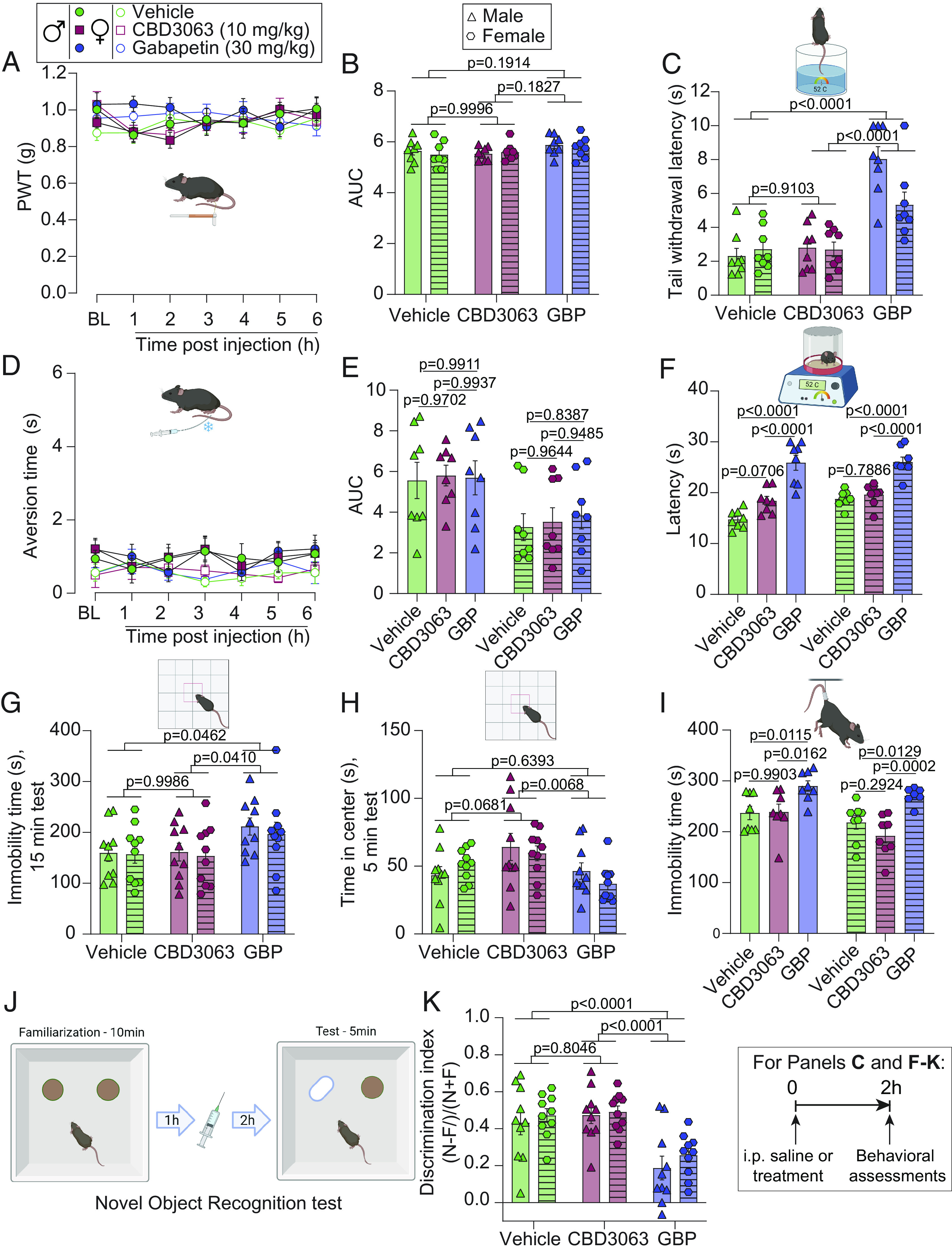
Transmembrane Cav2.2 (N-type) voltage-gated calcium channels are genetically and pharmacologically validated, clinically relevant pain targets. Clinical block of Cav2.2 (e.g., with Prialt/Ziconotide) or indirect modulation [e.g., with gabapentinoids such as Gabapentin (GBP)] mitigates chronic pain but is encumbered by side effects and abuse liability. The cytosolic auxiliary subunit collapsin response mediator protein 2 (CRMP2) targets Cav2.2 to the sensory neuron membrane and regulates their function via an intrinsically disordered motif. A CRMP2-derived peptide (CBD3) uncouples the Cav2.2-CRMP2 interaction to inhibit calcium influx, transmitter release, and pain. We developed and applied a molecular dynamics approach to identify the A1R2 dipeptide in CBD3 as the anchoring Cav2.2 motif and designed pharmacophore models to screen 27 million compounds on the open-access server ZincPharmer. Of 200 curated hits, 77 compounds were assessed using depolarization-evoked calcium influx in rat dorsal root ganglion neurons. Nine small molecules were tested electrophysiologically, while one (CBD3063) was also evaluated biochemically and behaviorally. CBD3063 uncoupled Cav2.2 from CRMP2, reduced membrane Cav2.2 expression and Ca2+ currents, decreased neurotransmission, reduced fiber photometry-based calcium responses in response to mechanical stimulation, and reversed neuropathic and inflammatory pain across sexes in two different species without changes in sensory, sedative, depressive, and cognitive behaviors. CBD3063 is a selective, first-in-class, CRMP2-based peptidomimetic small molecule, which allosterically regulates Cav2.2 to achieve analgesia and pain relief without negative side effect profiles. In summary, CBD3063 could potentially be a more effective alternative to GBP for pain relief.

Keywords: Cav2.2; analgesia; chronic pain; electrophysiology; peptidomimetic.


Conflict of interest statement

M.K. and R.K. are the co-founders of Regulonix LLC, a company developing non-opioids drugs for chronic pain.

Figures

Fig. 1.
Fig. 1.
Stability analysis of CBD3 dipeptides, pharmacophore model on average A1R2 dipeptide cluster center, and sample of matched hits. (A) Largest cluster of MD snapshots with rmsd less than 1 Å for dipeptides A1R2, R2S3, S3R4, and R4L5, with average percentage of cluster size in 3 independent simulations. (B) rmsd relative to cluster center and times the amino acid side chains are free of contacts (3.8 Å for hydrogen bond donors and 4 Å for hydrophobic side chains) within 3-ns windows in a representative MD trajectory. Gray regions highlight times for which RMSD of snapshots are less than 1 Å from cluster center and side chains are 80% or more free of contacts within 3 ns windows. (C) Pharmacophores radii used: guanidine group, 0.78 Å for positive ion and 1 Å for hydrogen bond donor; 0.78 Å for other hydrogen bond donors, and 1.0 Å for hydrophobic atoms. (D) Sample of compounds that matched all pharmacophores.
Fig. 2.
Fig. 2.
CBD3063 suppresses Cav2.2–CRMP2 interaction, surface trafficking of Cav2.2, and N-type (Cav2.2) calcium currents. (A) Schematic representation of CBD3063 modulation of CaV2.2–CRMP2 interaction. 1, CBD3063 disrupts CaV2.2–CRMP2 binding. 2, CBD3063 prevents CRMP2-mediated surface trafficking of CaV2.2 channels to the plasma membrane via CaV2.2 endocytosis (3). Representative immunoblots (B) and summary (C) of CRMP2 immunoprecipitation (IP) to detect Cav2.2 from CAD cells treated overnight with CBD3063 (20 µM) (n = 5 independent assays). P value as indicated; Mann-Whitney test. (D) Representative images of rat DRGs incubated overnight with 0.1% DMSO (control) or 20 µM CBD3063 following the PLA. PLA immunofluorescence labeled sites of interaction between CRMP2 and Cav2.2 (red puncta). In addition, nuclei are labeled with DAPI. (Scale bar: 10 µm.) (E) PLA analysis reveals fewer CaV2.2–CRMP2 interactions in CBD3063-treated DRGs than in controls (0.1% DMSO) (n = 102 to 141 cells). (F) Representative images of CaV2.2-labeled DRGs. (G) Overnight treatment with 20 µM CBD3063 affects CaV2.2 membrane/cytosol ratio in DRGs (n = 25 to 38 cells). (H) Schematic highlights bath solution for CaV2.2 current isolation. Graph summarizes peak N-type ICa2+ density in DRGs treated with CBD3063 or DMSO (n = 25 to 46 cells from five rats). (I) Example N-type calcium current traces from small-to-medium sized DRGs treated overnight with 20 µM CBD3063. Currents activated by a 200-ms pulse between −70 and +60 mV. (J) Double Boltzmann curve fits for current density–voltage. Asterisk (*) indicates P < 0.05. (K) Summary graph of peak N-type calcium current densities. (L) Boltzmann fits for voltage-dependent activation and inactivation. Half-maximal activation potential (V1/2) and slope values (k) detailed in SI Appendix, Table S2. Data derived from 32 to 36 cells across six rats. Error bars show mean ± SEM. See Dataset S1 for full statistics.
Fig. 3.
Fig. 3.
CBD3063 decreases spinal cord excitability and excitatory neurotransmitter release. (A) Hypothesis: In the DH of the spinal cord, CBD3063 interrupts the Cav2.2–CRMP2 interaction, leading to reduced pronociceptive transmitter release. (B) Representative traces of sEPSC recordings from substantia gelatinosa (SG) neurons in control conditions (Vehicle-ACSF) and after perfusing CBD3063 (20 µM) for 30 min. (C) Bar graph with scatter plot showing sEPSC frequency reduction after CBD3063 perfusion. (D) Bar graph with scatter plot showing that sEPSC amplitude is also decreased after perfusing spinal cord slices with CBD3063. P values as indicated; paired t test; n = 9 cells. (E) Cumulative distribution of sEPSC interevent intervals recorded from SG neurons. (F) Cumulative distribution of sEPSC amplitude intervals recorded from SG neurons. P values as indicated; Kolmogorov–Smirnov test; n = 9 cells. (G) KCl depolarization-evoked immunoreactive calcitonin gene–related peptide (CGRP) release was measured from spinal cords isolated from naive female rats following preincubation and incubation with 0.1% DMSO (control) or CBD3063 (20 μM) for 30 min. Graph showing iCGRP levels observed in bath solution normalized to the weight of each spinal cord tissue. P values as indicated; two-way ANOVA; n = 5 rats. (H) The bar graph shows that the peak evoked CGRP release of the treatment fraction was decreased by CBD3063 (20 µM) treatment compared with control (0.1% DMSO). P values as indicated; unpaired t test; n = 5 rats. For all data, error bars indicate mean ± SEM. Refer to Dataset S1 for full statistics.
Fig. 4.
Fig. 4.
CBD3063 reverses mechanical and cold allodynia in two models of neuropathic pain. (A) SNI model schematic. Timeline and treatment conditions indicated. (B) Baseline (BL) paw withdrawal threshold (PWT) measurements were conducted before (pre-SNI) and after (post-SNI) nerve injury. Dose–response curves of single i.p. injections of vehicle, CBD3063, or GBP were measured from 0 to 3 h post injection on day 21 post-SNI. n = 10 mice. Results were compared using two-way ANOVA, factors; time*treatment, and Tukey post hoc test. (C) Quantification of area under the curve (AUC) in panel B from 0 to 3 h. CBD3063 (1 to 10 mg/kg) reversed SNI-induced mechanical allodynia at a similar fashion as GBP. P values as indicated; one-way ANOVA with the Dunnett post hoc test. n = 10 mice. (D) Dose–response curve analysis for the antinociceptive activity induced by CBD3063. MPE (%) = Antinociception as a percentage of the maximum possible effect. (E) BL measurements of aversion time to acetone stimulation were conducted before (pre-SNI) and after (post-SNI) nerve injury. Aversion time responses were measured from 0 to 3 h post injection. n = 10 mice. Results were compared using two-way ANOVA, time*treatment, and Tukey post hoc test. (F) Quantification of AUC in panel E between 0 and 3 h. CBD3063 (1 to 10 mg/kg) and GBP exhibited comparable reversal of SNI-induced cold allodynia. P values as indicated; one-way ANOVA followed by the Dunnett post hoc test. n = 10 mice. (G) Dose–response curve analysis for cold aversion induced by CBD3063 as a percentage of the maximum possible effect. For all panels, error bars indicate mean ± SEM, groups composed of 50% males and 50% females. See Dataset S1 for full statistics.
Fig. 5.
Fig. 5.
CBD3063 reduces SNI-evoked increases in parabrachial glutamatergic neuron activity. (A) Timeline describing the order of in vivo fiber photometry experiments. (B) Representative GCamp6s viral expression and fiber optic track in the PBN. (CE) Summary of quantified AUC in response to (C) 0.07 g filament, (D) 1.0 g filament, and (E) pinprick stimuli at BL, post SNI, and following i.p. administration of CBD3063 (10 mg/kg) or GBP (30 mg/kg); Friedman tests followed by Dunn’s multiple comparisons (0.07 g, P = 0.0064, 1.0 g, P = 0.0064, pin prick, P = 0.0022. Dunn’s P values as indicated, N = 8 mice). (FI) Traces showing average normalized fluorescence calcium response to 0.07 g filament, (JM) 1.0 g filament, and (NQ) pin prick; black trace represents the mean of all animals’ responses, gray cloud represents SEM, vertical line at x = 0 represents stimulus application. Groups composed of 50% males and 50% females. See Dataset S1 for full statistics.
Fig. 6.
Fig. 6.
CBD3063 reverses PAC-induced mechanical and cold hypersensitivity. (A) PWT measurements were performed at BL before 4 doses of i.p. injection of PAC (8 mg/kg) or vehicle, and again 21 d after the first PAC-injection (time 0). Mice were then injected i.p. with CBD3063 (9 mg/kg) or vehicle (10% DMSO in saline) and tested for mechanical sensitivity at 1, 3, 6, and 24 h. CBD3063 reversed PAC-induced mechanical allodynia. P value as indicated; two-way ANOVA, factors; time*treatment, and Tukey post hoc test (*P < 0.05 vs. corresponding PAC/vehicle group). (B) Quantification of AUC in panel A between 0 and 24 h after i.p. injection. P value as indicated; one-way ANOVA followed by the Tukey post hoc test. (C) Pre-PAC BL measurements of aversion time were assessed before 4 i.p. injections of PAC (8 mg/kg), and again on day 21 after the first PAC-injection (time 0). Mice were then injected i.p. with CBD3063 (9 mg/kg) or vehicle and tested for cold sensitivity to an acetone drop stimulus as above. CBD3063 reversed PAC-induced cold allodynia. Results were compared using two-way ANOVA, factors; time*treatment, and Tukey post hoc test (*P < 0.05 vs. correspondent PAC/vehicle group). (D) Quantification of AUC in panel C between 0 and 24 h after i.p. injection. P value as indicated; one-way ANOVA followed by the Tukey post hoc test. N = 8 mice per condition. For all panels, error bars indicate mean ± SEM, and groups were composed of 50% males and 50% females. See Dataset S1 for full statistics.
Fig. 7.
Fig. 7.
CBD3063 effectively alleviates pain-like behavior through multiple routes of administration. Male (A) and female (B) rats developed significant hypersensitivity to von Frey monofilaments at days 17 and 21 post CION compared to BL. Intranasal CBD3063 resulted in a significant increase in the von Frey detection threshold in the ipsilateral side of male and female rats compared to vehicle (10% DMSO in saline) at 0.5 to 3 h post CBD3063 administration in males and from 0.5 to 2 h pot-CBD3063 in females. Male (C) and female (D) rats developed significant hypersensitivity as indicated by increased pinprick response scores at days 17 and 21 post CION compared to BL. Intranasal CBD3063 resulted in a significant decrease in pinprick response scores in the ipsilateral side compared to vehicle-treated rats at 30 min, 1, and 2 h post CBD3063 administration in males and up to 3 h in females. P value as indicated; Multiple Mann-Whitney tests. n = 10 rats. (E) Male and female mice developed significant hypersensitivity to von Frey filaments 2 d after intraplantar injection of CFA (5 µg) into the left hindpaw. (F) Two days after CFA injection, CDB3063 or vehicle (10% DMSO in saline) was administrated intraplantarly (25 µg/5 µL; i.pl.) in the inflamed paw and mechanical sensitivity was recorded for the following 4 h. Administration of CBD3063 resulted in a significant increase in the PWT compared to vehicle. n = 7 to 8 mice per group (~50% females). Results were compared using two-way ANOVA, factors; time*treatment, and Tukey post hoc test. P values as indicated. (G) Quantification of AUC in panel F between 2 d post-CFA and 4 h after i.pl. injection of CBD3063. P values as indicated; unpaired t test; n = 7 to 8 mice (~50% females). (H) Repeated i.t. injection of CBD3063 (0.3 µg/kg) caused a significant increase in PWTs in SNI-injured male rats compared to vehicle treatment (10% DMSO in saline) at 1, 3, 5, 7, 10, and 14 d after the first injection. n = 5 male rats per group. Results were compared using two-way ANOVA, factors; time*treatment, and Bonferroni post hoc test. P values as indicated. For all panels, error bars indicate mean ± SEM. See Dataset S1 for full statistics.
Fig. 8.
Fig. 8.
Intraperitoneal CBD3063 preserves adaptive pain without eliciting neurological side effects. (A) Time course of PWT after vehicle (10% DMSO in saline), CBD3063, or GBP in naive mice. BL measurements were taken before i.p. injection. (B) Quantification of AUC in panel A between BL and 6 h after i.p. injection. Neither CBD3063 nor GBP modified PWTs when compared to vehicle-treated mice. (C) Tail withdrawal latency measurements of mice. GBP, but not CBD3063, increased tail withdrawal latency compared to vehicle- and CBD3063-treated mice. (D) Time course of aversion time in response to acetone stimulation after vehicle, CBD3063, or GBP administration in mice. BL measurements were taken before injection. (E) Quantification of AUC in panel D between BL and 6 h after i.p. injection. Neither CBD3063 nor GBP modified aversion time when compared to vehicle-treated mice. (F) Withdrawal latency measurements revealed that GBP, but not CBD3063, increased latency compared to vehicle- or CBD3063-treated mice. (G) GBP produced sedative-like behaviors when compared with vehicle or CBD3063, assessed by duration of immobility in the Open Field during a 15-min test. (H) CBD3063 showed anxiolytic-like effects when compared with GBP and vehicle, indicated by time spent in the center of the Open Field during a 5-min test. (I) Immobility time evaluation in the Tail Suspension Test showing that GBP increased immobility time compared to vehicle and CBD3063-injected mice. CBD3063 did not affect immobility time compared to vehicle. (J) Cartoon depicting assessment of cognitive-like behaviors using the Novel Object Recognition (NOR) assay. Naive mice were first familiarized with two similar objects, followed by i.p. injection 1 h later (vehicle, CBD3063 or GBP), and 2 h after treatment tested for discrimination of a novel (N) vs. familiar (F) object. Discrimination-index = (N-F)/(N+F). (K) GBP showed a significant decrease in cognitive-like performance, suggested by decreased exploration of the novel object. CBD3063 did not compromise cognitive-like behavior. Injections were given intraperitoneally and 2 h before the test for panels B, C, EI, and K. N = 10 mice per group for panels G, H, and K. N = 8 mice per group for panels AF and I. Results from panels A and D were compared using two-way ANOVA, factors; time*treatment, and Tukey post hoc test. Quantification of AUC in panel B and E between 0 and 6 h after i.p. injection was analyzed by one-way ANOVA followed by the Tukey post hoc test. P values as indicated. Results from panels C, F, GI, and K were compared using one-way ANOVA followed by the Tukey post hoc test. P values as indicated. For all panels, error bars indicate mean ± SEM. Vehicle = 10% DMSO in saline, CBD3063 (10 mg/kg) and Gabapentin = GBP (30 mg/kg). See Dataset S1 for full statistics.

References

    1. Heinke B., Balzer E., Sandkühler J., Pre- and postsynaptic contributions of voltage-dependent Ca2+ channels to nociceptive transmission in rat spinal lamina I neurons Eur. J. Neurosci. 19, 103–111 (2004). - PubMed
    1. Kim C., et al. , Altered nociceptive response in mice deficient in the alpha(1B) subunit of the voltage-dependent calcium channel. Mol. Cell Neurosci. 18, 235–245 (2001). - PubMed
    1. Hatakeyama S., et al. , Differential nociceptive responses in mice lacking the alpha(1B) subunit of N-type Ca(2+) channels. Neuroreport 12, 2423–2427 (2001). - PubMed
    1. Saegusa H., et al. , Suppression of inflammatory and neuropathic pain symptoms in mice lacking the N-type Ca2+ channel. Embo. J. 20, 2349–2356 (2001). - PMC - PubMed
    1. Cizkova D., et al. , Localization of N-type Ca2+ channels in the rat spinal cord following chronic constrictive nerve injury. Exp. Brain Res. 147, 456–463 (2002). - PubMed