. 2023 Dec 18;58(24):2867-2880.e7.
doi: 10.1016/j.devcel.2023.10.012. Epub 2023 Nov 15.

Transcription factor NFYa controls cardiomyocyte metabolism and proliferation during mouse fetal heart development

Affiliations

Transcription factor NFYa controls cardiomyocyte metabolism and proliferation during mouse fetal heart development

Miao Cui et al. Dev Cell. .

Abstract

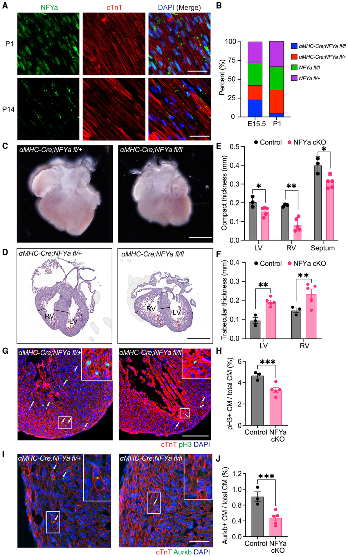
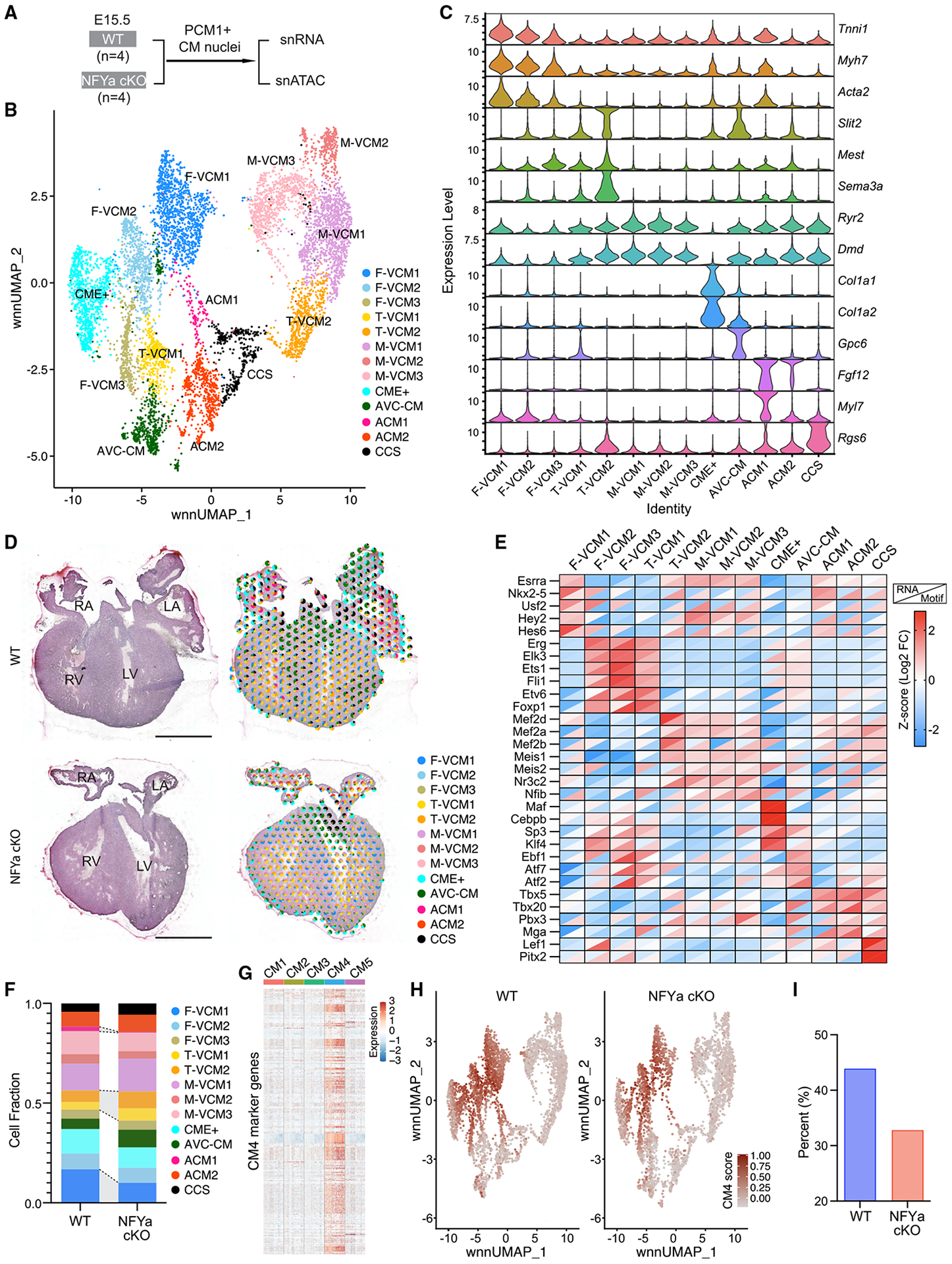
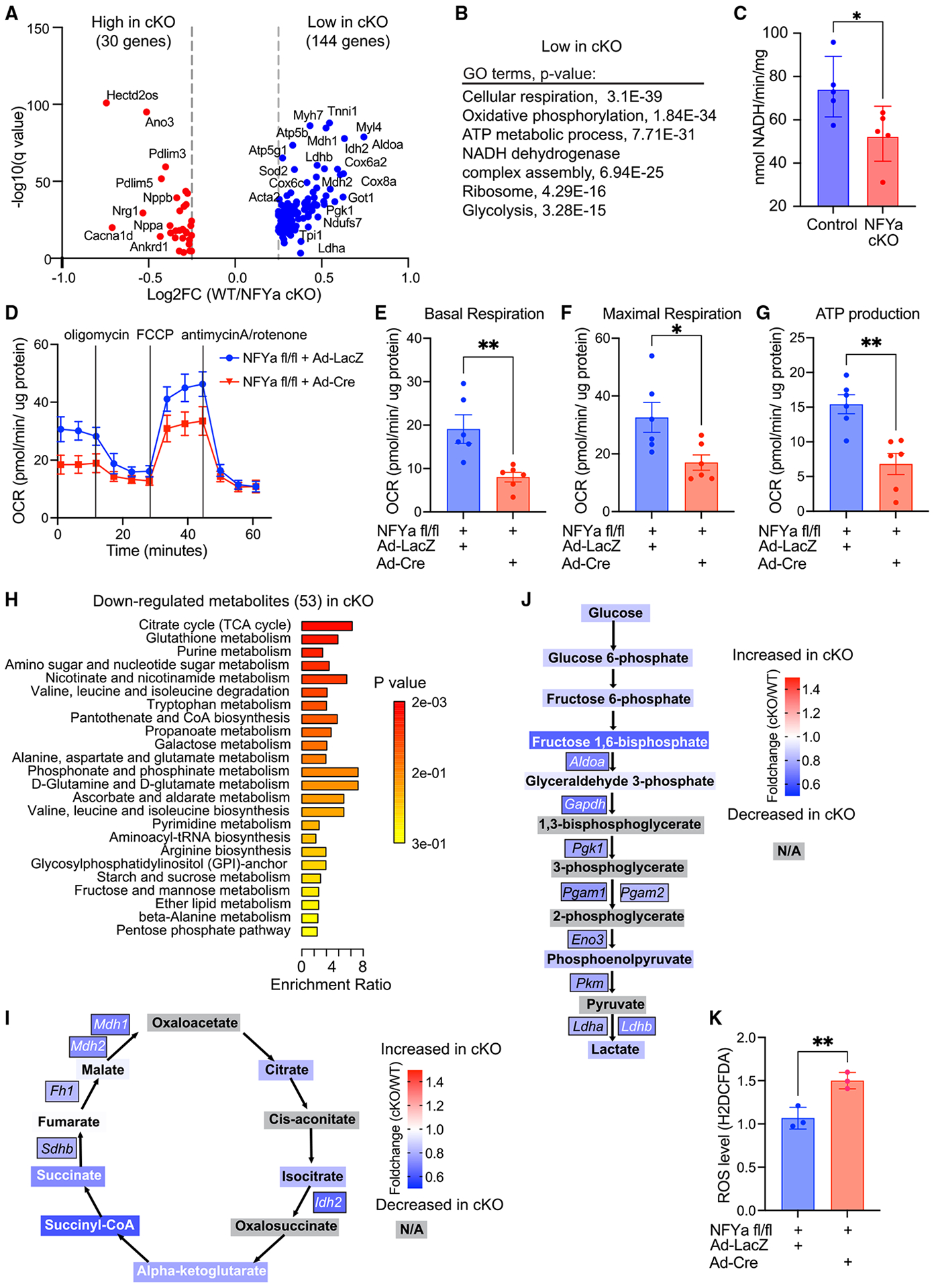
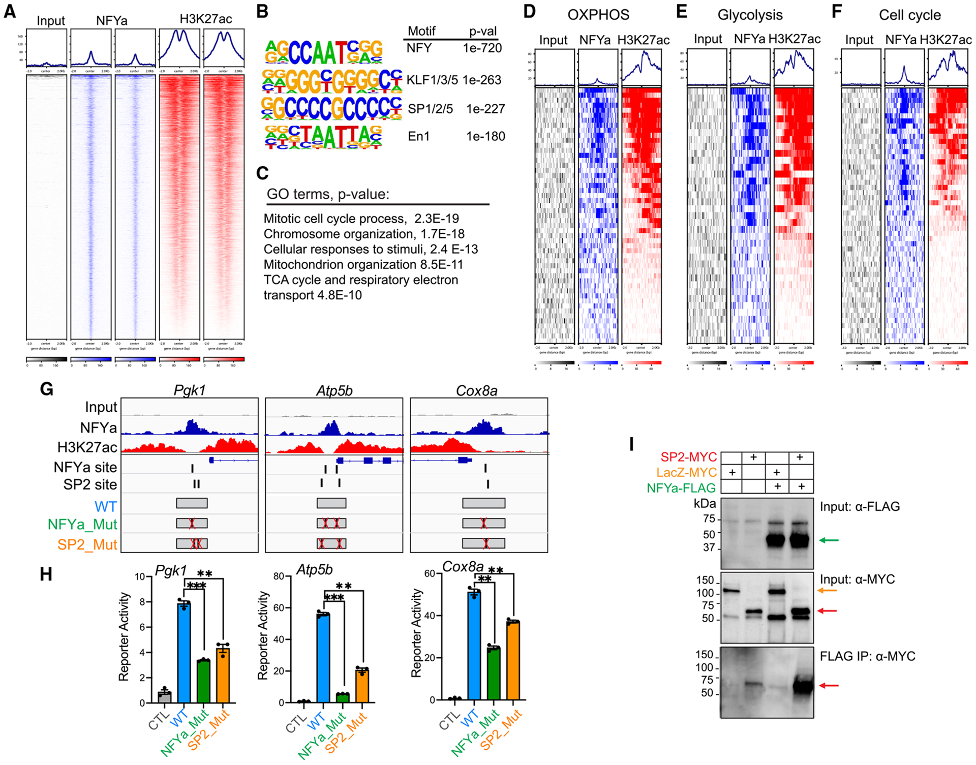
Cardiomyocytes are highly metabolic cells responsible for generating the contractile force in the heart. During fetal development and regeneration, these cells actively divide but lose their proliferative activity in adulthood. The mechanisms that coordinate their metabolism and proliferation are not fully understood. Here, we study the role of the transcription factor NFYa in developing mouse hearts. Loss of NFYa alters cardiomyocyte composition, causing a decrease in immature regenerative cells and an increase in trabecular and mature cardiomyocytes, as identified by spatial and single-cell transcriptome analyses. NFYa-deleted cardiomyocytes exhibited reduced proliferation and impaired mitochondrial metabolism, leading to cardiac growth defects and embryonic death. NFYa, interacting with cofactor SP2, activates genes linking metabolism and proliferation at the transcription level. Our study identifies a nodal role of NFYa in regulating prenatal cardiac growth and a previously unrecognized transcriptional control mechanism of heart metabolism, highlighting the importance of mitochondrial metabolism during heart development and regeneration.

Keywords: cardiac metabolism; cardiomyocyte proliferation; heart development; nuclear transcription factor Y.


Conflict of interest statement

Declaration of interests The authors declare no competing interests.

Figures

Figure 1.
Figure 1.. Cardiomyocyte-specific deletion of NFYa causes embryonic lethality
(A) Immunofluorescent staining of NFYa and cardiac troponin T (cTnT) on P1 and P14 hearts. Scale bars, 25 μm. (B) Percentage of genotypes observed at E15.5 and P1. NFYa cKO (αMHC-Cre;NFYafl/fl) is shown in blue. (C) Micrographs of representative control (αMHC-Cre;NFYafl/+) and NFYa cKO hearts at E15.5. Scale bars, 500 μm. (D) Hematoxylin and eosin (H&E) staining images of representative control and NFYa cKO hearts at E15.5. Black lines mark the compact myocardium zone, and red lines mark the trabecular zone. LV, Left ventricle; RV, right ventricle. Scale bars, 500 μm. (E) Thickness of the compact myocardium in the LV, RV, and septum. (F) Thickness of the LV and RV trabecular regions. (G) Immunofluorescence staining for pH3, cTnT, and DAPI of control and NFYa cKO hearts at E15.5. White arrows indicate pH3+ CMs. Scale bars, 100 μm. (H) Quantification of pH3+ CMs over total CMs. (I and J) Immunofluorescence staining for Aurkb, cTnT, and DAPI of control and NFYa cKO hearts at E15.5 (I) and quantification of Aurkb+ midbody frequency (J). Scale bars, 25 μm. White arrows indicate Aurkb+ midbodies between nuclei. All data are presented as mean ± SEM. *p < 0.05, **p < 0.01, ***p < 0.001 by Student’s t test (E and F) and z test (H and J).
Figure 2.
Figure 2.. Change of cardiomyocyte composition upon NFYa deletion
(A) Schematics of joint single-nucleus RNA and ATAC sequencing from WT and NFYa cKO hearts at E15.5. (B) UMAP shows CM populations from combined samples in (A). (C) Violin plots showing the expression of representative marker genes for each CM population. (D) Left, H&E images of WT and NFYa cKO hearts at E15.5. Right, cell type composition shown in pie charts for individual spatial spots sequenced from heart sections. Left and right ventricles (LV, RV) and atria (LA, RA) are indicated. Scale bars, 500 μm. (E) Heatmap showing RNA expression enrichment (upper triangle) from snRNA-seq and corresponding motif accessibility (lower triangles) from scATAC-seq for representative transcriptional regulators of each identified CM population. (F) Fraction of each CM population in WT and NFYa cKO hearts. (G) Heatmap showing expression of CM4 marker genes in CM1-5 cells, identified from our previous study. (H) Averaged expression of CM4 marker genes (CM4 score) in each CM from WT and NFYa cKO hearts. (I) Percentage of cells with high CM4 marker expression from WT and NFYa cKO hearts.
Figure 3.
Figure 3.. Metabolic dysregulation in NFYa cKO hearts
(A) Differentially expressed genes in NFYa cKO CMs compared with WT CMs. All CMs identified from the Multiome sequencing were analyzed. (B) GO terms for downregulated genes in NFYa cKO CMs. (C) NADH consumption by the electron transport chain at the mitochondrial membrane of NFYa cKO hearts and control hearts (αMHC-Cre;NFYafl/+ or αMHC-Cre;NFYa+/+). (D) Seahorse mitochondrial stress test showing oxygen consumption rates (OCRs) in neonatal NFYa fl/fl CMs infected with control (Ad-LacZ) or Cre (Ad-Cre) adenovirus. Drug addition is indicated. (E–G) OCR measurements of basal respiration (E), maximal respiration (F), and ATP production (G) in control (Ad-LacZ) and NFYa KO (Ad-Cre) CMs from (D). (H) Metabolic pathways downregulated in NFYa cKO hearts. (I and J) Fold-change of metabolites in the TCA cycle (I) and glycolysis (J) along with gene expression changes for key enzymes regulating these metabolic pathways in snRNA-seq data. Box color depicts the fold-change (cKO/WT), and blue indicates decrease in cKO and red indicates increase in cKO. Metabolites marked in gray were not included in the analysis. Only genes with significant changes from snRNA-seq data are shown. (K) Quantification of ROS levels in NFYafl/fl CMs infected with Ad-LacZ or Ad-Cre. All data are presented as mean ± SEM. *p < 0.05, **p < 0.01 by Student’s t test.
Figure 4.
Figure 4.. NFYa directly regulates the transcription of metabolic genes
(A) NFYa and H3K27ac ChIP-seq signals at NFYa-bound genomic regions in CMs. (B) Top enriched motifs at NFYa-bound genomic regions. (C) GO terms of the nearest genes associated with NFYa-bound regions. (D–F) NFYa and H3K27ac ChIP-seq signals at promoter regions of genes regulating OXPHOS (D), glycolysis (E), and cell cycle (F). (G) Schematics showing WT and mutant promoter regions of Pgk1, Atp5b, and Cox8a used for the reporter assay. NFYa and H3K27ac ChIP-seq signals are shown along with predicted NFYa and SP2 binding sites. (H) Reporter activity driven by WT and mutant promoters normalized to minimal promoter control (CTL). (I) Protein co-immunoprecipitation detects SP2 protein by NFYa pull-down. Green arrow marks NFYa-FLAG, yellow arrow marks LacZ-Myc, and red arrows mark SP2-Myc. All data are presented as mean ± SEM. **p < 0.01, ***p < 0.001, by Student’s t test.

Similar articles

Cited by

References

    1. Alkass K, Panula J, Westman M, Wu TD, Guerquin-Kern JL, and Bergmann O (2015). No evidence for cardiomyocyte number expansion in preadolescent mice. Cell 163, 1026–1036. 10.1016/j.cell.2015.10.035. - DOI - PubMed
    1. Guo Y, and Pu WT (2020). Cardiomyocyte maturation: new phase in development. Circ. Res 126, 1086–1106. 10.1161/CIRCRESAHA.119.315862. - DOI - PMC - PubMed
    1. Gan P, Wang Z, Morales MG, Zhang Y, Bassel-Duby R, Liu N, and Olson EN (2022). RBPMS is an RNA-binding protein that mediates cardiomyocyte binucleation and cardiovascular development. Dev. Cell 57, 959–973.e7. 10.1016/j.devcel.2022.03.017. - DOI - PMC - PubMed
    1. Tomita-Mitchell A, Stamm KD, Mahnke DK, Kim MS, Hidestrand PM, Liang HL, Goetsch MA, Hidestrand M, Simpson P, Pelech AN, et al. (2016). Impact of MYH6 variants in hypoplastic left heart syndrome. Physiol. Genomics 48, 912–921. 10.1152/physiol-genomics.00091.2016. - DOI - PMC - PubMed
    1. Porrello ER, Johnson BA, Aurora AB, Simpson E, Nam YJ, Matkovich SJ, Dorn GW 2nd, van Rooij E, and Olson EN (2011). MiR-15 family regulates postnatal mitotic arrest of cardiomyocytes. Circ. Res 109, 670–679. 10.1161/CIRCRESAHA.111.248880. - DOI - PMC - PubMed

Publication types