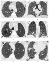
Mortality surrogates in combined pulmonary fibrosis and emphysema
- PMID: 37973176
- PMCID: PMC7616106
- DOI: 10.1183/13993003.00127-2023
Mortality surrogates in combined pulmonary fibrosis and emphysema
Abstract
Background: Idiopathic pulmonary fibrosis (IPF) with coexistent emphysema, termed combined pulmonary fibrosis and emphysema (CPFE) may associate with reduced forced vital capacity (FVC) declines compared to non-CPFE IPF patients. We examined associations between mortality and functional measures of disease progression in two IPF cohorts.
Methods: Visual emphysema presence (>0% emphysema) scored on computed tomography identified CPFE patients (CPFE/non-CPFE: derivation cohort n=317/n=183, replication cohort n=358/n=152), who were subgrouped using 10% or 15% visual emphysema thresholds, and an unsupervised machine-learning model considering emphysema and interstitial lung disease extents. Baseline characteristics, 1-year relative FVC and diffusing capacity of the lung for carbon monoxide (D LCO) decline (linear mixed-effects models), and their associations with mortality (multivariable Cox regression models) were compared across non-CPFE and CPFE subgroups.
Results: In both IPF cohorts, CPFE patients with ≥10% emphysema had a greater smoking history and lower baseline D LCO compared to CPFE patients with <10% emphysema. Using multivariable Cox regression analyses in patients with ≥10% emphysema, 1-year D LCO decline showed stronger mortality associations than 1-year FVC decline. Results were maintained in patients suitable for therapeutic IPF trials and in subjects subgrouped by ≥15% emphysema and using unsupervised machine learning. Importantly, the unsupervised machine-learning approach identified CPFE patients in whom FVC decline did not associate strongly with mortality. In non-CPFE IPF patients, 1-year FVC declines ≥5% and ≥10% showed strong mortality associations.
Conclusion: When assessing disease progression in IPF, D LCO decline should be considered in patients with ≥10% emphysema and a ≥5% 1-year relative FVC decline threshold considered in non-CPFE IPF patients.
Copyright ©The authors 2024. For reproduction rights and permissions contact permissions@ersnet.org.
Conflict of interest statement
Conflict of interest: J. Jacob reports fees from Boehringer Ingelheim, Roche, NHSX, Takeda and GlaxoSmithKline, unrelated to the submitted work, and was supported by Wellcome Trust Clinical Research Career Development Fellowship 209553/Z/17/Z and the NIHR Biomedical Research Centre at University College London. N. Mogulkoc reports grant TUBITAK (EJP Rare Disease project “COCOS-IPF”), fees from Boehringer Ingelheim, Roche, and Nobel Turkey unrelated to the submitted work, and received support for travel to meetings from Roche and Actelion. T.J. Corte reports unrestricted educational grants from Boehringer Ingelheim, Roche, Biogen and Galapagos, fees from Roche, BMS, Boehringer Ingelheim, Vicore and DevPro, assistance for travel to meetings from Boehringer Ingelheim, and participation on a data safety monitoring board or advisory board for Roche, BMS, Boehringer Ingelheim, Vicore, Ad Alta, Bridge Biotherapeutics and DevPro. P. Vasudev reports financial interests from Blackford Analysis. T. Goos is supported by Research Foundation Flanders (1S73921N). L.J. De Sadeleer is supported by Marie Sklodowska-Curie actions postdoctoral fellowship within the European Union's Horizon Europe research and innovation programme. H. Jo reports fees from Boehringer Ingelheim and Roche, and received assistance for travel to meetings from Boehringer Ingelheim and Roche. S. Verleden reports consultancy fees from Boehringer Ingelheim and Sanofi. M. Vermant is supported by an FWO (Research Flanders Foundation) fellowship. S.M. Janes reports fees from AstraZeneca, Bard1 Bioscience, Achilles Therapeutics and Jansen unrelated to the submitted work, received assistance for travel to meetings from AstraZeneca and Takeda, and is the investigator lead on grants from GRAIL Inc., GlaxoSmithKline plc and Owlstone. A.U. Wells reports personal fees and non-financial support from Boehringer Ingelheim, Bayer and Roche Pharmaceuticals, and personal fees from Blade, outside of the submitted work. The remaining authors report no relevant conflicts of interest.
Figures


Comment in
-
Combined pulmonary fibrosis and emphysema syndrome: the age of majority.Eur Respir J. 2024 Apr 4;63(4):2400353. doi: 10.1183/13993003.00353-2024. Print 2024 Apr. Eur Respir J. 2024. PMID: 38575167 No abstract available.
References
-
- King CS, Nathan SD. Idiopathic pulmonary fibrosis: effects and optimal management of comorbidities. Lancet Respir Med. 2017;5:72–84. - PubMed
-
- Cottin V, Nunes H, Brillet P, et al. Combined pulmonary fibrosis and emphysema: a distinct underrecognised entity. Eur Respir J. 2005;26:586–593. - PubMed
-
- Cottin V, Hansell DM, Sverzellati N, et al. Effect of Emphysema Extent on Serial Lung Function in Patients with Idiopathic Pulmonary Fibrosis. Am J Respir Crit Care Med. 2017;196:1162–1171. - PubMed
MeSH terms
Grants and funding
LinkOut - more resources
Full Text Sources
Medical
