. 2023 Nov 16;14(11):750.
doi: 10.1038/s41419-023-06279-w.

Specific intracellular retention of circSKA3 promotes colorectal cancer metastasis by attenuating ubiquitination and degradation of SLUG

Affiliations

Specific intracellular retention of circSKA3 promotes colorectal cancer metastasis by attenuating ubiquitination and degradation of SLUG

Jingwen Deng et al. Cell Death Dis. .

Abstract

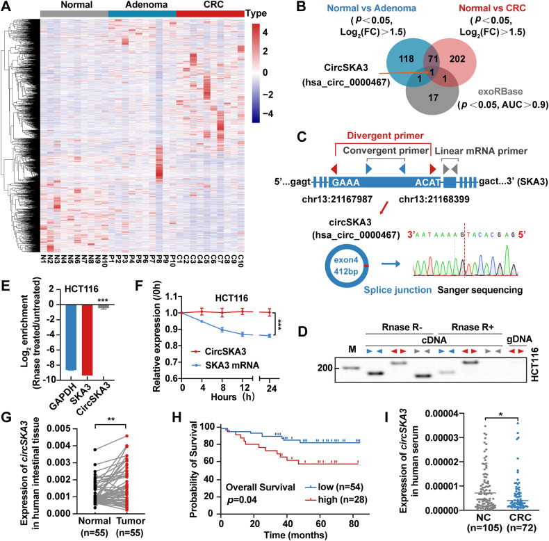
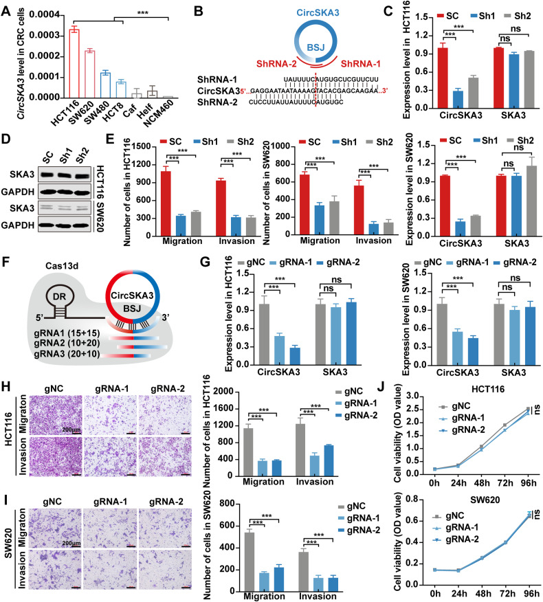
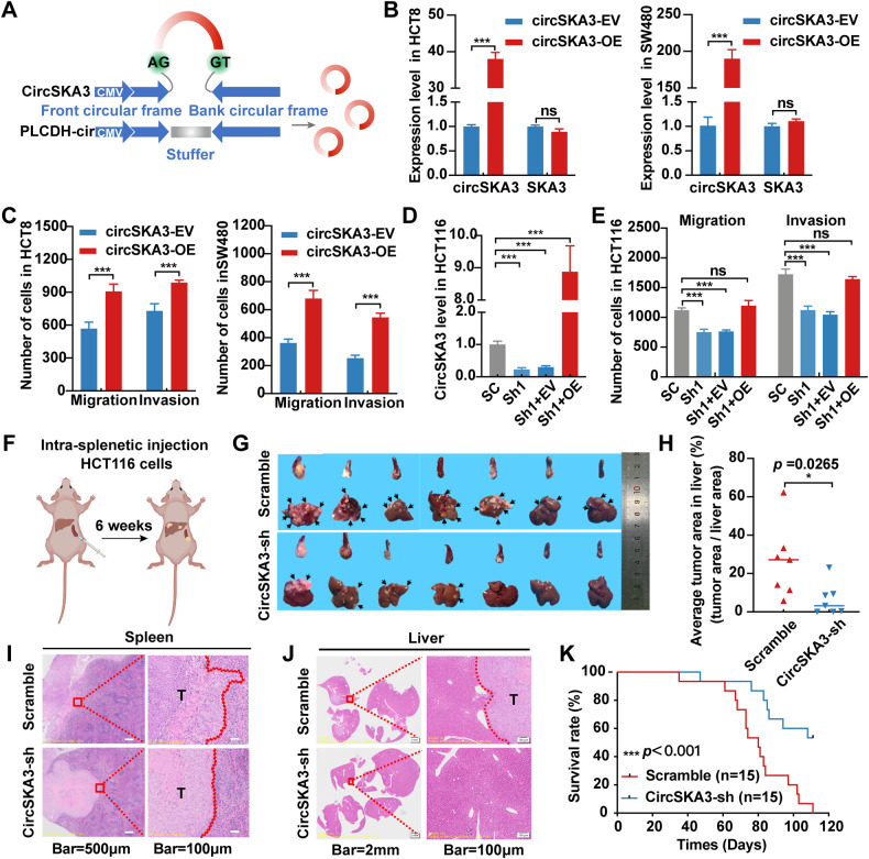
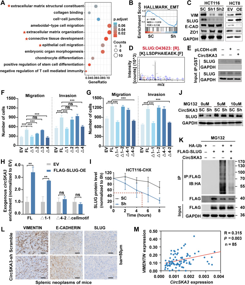
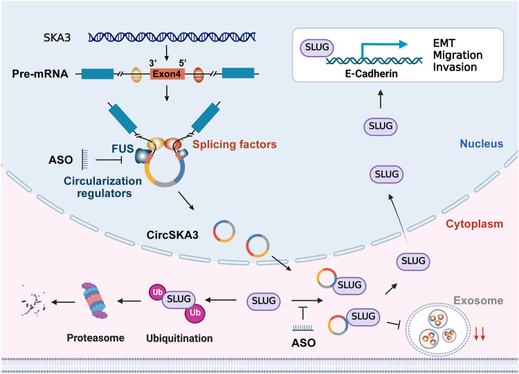
Our previous study demonstrated that tumor-suppressor circular RNAs (circRNAs) can be specifically secreted outside of colorectal cancer (CRC) cells within exosomes to maintain tumor cell fitness. However, whether tumor-driving circRNAs can be specifically retained in cells to facilitate tumor progression remains unknown. In this study, circRNA-seq showed that circSKA3 was significantly upregulated in CRC tissues but downregulated in serum samples from CRC patients. In addition, circSKA3 promoted CRC progression in vitro and in vivo and was retained in CRC cells via a specific cellmotif element. Interestingly, the cellmotif element was also the site of interaction of circSKA3 with SLUG, which inhibited SLUG ubiquitination degradation and promoted CRC epithelial-mesenchymal transition (EMT). Moreover, FUS was identified as a key circularization regulator of circSKA3 that bound to the key element. Finally, we designed and synthesized specific antisense oligonucleotides (ASOs) targeting circularization and cellmotif elements, which repressed circSKA3 expression, abolished the SLUG-circSKA3 interaction, and further inhibited CRC EMT and metastasis in vitro and in vivo.


Conflict of interest statement

The authors declare no competing interests.

Figures

Fig. 1
Fig. 1. CircSKA3 characteristics and expression in CRC.
A CircRNA-sequencing heatmap of 10 paired normal intestinal mucosa–adenoma/adenocarcinoma tissue samples. B Venn diagram of upregulated circRNAs in CRC, adenoma and serum exosomes from CRC patients in the GEO database with AUC > 0.9. E Schematic illustration of circSKA3 circularization from exon 4 of the SKA3 gene and Sanger sequencing of the BSJ site of circSKA3. C, D The head-to-tail splicing of circSKA3 was clarified in cDNA and gDNA of HCT116 cells by RT‒PCR with different primers and RNase R treatment. E The stability of circSKA3 and linear SKA3 in HCT116 was assessed by RNase R treatment followed by RT‒qPCR. F Evaluation of the stability of circSKA3 and SKA3 mRNA in HCT116 treated with actinomycin D. G Expression levels of circSKA3 in 55 paired normal and CRC samples as determined by RT‒qPCR (normalized to GAPDH). H K–M survival analysis of overall survival in CRC patients according to the circSKA3 expression level. I Detection of circSKA3 in the serum of 105 normal controls and 72 CRC patients. All results are representative of three independent experiments, and shown as mean ± SD. Statistical significance was accessed by paired-samples Student’s t-tests (G), Student’s t-test (E, F, I), and log-rank test (H). *p < 0.05, **p < 0.01, ***p < 0.001.
Fig. 2
Fig. 2. CircSKA3 promotes CRC migration and invasion in vitro.
A Expression of circSKA3 in colorectal cell lines. B Schematic design of the shRNA sequence targeting the BSJ site of circSKA3. C Knockdown of circSKA3 via shRNA in HCT116 and SW620 cells. D Detection of the protein level of the host gene SKA3 after circSKA3 knockdown by immunoblotting. E Quantitative analysis of the migration and invasion abilities of HCT116 and SW620 cells after circSKA3 knockdown by Transwell assay. F Schematic of sgRNA design for the circSKA3 BSJ site. G Efficiency of circSKA3 knockdown by RfxCas13d-BSJ-gRNA in HCT116 and SW620 cells. H, I Transwell assay for the migration and invasion abilities of HCT116 and SW620 cells after circSKA3 knockdown by RfxCas13d-BSJ-gRNA. The right graph shows the quantitative analysis results (scale bar = 200 μm). J Effects of circSKA3-specific knockdown by RfxCas13d-BSJ-gRNA on HCT116 and SW620 cell proliferation. All results are representative of three independent experiments, and shown as mean ± SD (C, G, J) or mean ± SEM (A, E, H, I). Statistical significance was accessed by one-way ANOVA (A, C, E, G, H, I, J). ns, not significant with p > 0.05, **p < 0.01, ***p < 0.001.
Fig. 3
Fig. 3. CircSKA3 promotes CRC metastasis in vitro and in vivo.
A Schematic of circSKA3 overexpression plasmid construction based on the pLCDH-ciR vector. B Overexpression of circSKA3 via lentiviral expression vector in HCT8 and SW480 cells. C Quantitative analysis of the migration and invasion abilities of HCT8 and SW480 cells after circSKA3 overexpression by Transwell assay. D Overexpression of circSKA3 in circSKA3-knockdown HCT116 cells. E Quantitative analysis of the migration and invasion abilities of circSKA3-knockdown HCT116 cells after re-expression of circSKA3 by Transwell assay. F Schematic diagram of mouse spleen-liver metastasis model construction in nude mice. G Images of nude mouse spleens and livers. The arrows indicate metastases (n = 7). H Quantitative analysis of the average tumor area in the distant liver metastasis model. I H&E staining of the spleen in situ tumors in nude mice. J H&E staining of liver metastatic foci. K Survival analysis of nude mice with distant liver metastases. All results are representative of three independent experiments, and shown as mean ± SD (B, D, H, K) or mean ± SEM (C, E). Statistical significance was accessed by Student’s t-test (B, C, H), one-way ANOVA (D, E), and log-rank test (K). ns, not significant with p > 0.05, *p < 0.05, ***p < 0.001.
Fig. 4
Fig. 4. CircSKA3 retention in CRC cell cytoplasm.
A Detection of the distribution of circSKA3 in HCT116 and HCT8 cells by RT‒qPCR. B, C Identification of exosomes in HCT116 and HCT8 cells using transmission electron microscopy and immunoblotting with the exosome-associated proteins TSG101 and CD63 (calnexin and cell lysates were used as a negative control and positive loading control, respectively) (scale bar = 500 μm). D Exosome secretion assay for CRC cells, NCM460 normal intestinal epithelial cells and 293T cells transfected with the circSKA3 overexpression vector. E Schematic diagram for circSKA3 truncation plasmid construction. F Exosome secretion assay for HCT8 cells transfected with truncated circSKA3 overexpression vectors (Δ1, Δ2, Δ3 and Δ4). G Exosome secretion assay for HCT8 cells transfected with truncated circSKA3 overexpression vectors (Δ4-1 and Δ4-2). H Schematic of the circSKA3 FL and cellmotif (AGAAC) truncation constructs. I Exosome secretion assay for HCT8 cells transfected with the above two vectors. All results are representative of three independent experiments, and shown as mean ± SD (A, D, F, G, I).
Fig. 5
Fig. 5. CircSKA3 promotes EMT in CRC by stabilizing SLUG.
A Top biological processes regulated by circSKA3 as determined by GO enrichment analysis of the differentially expressed genes (DEGs) based on the Database for Annotation, Visualization and Integrated Discovery (DAVID) online tools (|log2(fold-change)| >1 and p ≤ 0.05). B GSEA of circSKA3-knockdown HCT116 cells. C Detection of EMT-related markers in circSKA3-knockdown HCT116 cells and circSKA3-overexpressing HCT8 cells by immunoblotting. D Amino acid sequence of SLUG as detected by MS. E Detection of SLUG in RNA pulldown assays by immunoblotting. F, G Quantitative analysis of the results of a transwell assay for the migration and invasion abilities of HCT8 cells after overexpression of truncated circSKA3 and FL circSKA3. H Detection of the enrichment of FL, Δ1-1, Δ4-2, and Δcellmotif-truncated circSKA3 in the RIP assay by RT‒qPCR. I Statistical chart of SLUG protein levels at different time points after CHX treatment of control and knockdown HCT116 cells. J Detection of SLUG protein levels in circSKA3-knockdown and control HCT116 cells treated with different concentrations of MG132 by immunoblotting. K Detection of the ubiquitination level of SLUG by IP assay. L Detection of E-CADHERIN, VIMENTIN, and SLUG expression in a mouse model of CRC spleen-liver metastasis (scale bar=50 μm). M Correlation between circSKA3 and VIMENTIN mRNA levels in human CRC samples. All results are representative of three independent experiments, and shown as mean ± SD (H) or mean ± SEM (F, G, I). Statistical significance was accessed by Student’s t-test (H), one-way ANOVA (F, G), and Pearson’s correlation analysis (M). ns, not significant with p > 0.05, *p < 0.05, **p < 0.01, ***p < 0.001.
Fig. 6
Fig. 6. FUS regulates the circularization of circSKA3 via a specific motif.
A Schematic diagram of truncated circSKA3 plasmid construction. B, C Circularization efficiency of circSKA3 and the linear SKA3 level. D Amino acid sequence of FUS identified by MS. E Detection of FUS in the GST-RNA pulldown assay by immunoblotting. F Detection of SLUG in the RIP assay by immunoblotting. G Detection of circSKA3 enrichment in the RIP assay by RT‒qPCR. H Detection of the mRNA expression of FUS, circSKA3, and SKA3 by RT‒qPCR. I Detection of FLAG in the RIP assay by immunoblotting. J Detection of truncated circSKA3 enrichment in the RIP assay by RT‒qPCR. K Correlation between circSKA3 and FUS mRNA levels in human CRC samples. All results are representative of three independent experiments, and shown as mean ± SD (B, C, G, H, J, K). Statistical significance was accessed by Student’s t-test (G), one-way ANOVA (B, C, H, J), and Pearson’s correlation analysis (K). ns, not significant with p > 0.05, *p < 0.05, **p < 0.01, ***p < 0.001.
Fig. 7
Fig. 7. The ASOs inhibit CRC metastasis in vitro and in vivo by regulating circSKA3 circularization and blocking the functional motif.
A Schematic diagram of the functional mechanism and chemical modification of ASOs. B Expression levels of circSKA3 and SKA3 in the ASO-NC group and the groups treated with ASOs targeting specific circSKA3 motifs in HCT116 cells. C, D Transwell assay and quantitative analysis of the migration and invasion abilities of ASO-treated HCT116 cells (scale bar = 400 μm). E Schematic diagram of ASO treatment in the spleen-liver metastasis model via tail vein injection (n = 3). Blue arrows represent the time point of tumor modeling, black represents the time point of administration of ASO and the time point of detection of mouse body weight, and red represents the time point of sacrificing mice (Created with BioRender.com). F Images of the spleen and liver after in situ injection of HCT116 cells for 15 days. G Body weights of nude mice after ASO treatment. H Images of the spleens and livers of nude mice treated with ASOs six times. I Number of distant liver metastatic lesions. J H&E staining of splenic neoplasms and liver metastatic foci. K, L E-CADHERIN and VIMENTIN and SLUG expression in xenograft tumors after ASO treatment (scale bar = 50 μm). All results are representative of three independent experiments, and shown as mean ± SD (B, G, I) or mean ± SEM (D). Statistical significance was accessed by one-way ANOVA (B, D, G, I). ns, not significant with p > 0.05, **p < 0.01, ***p < 0.001.
Fig. 8
Fig. 8
CircSKA3 promotes CRC progression by attenuating the ubiquitination and degradation of SLUG.

Similar articles

Cited by

References

    1. Kristensen LS, Jakobsen T, Hager H, Kjems J. The emerging roles of circRNAs in cancer and oncology. Nat Rev Clin Oncol. 2022;19:188–206. doi: 10.1038/s41571-021-00585-y. - DOI - PubMed
    1. Li X, Yang L, Chen LL. The biogenesis, functions, and challenges of circular RNAs. Mol Cell. 2018;71:428–42. doi: 10.1016/j.molcel.2018.06.034. - DOI - PubMed
    1. Chen S, Cao X, Zhang J, Wu W, Zhang B, Zhao F. circVAMP3 drives CAPRIN1 phase separation and inhibits hepatocellular carcinoma by suppressing c-Myc translation. Adv Sci (Weinh) 2022;9:e2103817. doi: 10.1002/advs.202103817. - DOI - PMC - PubMed
    1. Shi X, Yang J, Liu M, Zhang Y, Zhou Z, Luo W, et al. Circular RNA ANAPC7 inhibits tumor growth and muscle wasting via PHLPP2-AKT-TGF-β signaling axis in pancreatic cancer. Gastroenterology. 2022;162:2004–17.e2002. doi: 10.1053/j.gastro.2022.02.017. - DOI - PMC - PubMed
    1. Chen C, Yu H, Han F, Lai X, Ye K, Lei S, et al. Tumor-suppressive circRHOBTB3 is excreted out of cells via exosome to sustain colorectal cancer cell fitness. Mol Cancer. 2022;21:46. doi: 10.1186/s12943-022-01511-1. - DOI - PMC - PubMed

Publication types