. 2023 Nov 16;12(1):55.
doi: 10.1038/s41389-023-00496-w.

NF-κB signaling activation and roles in thyroid cancers: implication of MAP3K14/NIK

Affiliations

NF-κB signaling activation and roles in thyroid cancers: implication of MAP3K14/NIK

Françoise Cormier et al. Oncogenesis. .

Abstract

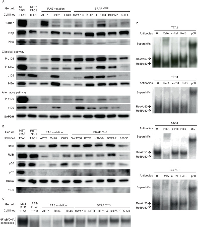
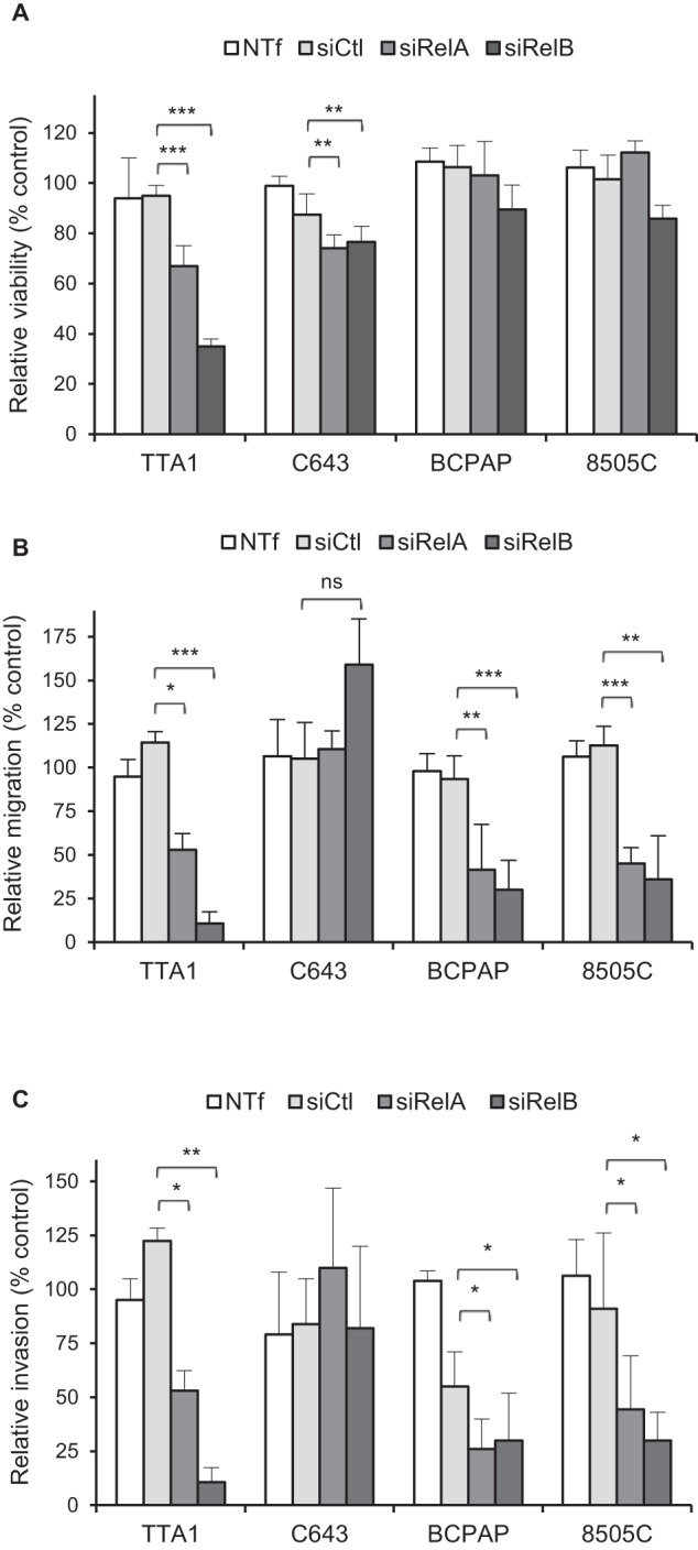
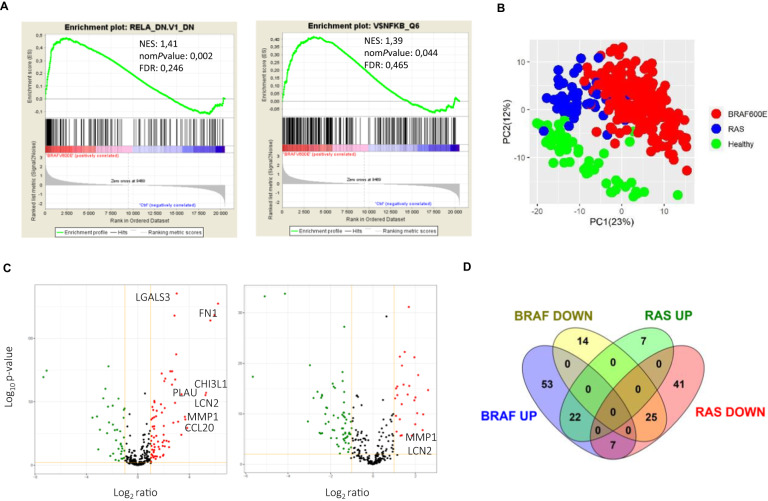
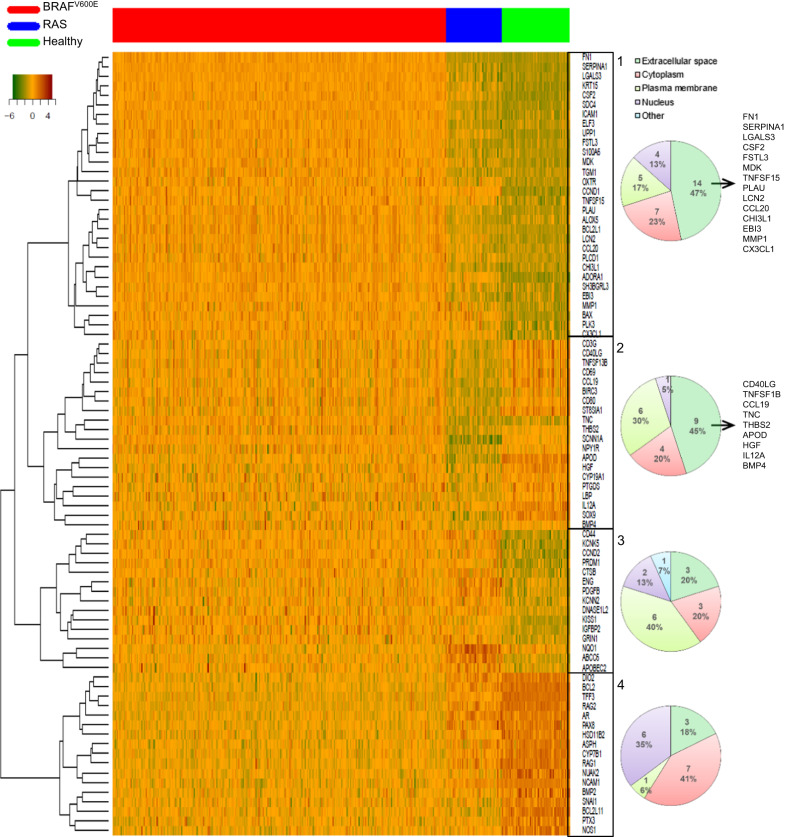
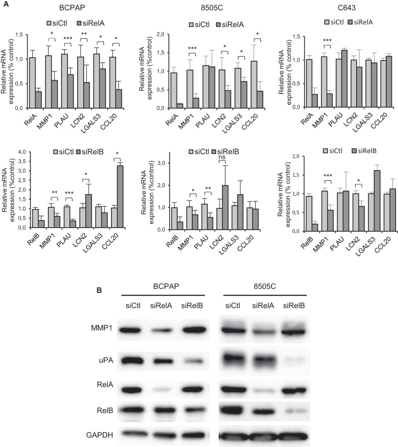
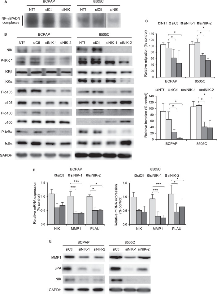
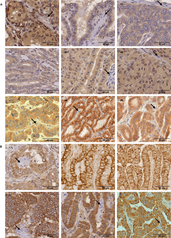
Among follicular-derived thyroid cancers (TC), those with aggressive behavior and resistance to current treatments display poor prognosis. NF-κB signaling pathways are involved in tumor progression of various cancers. Here, we finely characterize the NF-κB pathways and their involvement in TC. By using immunoblot and gel shift assays, we demonstrated that both classical and alternative NF-κB pathways are activated in ten TC-derived cell lines, leading to activated RelA/p50 and RelB/p50 NF-κB dimers. By analyzing the RNAseq data of the large papillary thyroid carcinoma (PTC) cohort from The Cancer Genome Atlas (TCGA) project, we identified a tumor progression-related NF-κB signature in BRAFV600E mutated-PTCs. That corroborated with the role of RelA and RelB in cell migration and invasion processes that we demonstrated specifically in BRAFV600E mutated-cell lines, together with their role in the control of expression of genes implicated in invasiveness (MMP1, PLAU, LCN2 and LGALS3). We also identified NF-κB-inducing kinase (NIK) as a novel actor of the constitutive activation of the NF-κB pathways in TC-derived cell lines. Finally, its implication in invasiveness and its overexpression in PTC samples make NIK a potential therapeutic target for advanced TC treatment.


Conflict of interest statement

The authors declare no competing interests.

Figures

Fig. 1
Fig. 1. The NF-κB pathways are constitutively activated in thyroid carcinoma cell lines.
A The expression and site-specific phosphorylation of components of the classical and alternative NF-κB pathways were analyzed in immunoblot assay of cytoplasmic proteins with appropriate antibodies. GAPDH level was used as a protein loading control. *The P-IKK upper band corresponds to IKKβ and the lower band to IKKα. B The nuclear localization of RelA, RelB, p50 and p52 proteins was analyzed in immunoblot assay of nuclear protein extracts with the appropriate antibodies. HDAC level was used as a nuclear protein loading control, and p100 level was led as an indicator of nuclear extraction quality. C The baseline NF-κB activity in total protein extracts of thyroid carcinoma cell lines was assessed in gel shift experiment. D The NF-κB subunits contributing to the NF-κB activity in 4 thyroid carcinoma cell lines were identified in supershift experiments with anti-RelA, -c-Rel, -RelB and -p50 antibodies. Data of representative experiments are shown. Gen.Alt. : Genomic Alteration. ampl: amplification.
Fig. 2
Fig. 2. RelA and RelB are differently implicated in the growth, motile and invasive potentials of thyroid cell lines.
After transfection with control siRNA (siCtl), RelA siRNA (siRelA), or RelB siRNA (siRelB), the indicated cell lines were plated for viability (A), migration (B) or invasion (C) assay. (A) The mean results of relative viability ±SD compared to non transfected (NTf) cells of eight replicates in two independent experiments are reported. B, C The results are means ± SD of relative motility or invasion compared to NTf cells in two or three independent migration and invasion experiments carried out in duplicate. ns: not significative.
Fig. 3
Fig. 3. The PTC transcriptional program includes NF-κB signatures.
A GSEA enrichment plots for the oncogenic signature RelA_DN.V1_DN and for the gene set NFKB_Q6 (set of genes with the transcription factor binding site V$NFKB_6 which is bound by NF-κB factors members in the regions up to 4 kb around their transcription starting sites) in BRAF-mutated PTCs. B Principal component analysis of the NF-κB target genes set expression in BRAF-, RAS- mutated PTCs and healthy thyroid tissues. C Volcano plots of the differential expression of the NF-κB target genes in BRAF- (left panel) and RAS- (right panel) mutated PTCs. Red and green dots represent significantly upregulated or downregulated genes, respectively, and gray dots represent not significantly deregulated genes (P value < 0.01 and fc >2). Selected genes from the tumor-progression associated signature are highlighted. D Venn diagram representation of the differentially expressed NF-κB target genes in BRAF-mutated PTCs and RAS-mutated PTCs.
Fig. 4
Fig. 4. A tumor progression NF-κB related signature characterizes the BRAF-mutated PTCs.
Heatmap for relative expression of the top 50 more significantly differentially expressed NF-κB target genes between BRAF-mutated PTCs, or RAS-mutated PTCs and healthy thyroid tissues in BRAF-mutated PTCs (N = 286), RAS-mutated PTCs (N = 48) and healthy thyroid tissues (N = 58) from the TCGA database. Pie charts represent the distribution of the localization of gene products comprised in clusters 1, 2, 3 and 4, as indicated. The genes from clusters 1 and 2 whose the product is expressed in the extracellular space are listed in right.
Fig. 5
Fig. 5. RelA and RelB regulate the expression of cell invasion-related genes of the NF-κB signature of BRAF-mutated PTCs.
The BCPAP, 8505C and C643 cell lines were transfected with control siRNA (siCtl), RelA siRNA (siRelA), or RelB siRNA (siRelB). A Two days after transfection, mRNA expression of the indicated genes was analyzed by qRT-PCR. Results are means ± SD of relative mRNA expression level compared to control in four or five independent experiments. B Protein expression was analyzed in immunoblot assay with the appropriate antibodies. Data of representative experiments are shown.
Fig. 6
Fig. 6. The MAP3K NIK contributes to the NF-κB pathways activation and regulates the cell invasion process in BRAF-mutated cell lines.
The BCPAP and 8505C cell lines were transfected with control (siCtl) or NIK siRNA (siNIK, siNIK-1 or siNIK-2). Not transfected cells (NTf) were included as another negative control. A Two days after transfection, NF-κB activity analysis was performed in EMSA experiment as in Fig. 1C. B. Expression and site-specific phosphorylation of components of the classical and alternative NF-κB pathways were analyzed in immunoblotting assay of whole cell proteins. (*The P-IKK upper band corresponds to IKKβ and the lower band to IKKα). C Assessment of migration (upper panel) and invasion (bottom panel) capacities. The results are means ± SD of two independent migration and invasion experiments carried out in duplicated. D MMP1 and PLAU mRNA expression analysis were performed by qRT-PCR. The results are means ± SD of relative mRNA expression level compared to control in two or three experiments. E MMP1 and uPA protein expression analysis was analyzed in immunoblot assay. Data of representative experiments are shown in (B, E).
Fig. 7
Fig. 7. NIK and RelB expression in primary papillary thyroid cancer.
A Paraffin-embedded sections of nine PTC samples were immunostained using anti-NIK antibody and counterstained with hematoxylin. Representative views showing positive immunoreactivity in neoplastic papillae compared to stromal cells (indicated by arrows) with no immunoreactivity, are presented. B Paraffin-embedded sections of six PTC samples were immunostained using anti-RelB antibody and counterstained with hematoxylin (samples #1, #4, #5 and #7) or only immunostained using anti-RelB antibody (samples #2 and #3). Representative views showing positive cytoplasmic and nuclear immunoreactivity in tumoral cells are presented. Nuclear immunoreactivity is indicated by a slight gray to brown staining of nuclei in samples #1, #4, #5 and #7, compared to nuclei multinucleated giant cells (indicated by arrows in #1, #4 and #5) or stromal cells (indicated by an arrow in #7) which displayed blue staining. No hematoxylin staining in samples #2 and 3 allowed to clearly detect RelB nuclear immunopositivity in tumoral cells through a slight peroxydase-positive staining.

Similar articles

Cited by

References

    1. Kim J, Gosnell JE, Roman SA. Geographic influences in the global rise of thyroid cancer. Nat Rev Endocrinol. 2020;16:17–29. doi: 10.1038/s41574-019-0263-x. - DOI - PubMed
    1. Carneiro RM, Carneiro BA, Agulnik M, Kopp PA, Giles FJ. Targeted therapies in advanced differentiated thyroid cancer. Cancer Treat Rev. 2015;41:690–8. doi: 10.1016/j.ctrv.2015.06.002. - DOI - PubMed
    1. Molinaro E, Romei C, Biagini A, Sabini E, Agate L, Mazzeo S, et al. Anaplastic thyroid carcinoma: from clinicopathology to genetics and advanced therapies. Nat Rev Endocrinol. 2017;13:644–60. doi: 10.1038/nrendo.2017.76. - DOI - PubMed
    1. Crispo F, Notarangelo T, Pietrafesa M, Lettini G, Storto G, Sgambato A, et al. BRAF inhibitors in thyroid cancer: clinical impact, mechanisms of resistance and future perspectives. Cancers. 2019;11:1388. doi: 10.3390/cancers11091388. - DOI - PMC - PubMed
    1. Ferrari SM, Elia G, Ragusa F, Ruffilli I, La Motta C, Paparo SR, et al. Novel treatments for anaplastic thyroid carcinoma. Gland Surg. 2020;9:S28–S42. doi: 10.21037/gs.2019.10.18. - DOI - PMC - PubMed