. 2023 Nov 16;13(1):20058.
doi: 10.1038/s41598-023-47498-7.

Continuous detrimental activity of intra-articular fibrous scar tissue in correlation with posttraumatic ankle osteoarthritis

Affiliations

Continuous detrimental activity of intra-articular fibrous scar tissue in correlation with posttraumatic ankle osteoarthritis

Nhat Tien Tran et al. Sci Rep. .

Abstract

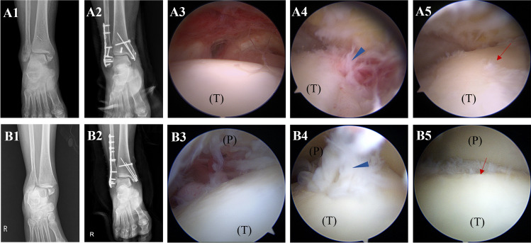
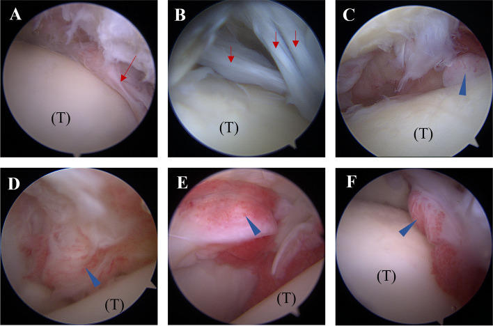
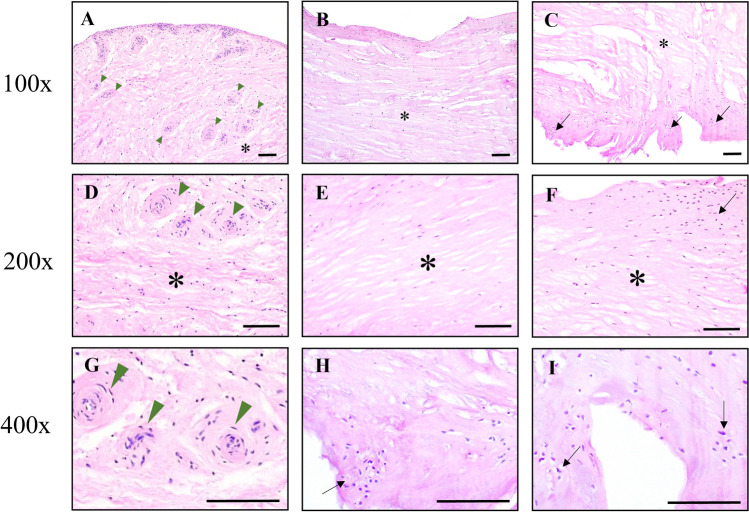
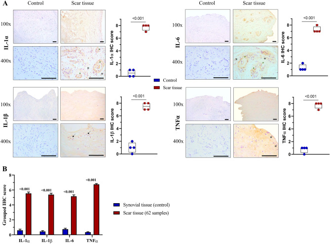
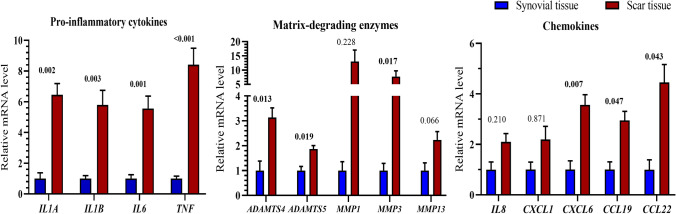
Posttraumatic osteoarthritis is primarily characterized by articular cartilage destruction secondary to trauma or fracture events. Even while intra-articular scar tissue can be observed following ankle fractures, little is known about its nature and molecular events linking its biological activity and cartilage deterioration. Here, we investigated scar tissue's histological and molecular characteristics, and its relationship with localized articular cartilage alterations consistent with early osteoarthritic degeneration. Intra-articular scar tissues from sixty-two patients who underwent open reduction internal fixation for ankle fracture were obtained at hardware removal time (6-44 months after fracture). Histological analysis demonstrated that scar tissue has the nature of fibrosis with fibrous tissue hyperplasia, fibroblast proliferation, and chondrometaplasia. These fibrous scar tissues showed overexpressed pro-inflammatory cytokines and high mRNA expression levels of osteoarthritis-related markers (cytokines, chemokines, and enzymes) compared to the normal synovium. Furthermore, those transcriptional levels were significantly correlated with the grade of talar chondral degeneration. Our findings suggest that following an ankle fracture, the intra-articular fibrous scar tissue exhibits high catabolic and inflammatory activity, which has a long-lasting negative impact correlated to cartilage deterioration in the development of posttraumatic osteoarthritis.


Conflict of interest statement

The authors declare no competing interests.

Figures

Figure 1
Figure 1
The second-look arthroscopic pictures show the growth of intra-articular scar tissue following trimalleolar fracture (A1A5) and bimalleolar fracture (B1B5). (A1, B1) Preoperative X-ray. (A2, B2) Postoperative X-ray shows good reduction and internal fixation. (A3, B3) The initial intact cartilage at fractured time. (A4, B4) The second-look arthroscopic finding depicts scar tissue (arrowhead) formed extensively and adhered to the talar dome. (A5, B5) The underlying degenerative cartilage damage (arrow) is exposed after removing scar tissue. (P): Tibial plafond, (T): Talus.
Figure 2
Figure 2
The arthroscopic images demonstrate arthrofibrosis and synovitis formation following ankle fracture. (A, B) Arthrofibrosis (red arrow). (C, D) Mild-moderate synovitis. (E, F) Severe and diffuse synovitis. Blue arrowhead: synovitis, (T): talus.
Figure 3
Figure 3
Hematoxylin and Eosin staining of intra-articular scar tissues demonstrated fibrous tissue hyperplasia, clarified by the fibrosis (asterisk), vascular proliferation (arrowhead), and chondrometaplasia (transformation of synovial tissue into hyaline-appearing cartilage). These areas of chondrocytes (black arrow) simultaneously appeared with fibrous tissue (asterisk), showing the chondrometaplasia evident in these intra-articular scar tissue specimens. Scale bars represent 100 μm. Low to high magnification by optical microscopy: (AC) 100x, (DF) 200x, (GI) 400x.
Figure 4
Figure 4
The expression of inflammatory cytokines in intra-articular fibrous scar tissue. Representative immunohistochemical staining for pro-inflammatory cytokines (IL-1α, IL1-β, IL6, TNFα) was conducted in both scar tissue and synovial tissue (control group). (A) Positive cells are depicted with cytoplasmic brown staining, with nuclei counterstained using hematoxylin. The high-magnification view highlights positive staining in various cell types, including fibroblasts (asterisk), proliferative vascular endothelial cells (arrowhead), and chondrocytes (arrow). Scale bars indicate 100 μm. The quantification of immunohistochemical staining involves calculating the IHC score for each sample based on the average of four randomly selected fields at 400 × magnification, both for the scar tissue and control samples. The corresponding p-values are shown above the horizontal bars. (B) The IHC scores for scar tissue (62 samples) are presented, and the associated p-values are displayed above each pair of bars. All data were analyzed using the Mann–Whitney U test.
Figure 5
Figure 5
Messenger RNA gene expression levels of pro-inflammatory cytokines (IL1A, IL1B, IL6, and TNF), matrix-degrading enzymes (ADAMTS4, ADAMTS5, MMP1, MMP3, and MMP13), and chemokines (IL8, CXCL1, CXCL6, CCL19, and CCL22) in intra-articular scar tissues procured from 62 ankles underwent fracture compared with those in normal synovial tissues. Expression levels were determined using quantitative real-time polymerase chain reaction with GAPDH (glyceraldehyde 3-phosphate dehydrogenase) as the reference. The p-values are indicated above each bar pair. Data were analyzed by the Mann–Whitney U test.
Figure 6
Figure 6
Heat map visualization of the mRNA expression of osteoarthritis-related markers in correlation with the grade of cartilage degradation. (A) Relative mRNA levels of the 14 target genes change response to Outerbridge classification of cartilage degeneration. The genes are represented in the columns, the Outerbridge grade (I-IV) is represented in the rows, and the gene expression levels are normalized in percentage (the largest value of relative mRNA expression level in each target gene is defined as 100%). (B) The correlation between relative mRNA level in intra-articular fibrous scar tissue versus chondral damage grade using Spearman's rank correlation coefficient. Rho-value is indicated inside the cells (*p < 0.05, **p < 0.01, ***p < 0.001).

Similar articles

References

    1. Horisberger M, Valderrabano V, Hintermann B. Posttraumatic ankle osteoarthritis after ankle-related fractures. J. Orthop. Trauma. 2009;23:60–67. doi: 10.1097/BOT.0b013e31818915d9. - DOI - PubMed
    1. Barg A, et al. Ankle osteoarthritis: Etiology, diagnostics, and classification. Foot Ankle Clin. 2013;18:411–426. doi: 10.1016/j.fcl.2013.06.001. - DOI - PubMed
    1. Valderrabano V, Horisberger M, Russell I, Dougall H, Hintermann B. Etiology of ankle osteoarthritis. Clin. Orthop. Relat. Res. 2009;467:1800–1806. doi: 10.1007/s11999-008-0543-6. - DOI - PMC - PubMed
    1. Nwankwo EC, Labaran LA, Athas V, Olson S, Adams SB. Pathogenesis of posttraumatic osteoarthritis of the ankle. Orthop. Clin. North Am. 2019;50:529–537. doi: 10.1016/j.ocl.2019.05.008. - DOI - PubMed
    1. Delco ML, Kennedy JG, Bonassar LJ, Fortier LA. Post-traumatic osteoarthritis of the ankle: A distinct clinical entity requiring new research approaches. J. Orthop. Res. 2017;35:440–453. doi: 10.1002/jor.23462. - DOI - PMC - PubMed

Publication types