. 2023 Nov 16;13(1):20005.
doi: 10.1038/s41598-023-45949-9.

BubbleDrive, a low-volume incubation chamber for acute brain slices

Affiliations

BubbleDrive, a low-volume incubation chamber for acute brain slices

Aditi Naik et al. Sci Rep. .

Erratum in

Abstract

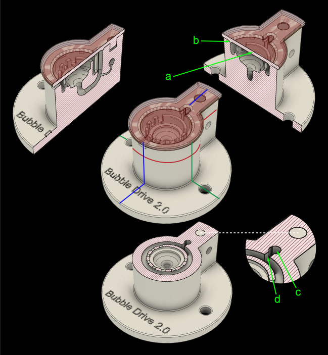
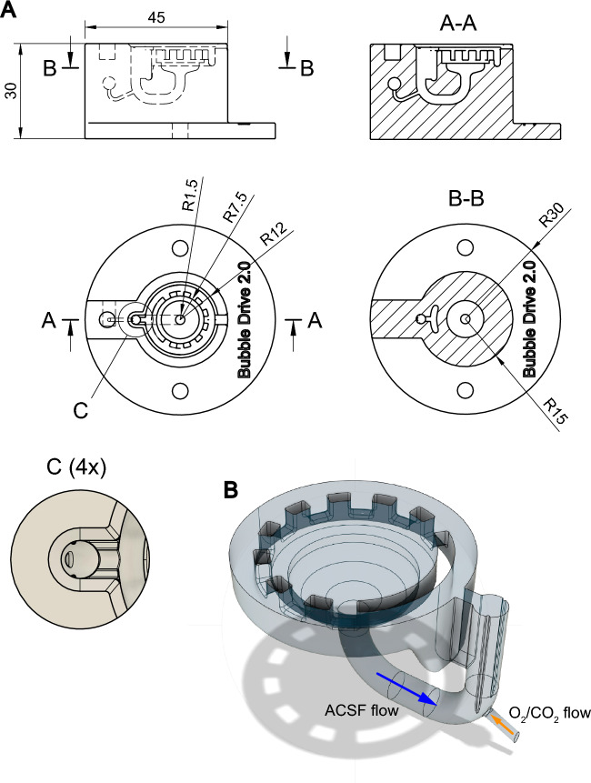
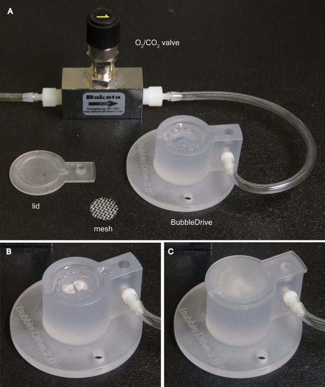
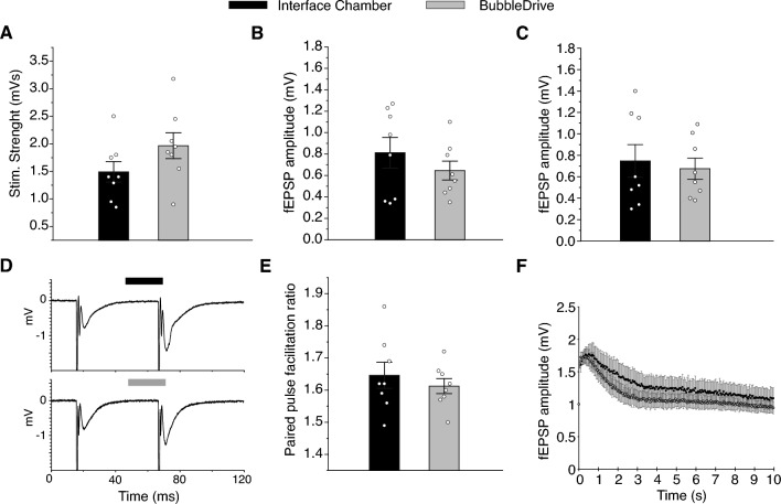
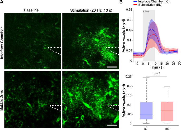
Acute brain slices are a common and useful preparation in experimental neuroscience. A wide range of incubation chambers for brain slices exists but only a few are designed with very low volumes of the bath solution in mind. Such chambers are necessary when high-cost chemicals are to be added to the solution or when small amounts of substances released by the slice are to be collected for analysis. The principal challenge in designing a very low-volume incubation chamber is maintaining good oxygenation and flow without mechanically disturbing or damaging the slices. We designed and validated BubbleDrive, a 3D-printed incubation chamber with a minimum volume of 1.5 mL which can hold up to three coronal mouse slices from one hemisphere. It employs the carbogen gas bubbles to drive the flow circulation in a consistent and reproducible manner, and without disturbing the brain slices. The BubbleDrive design and construction were successfully validated by comparison to a conventional large-volume incubation chamber in several experimental designs involving measurements of extracellular diffusion parameters, the electrophysiology of neuronal and astrocytic networks, and the effectiveness of slice incubation with hyaluronidase enzyme.


Conflict of interest statement

The authors declare no competing interests.

Figures

Figure 1
Figure 1
BubbleDrive model shows three cross sections along the planes marked with colored lines on the centrally positioned perspective view. (a) lid, (b) triangular lip, (c) chimney, (d) slot.
Figure 2
Figure 2
(A) Technical drawing of the BubbleDrive. The top and side orthographic views (middle left and upper left placements in the figure, respectively) are complemented with two cross-sectional orthographic views marked A-A and B-B, and one detailed view (4x magnification) marked (C). All dimensions (linear and radii) are in mm. (B) A 3D model of the ACSF volume inside the BubbleDrive. The direction of gas supply is marked with an orange arrow while the direction of ACSF circulation is marked with a blue arrow. The gas bubbles rise to break the surface at the top of the chimney while the heavier ACSF liquid is forced sideways through the adjacent slot and into the circular chanel. This channel is quite deep near the slot but gradually shallows further away from it to keep the overall liquid volume as small as possible.
Figure 3
Figure 3
BubbleDrive in use. (A) A photograph of BubbleDrive with all its accessories: the gas valve and tubing, the mesh, and the (inverted) lid. The chamber with two (B) and three (C) coronal slices, shown without and with the lid in place, respectively.
Figure 4
Figure 4
Liquid circulation in the BubbleDrive. The figure represents one frame of a Supplemental Video 1 which visualizes the movement of solution in the BubbleDrive using the Fast Green Dye. The dye was added at the center of the slice incubation area before turning on the gas flow. Notice the efficient mixing of the dye with the solution and the flow established immediately after turning on the gas flow.
Figure 5
Figure 5
Structure of ECS and extracellular diffusion. (A) Timeline showing the incubation of slices in the BubbleDrive (BD) or conventional chamber (CC), and the ECS diffusion experiments. (B) Representative RTI experiment showing TMA+ diffusion records in the somatosensory cortex of slices incubated in BD and CC, and in dilute agarose gel before and after brain measurements. The horizontal bar on the x-axis shows the duration of TMA+ release for each record. For this experiment, the distance between the source and the detector microelectrodes was 130 μm and the average transport number was 0.318. For the slice from CC (an average from 2 records): α=0.215, θ=0.431, κ=0.009 s-1; for the slice from BD (an average of 2 records): α=0.214, θ=0.479, κ=0.007 s-1. (C, left) Representative dex3 diffusion records in the somatosensory cortex of slices incubated in each chamber. (C, right) Change with time in the intensity profiles along the highlighted axis. θ=0.312 for BD and 0.334 for CC. (D) Summary of the ECS volume fraction and diffusion permeability data for TMA+. Data from each slice (circles), mean values (bars) and SEM (error bars) are shown. (E) Summary of dex3 diffusion permeability data. Data from each slice (circles), mean values (bars) and SEM (error bars) are shown.
Figure 6
Figure 6
Synaptic transmission, excitability, paired-pulse facilitation (PPF) and 20 Hz stimulation in stratum radiatum. (A) Stimulation strengths necessary to elicit a pre-volley of 1 mV in amplitude. (B) Field EPSP amplitude (fEPSP amplitude) as a function of the same pre-volley amplitude. (C) The fEPSP amplitude necessary to elicit a just detectable population spike on the fEPSP. (D) Example fEPSPs from slices incubated in the two incubation chambers. A mean of six consecutive synaptic responses which elicited synaptic stimulation. (E) Paired pulse facilitation ratio at an interstimulus interval of 50 ms. (F) Dynamic fEPSP amplitude as a result of 20 Hz stimulation for 10 s. Data are shown as mean ± SEM.
Figure 7
Figure 7
Astrocytic Ca2+ signaling responses to electrophysiological stimulation in hippocampal brain slices. (A) Representative mean projection of the baseline period and the stimulation period. Stippled line denotes placement of stimulation electrode. Scale bar 20 μm. (B, top) Mean active x-y-t voxels (detected with the ROA method) with astrocytic Ca2+ signals over time from slices incubated in an interface chamber and in the BubbleDrive. (B, bottom) Mean percentage of active x-y-t voxels (detected with the ROA method) in the stimulation period. Statistical analysis using Wilcoxon Rank Sum test.
Figure 8
Figure 8
Incubation of brain slices with hyaluronidase. Representative confocal images of immunohistochemistry in somatosensory cortex showing HA in green and cell nuclei in blue (DAPI staining) after 1 hour and 3 hours of incubation with hyaluronidase in BubbleDrive. Loss of HA intensity (green) due to enzymatic cleavage is apparent at both incubation times when compared to control slices. Cleavage of HA at the center of the slice even with 1 hour incubation in BubbleDrive shows a time-efficient penetration of the enzyme into the tissue. Scale bar 100 μm.

Similar articles

Cited by

References

    1. Dorris DM, Hauser CA, Minnehan CE, Meitzen J. An aerator for brain slice experiments in individual cell culture plate wells. J. Neurosci. Methods. 2014;238:1–10. doi: 10.1016/j.jneumeth.2014.09.017. - DOI - PubMed
    1. Takano T, et al. Rapid manifestation of reactive astrogliosis in acute hippocampal brain slices. Glia. 2014;62:78–95. doi: 10.1002/glia.22588. - DOI - PMC - PubMed
    1. Fiala JC, et al. Timing of neuronal and glial ultrastructure disruption during brain slice preparation and recovery in vitro. J. Comp. Neurol. 2003;465:90–103. doi: 10.1002/cne.10825. - DOI - PubMed
    1. Tang W, et al. Stimulation-evoked Ca2+ signals in astrocytic processes at hippocampal CA3-CA1 synapses of adult mice are modulated by glutamate and ATP. J. Neurosci. 2015;35:3016–3021. doi: 10.1523/JNEUROSCI.3319-14.2015. - DOI - PMC - PubMed
    1. Szokol K, et al. Augmentation of Ca2+ signaling in astrocytic endfeet in the latent phase of temporal lobe epilepsy. Front. Cell. Neurosci. 2015;9:49. doi: 10.3389/fncel.2015.00049. - DOI - PMC - PubMed

Publication types