. 2023 Nov 16;13(1):20029.
doi: 10.1038/s41598-023-47485-y.

Rescue of murine hind limb ischemia via angiogenesis and lymphangiogenesis promoted by cellular communication network factor 2

Affiliations

Rescue of murine hind limb ischemia via angiogenesis and lymphangiogenesis promoted by cellular communication network factor 2

Masayuki Shimizu et al. Sci Rep. .

Abstract

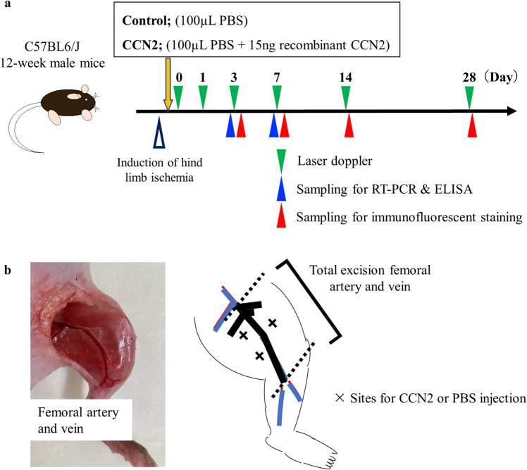
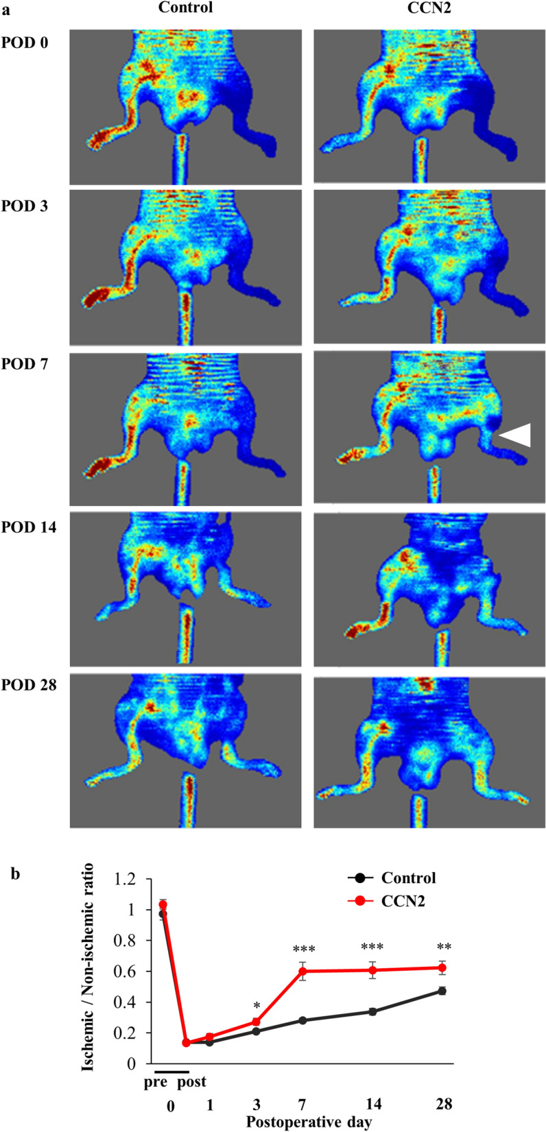
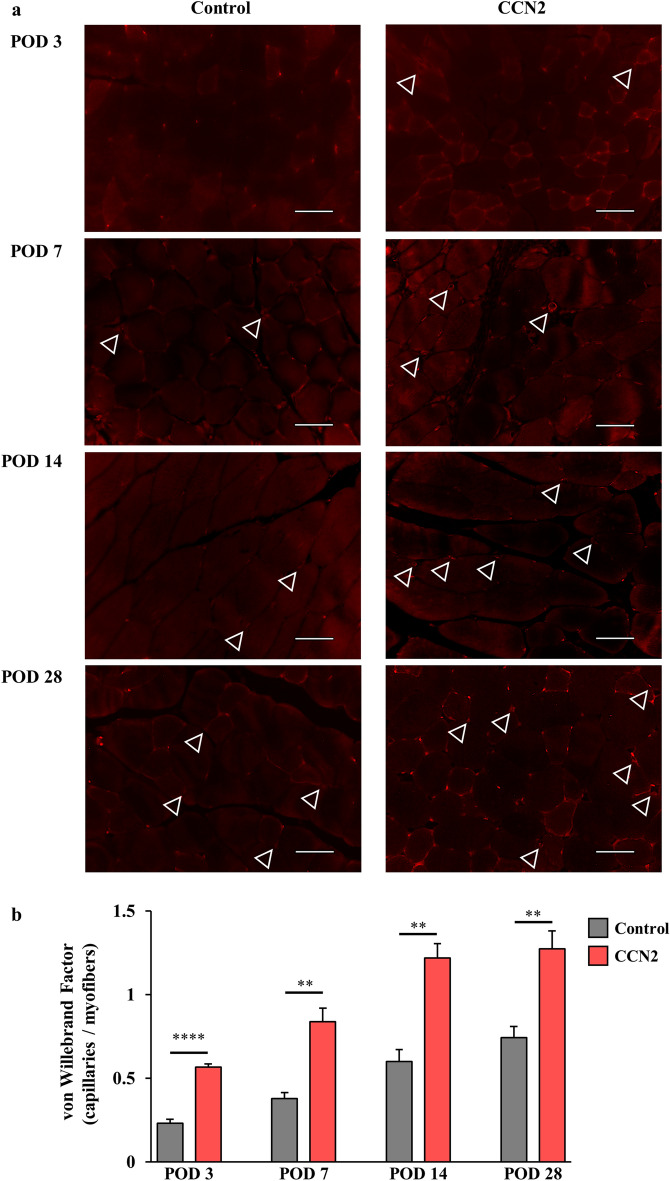
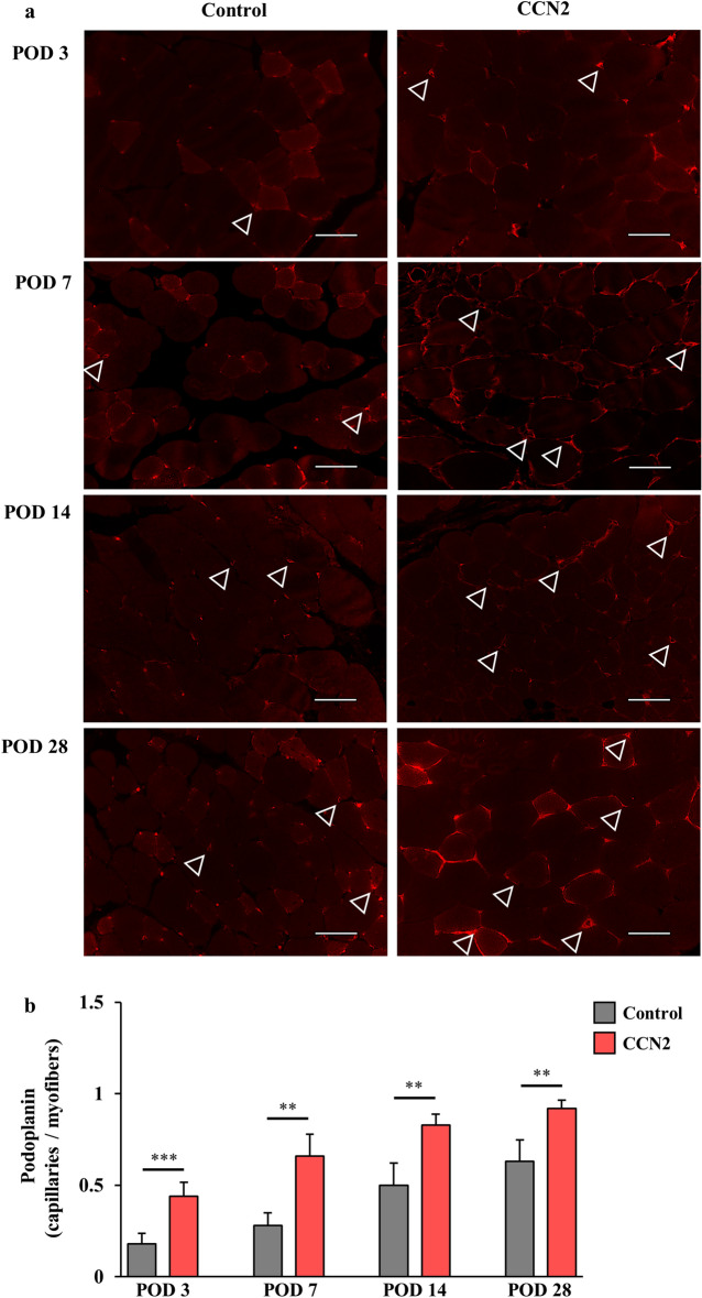
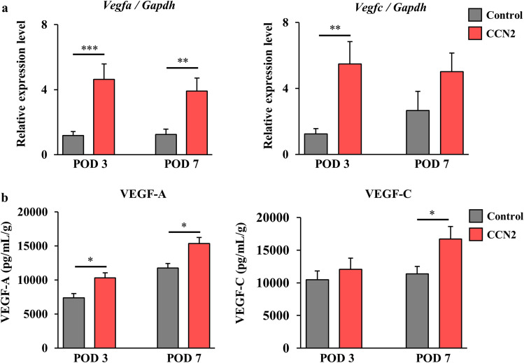
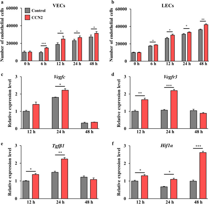
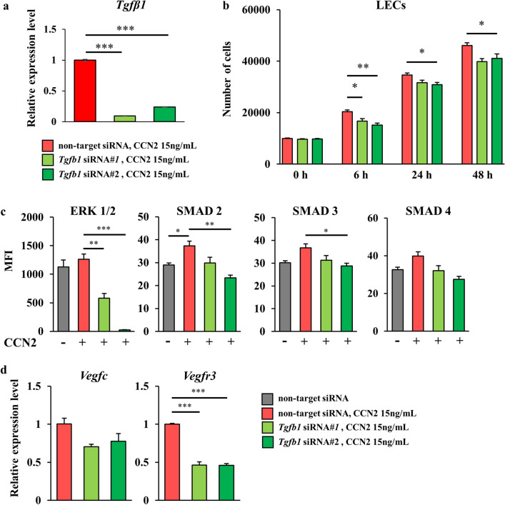
Critical limb ischemia (CLI) is caused by severe arterial blockage with reduction of blood flow. The aim of this study was to determine whether therapeutic angiogenesis using cellular communication network factor 2 (CCN2) would be useful for treating CLI in an animal model. Recombinant CCN2 was administered intramuscularly to male C57BL/6J mice with hind limb ischemia. The therapeutic effect was evaluated by monitoring blood flow in the ischemic hind limb. In an in vivo assay, CCN2 restored blood flow in the ischemic hind limb by promoting both angiogenesis and lymphangiogenesis. VEGF-A and VEGF-C expression levels increased in the ischemic limb after treatment with CCN2. In an in vitro assay, CCN2 promoted proliferation of vascular and lymphatic endothelial cells, and it upregulated expression of Tgfb1 followed by expression of Vegfc and Vegfr3 in lymphatic endothelial cells under hypoxia. Suppression of Tgfb1 did not affect the activity of CCN2, activation of the TGF-β/SMAD signaling pathway, or expression of Vegfr3 in lymphatic endothelial cells. In summary, treatment using recombinant CCN2 could be a promising therapeutic strategy for CLI.


Conflict of interest statement

The authors declare no competing interests.

Figures

Figure 1
Figure 1
Study design. (a) Study design. Twelve-week-old male C57BL6/J mice with induced hind limb ischemia received an injection of CCN2 15 ng/PBS 100 μL (CCN2 group) or PBS 100 μL alone (control group). Blood flow in the hind limb was monitored by laser Doppler on postoperative days 0, 1, 3, 7, 14, and 28 (green triangles). Samples for RT-PCR, ELISA, and immunofluorescent staining were obtained from some mice on the scheduled days (blue and red triangles). (b) Diagram showing injection of CCN2 into the thigh muscles of mice with induced hind limb ischemia. CCN2 cellular communication network factor 2, ELISA enzyme-linked immunosorbent assay, PBS phosphate-buffered saline, RT-PCR real-time polymerase chain reaction.
Figure 2
Figure 2
Blood flow in ischemic hind limbs after treatment with CCN2. (a) Representative laser Doppler images of mice in the control and CCN2 groups on postoperative days 0, 1, 3, 7, 14, and 28. Blood flow is visualized and differentiated by the colors red (rich blood flow) and blue (poor blood flow). The white triangle in the figure on postoperative day 7 in the CCN2 group indicates recovery of blood flow. (b) Change in the ischemic/non-ischemic ratio in the control group (n = 10) and the CCN2 group (n = 11). The data are expressed as the mean ± standard error. *P < 0.05; **P < 0.01; ***P < 0.001. CCN2 cellular communication network factor 2, POD postoperative day.
Figure 3
Figure 3
Blood vessels in the ischemic hind limb after treatment with CCN2. (a) von Willebrand factor (vWF)-positive capillaries (blood vessels) in myofibers of the ischemic hind limb in the control group (left) and the CCN2 group (right). The capillaries (stained in red) are indicated by white triangles. Original magnification, × 400; scale bar, 50 µm. (b) Change in density of vWF-positive capillaries per myofiber in the control group (n = 5; gray) and the CCN2 group (n = 5; light blue). The data are expressed as the mean ± standard error. *P < 0.05; **P < 0.01; ***P < 0.001; ****P < 0.0001. CCN2 cellular communication network factor 2, POD postoperative day.
Figure 4
Figure 4
Lymphatic vessels in the ischemic hind limb after treatment with CCN2. (a) Podoplanin-positive capillaries (lymphatic vessels) in myofibers of the ischemic hind limb in the control group (left) and the CCN2 group (right). The capillaries (stained in red) are indicated by white triangles. Original magnification, × 400; scale bar, 50 µm. (b) Change in density of podoplanin-positive capillaries per myofiber in the control group (n = 5; gray) and the CCN2 group (n = 5; light blue). The data are expressed as the mean ± standard error. *P < 0.05; **P < 0.01; ***P < 0.001; ****P < 0.0001. CCN2 cellular communication network factor 2, POD postoperative day.
Figure 5
Figure 5
Gene expression and protein levels of angiogenesis and lymphangiogenesis markers in the thigh muscles after treatment with CCN2. (a) Vegfa (left) and Vegfc (right) expression levels in the ischemic hind limb on postoperative days 3 and 7 in the control group (gray) and the CCN2 group (light blue), n = 7–8 per group. (b) VEGF-A (left) and VEGF-C (right) levels in the ischemic hind limb on postoperative days 3 and 7 in the control group (gray) and the CCN2 group (light blue), n = 6–7 per group. The data are expressed as the mean ± standard error, n = 6–7 per group. *P < 0.05; **P < 0.01; ***P < 0.001. ns not significant, CCN2 cellular communication network factor 2, POD postoperative day, VEGF vascular endothelial growth factor.
Figure 6
Figure 6
Effects of CCN2 on proliferation of VECs and LECs and lymphangiogenesis-correlated gene expression under hypoxia. The number of murine VECs [passage 6, n = 5; (a)] and LECs [passage 6, n = 5; (b)] cultured with and without CCN2 (CCN2 group and control group, respectively) under hypoxia after 0, 6, 12, 24, and 48 h of incubation. Cellular proliferation was assessed by the MTS assay. The data are expressed as the mean ± standard error. *P < 0.05; **P < 0.01; ***P < 0.001. ns not significant. (cf) Changes in expression of Vegfc (c), Vegfr3 (d), Tgfβ1 (e), and Hif1α (f) in LECs cultured with and without CCN2 (CCN2 group and control group, respectively) under hypoxia. Gapdh was used as an internal control, and gene expression levels were expressed relative to Gapdh mRNA. The data are expressed as the mean ± standard error, n = 3/group. *P < 0.05; **P < 0.01; ***P < 0.001. CCN2 cellular communication network factor 2, LECs lymphatic endothelial cells, VECs vascular endothelial cells.
Figure 7
Figure 7
Influence of Tgfβ1 knockdown on lymphangiogenesis in LECs under hypoxia. Tgfb1 knockdown were performed by transfection with siRNA against Tgfb1 using two types of Tgfb1 siRNA (siRNA#1 and siRNA#2). (a) Expression level of Tgfβ1 in LECs after administration of CCN2 under hypoxia. The data are expressed as the mean ± standard error, n = 3/group. (b) Cellular proliferation of LECs after administration of CCN2 under hypoxia. The data are expressed as the mean ± standard error. The LECs were in passage 6, n = 5/group. (c) Levels of ERK 1/2, SMAD 2, SMAD 3, and SMAD 4 in LECs after administration of CCN2 under hypoxia. The median fluorescence intensity of these proteins was measured using the Luminex® system, n = 3/group. (d) Expression levels of Vegfc and Vegfr3 in LECs after administration of CCN2 under hypoxia. The data are expressed as the mean ± standard error, n = 3/group. *P < 0.05; **P < 0.01; ***P < 0.001. ns not significant, CCN2 cellular communication network factor 2, ERK extracellular signal-regulated kinase, LECs lymphatic endothelial cells, SMAD suppressor of mothers against decapentaplegic.

Similar articles

Cited by

References

    1. Roth GA, Mensah GA, Johnson CO, Addolorato G, Ammirati E, Baddour LM, et al. Global burden of cardiovascular diseases and risk factors, 1990–2019: Update from the GBD 2019 study. J. Am. Coll. Cardiol. 2020;76(25):2982–3021. doi: 10.1016/j.jacc.2020.11.010. - DOI - PMC - PubMed
    1. Barnes JA, Eid MA, Creager MA, Goodney PP. Epidemiology and risk of amputation in patients with diabetes mellitus and peripheral artery disease. Arterioscler. Thromb. Vasc. Biol. 2020;40(8):1808–1817. doi: 10.1161/ATVBAHA.120.314595. - DOI - PMC - PubMed
    1. Baubeta Fridh E, Andersson M, Thuresson M, Sigvant B, Kragsterman B, Johansson S, et al. Amputation rates, mortality, and pre-operative comorbidities in patients revascularised for intermittent claudication or critical limb ischaemia: A population based study. Eur. J. Vasc. Endovasc. Surg. 2017;54(4):480–486. doi: 10.1016/j.ejvs.2017.07.005. - DOI - PubMed
    1. Caro J, Migliaccio-Walle K, Ishak KJ, Proskorovsky I. The morbidity and mortality following a diagnosis of peripheral arterial disease: Long-term follow-up of a large database. BMC Cardiovasc. Disord. 2005;5:14. doi: 10.1186/1471-2261-5-14. - DOI - PMC - PubMed
    1. Adam DJ, et al. Bypass versus angioplasty in severe ischaemia of the leg (BASIL): Multicentre, randomised controlled trial. Lancet. 2005;366(9501):1925–1934. doi: 10.1016/S0140-6736(05)67704-5. - DOI - PubMed

Substances