. 2023 Dec;26(12):2073-2080.
doi: 10.1038/s41593-023-01484-4. Epub 2023 Nov 16.

Cryo-EM of Aβ fibrils from mouse models find tg-APPArcSwe fibrils resemble those found in patients with sporadic Alzheimer's disease

Affiliations

Cryo-EM of Aβ fibrils from mouse models find tg-APPArcSwe fibrils resemble those found in patients with sporadic Alzheimer's disease

Mara Zielinski et al. Nat Neurosci. 2023 Dec.

Abstract

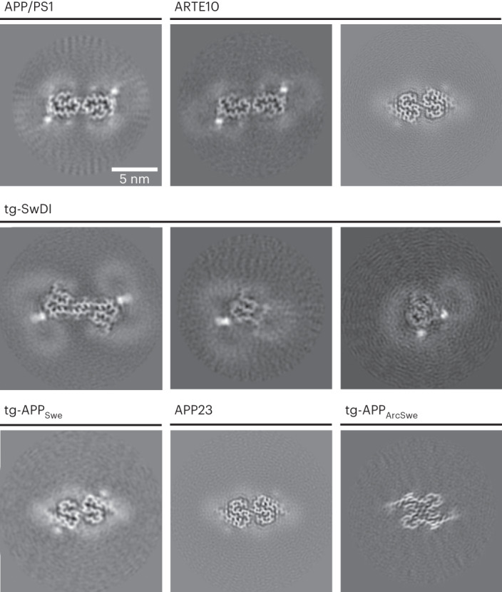
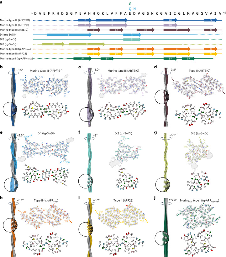
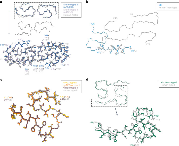
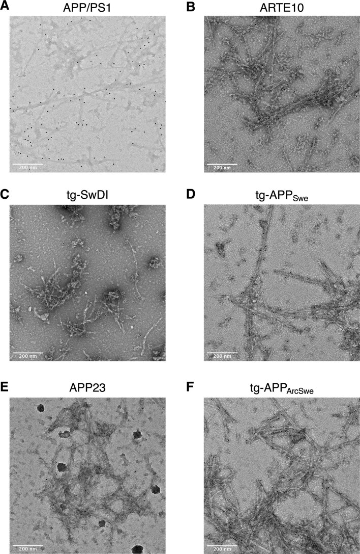
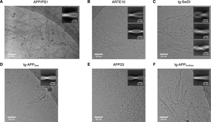
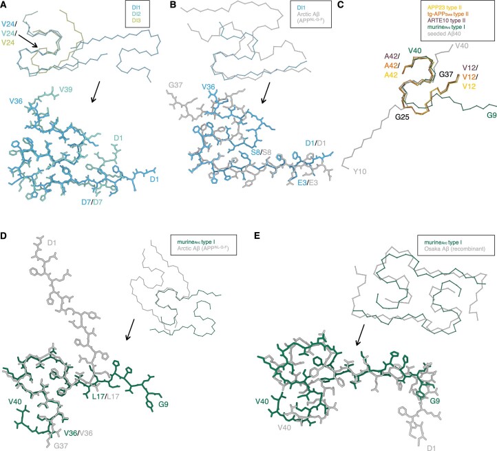
The use of transgenic mice displaying amyloid-β (Aβ) brain pathology has been essential for the preclinical assessment of new treatment strategies for Alzheimer's disease. However, the properties of Aβ in such mice have not been systematically compared to Aβ in the brains of patients with Alzheimer's disease. Here, we determined the structures of nine ex vivo Aβ fibrils from six different mouse models by cryogenic-electron microscopy. We found novel Aβ fibril structures in the APP/PS1, ARTE10 and tg-SwDI models, whereas the human type II filament fold was found in the ARTE10, tg-APPSwe and APP23 models. The tg-APPArcSwe mice showed an Aβ fibril whose structure resembles the human type I filament found in patients with sporadic Alzheimer's disease. A detailed assessment of the Aβ fibril structure is key to the selection of adequate mouse models for the preclinical development of novel plaque-targeting therapeutics and positron emission tomography imaging tracers in Alzheimer's disease.


Conflict of interest statement

L.N.G.N. is on the scientific advisory board and receives a research grant from BioArctic. M.I. is a paid consultant to BioArctic. D.W. is a founder and shareholder of the company Priavoid and a member of its supervisory board. D.W. is co-inventor of patents related to the compound RD2. D.W. is a founder and shareholder of attyloid. D.W. is a member of attyloid’s supervisory board. These had no influence on the interpretation of the data. Benedikt Frieg is now an AstraZeneca employee. All other authors declare no competing interests.

Figures

Fig. 1
Fig. 1. Cryo-EM reconstructions of Aβ fibrils extracted from APP/PS1, ARTE10, tg-SwDI, tg-APPSwe, APP23 and tg-APPArcSwe mouse brain tissue.
For every reconstructed fibril, a projection of the reconstructed density including approximately one β-rung is shown. The scale bar in the top left panel applies to all shown panels. From upper left to lower right: murine type III (APP/PS1), murine type III (ARTE10), murine type II (ARTE10), DI1, DI2, DI3, murine type II (tg-APPSwe), murine type II (APP23) and murineArc type I. The number of fibril segments used to generate the reconstructions is given in Extended Data Table 2.
Fig. 2
Fig. 2. Overview of all murine Aβ fibril structures.
a, Amino acid sequence of Aβ42. The sequence contains the following mutations for tg-SwDI: E22Q and D23N; and for tg-APPArcSwe: E22G. Solid lines indicate the part of the sequence for which atomic model building was possible (accordingly, dotted lines represent parts of the sequence that were not modeled). Arrows indicate β-strands. bj, Each panel shows the reconstructed cryo-EM density along the helical axis with a close-up and a label denoting the helical twist (left); the cryo-EM density map (in transparent gray) with the corresponding atomic model (top right); a schematic of the fold, produced with atom2svg.py (red, acidic side chain; blue, basic side chain; green, hydrophilic side chain; white, hydrophobic side chain; pink, glycine; yellow, sulfur-containing) (bottom right). Cryo-EM structure of murine type III Aβ42 fibrils from APP/PS1 mouse brain (b), murine type III Aβ fibrils from ARTE10 mouse brain (c), type II Aβ42 fibrils from ARTE10 mouse brain (d), DI1 Aβ fibrils from tg-SwDI mouse brain (e), DI2 Aβ fibrils from tg-SwDI mouse brain (f), DI3 Aβ fibrils from tg-SwDI mouse brain (g), type II Aβ42 fibrils from tg-APPSwe mouse brain (h), type II Aβ42 fibrils from APP23 mouse brain (i) and murineArc type I fibrils from tg-APPArcSwe mouse brain (j). For f and g (DI2 and DI3 fibrils from tg-SwDI mouse brain), the displayed atomic models have limited accuracy owing to the medium resolution.
Fig. 3
Fig. 3. Comparison of brain-derived murine Aβ fibrils to brain-derived human extracted Aβ fibrils.
a, Comparison of murine type III Aβ fibrils (blue, APP/PS1; lavender, ARTE10) with the cryo-EM structure of human brain-extracted Aß filaments with the E693G (E22G) mutation (gray; PDB 8BG0). b, Comparison of the DI1 Aβ fibril (light blue) with the cryo-EM structure of Aβ40 fibrils extracted from the meninges of human brain tissue from a patient with Alzheimer’s disease (gray; PDB 6SHS). c, Comparison of the APP23 (yellow), tg-APPSwe (orange) and ARTE10 (burgundy) Aβ42 fibril fold with human type II Aβ42 filaments (gray; PDB 7Q4M). d, Comparison of the murineArc type I (green) Aβ40 fibril fold with human type I Aβ42 filament fold (gray; PDB 7Q4B).
Extended Data Fig. 1
Extended Data Fig. 1. Immunohistochemical characterisation of mouse brain tissue.
Immunohistochemical staining showing Aβ plaques in the hippocampus (left images) and cortex (right images) of APP23 (upper panel), APP/PS1 (middle panel) and tg-SwDI mice (lower panel). Two different stainings were conducted (6E10 labelled in red and 4G8 labelled in green). Nuclear staining was done with DAPI (blue). Representative images are shown (n = 3). Scale bar= 500 μm.
Extended Data Fig. 2
Extended Data Fig. 2. Immunogold negative stain electron microscopy images of purified Aβ fibrils.
Immunogold negative-stain electron microscopy images of the purified Aβ fibrils from (A) APP/PS1 (n = 30), (B) ARTE10 (n = 6), (C) tg-SwDI (n = 21), (D) tg-APPSwe (n = 10), (E) APP23 (n = 8) and (F) tg-APPArcSwe (n = 15) mouse models. NAB228 was used as primary antibody. A goat anti-mouse gold-conjugated antibody with a gold particle diameter of 6 nm was used as secondary antibody.
Extended Data Fig. 3
Extended Data Fig. 3. Cryo-EM micrographs and 2D classes of purified Aβ fibrils.
One exemplary cryo-EM micrograph and 2D class averages of (A) APP/PS1, (B) ARTE10, (C) tg-SwDI, (D) tg-APPSwe, (E) APP23 and (F) tg-APPArcSwe. The number of micrographs recorded for each sample is given in Extended Data Table 2. The number of fibril segments used to generate the displayed 2D classes are given in Extended Data Table 2 (n=final particle images).
Extended Data Fig. 4
Extended Data Fig. 4. Comparison of murine Aβ fibrils with other structures.
(A) Comparison of the main chain trace of DI1 (light blue) with DI2 (teal) and DI3 (light green) (top) and comparison of DI1 and DI2 (bottom). (B) Comparison of DI1 (light blue) and murine Aβ42(E22G) filaments extracted from knock-in APPNL-G-F mice (gray, PDB code: 8BG9). (C) Comparison of APP23 type II (yellow), tg-APPSwe type II (orange), ARTE10 type II (burgundy), tg-APPArcSwe murineArc type I (green) fibrils with the cryo-EM structure of Aβ40 fibrils seeded from brain homogenates from cortical tissue of an AD patient (gray, PDB code: 6W0O). (D) Comparison of murineArc type I Aβ40 (green) with murine Aβ42(E22G) filaments extracted from knock-in APPNL-G-F mice (gray, PDB code: 8BG9). (E) Comparison of murineArc type I Aβ40 (green) with an NMR structure of recombinant Aβ40 E22Δ fibrils (gray, PDB code: 2MVX).
Extended Data Fig. 5
Extended Data Fig. 5. Additional densities bound to murine Aβ fibrils.
(A,B) Reconstruction of Aβ fibrils extracted from (A) APP/PS1 and (B) tg-SwDI mice brain. Red arrows indicate localized, strong density, blue arrows indicate micelle-like, weak densities. (C,D) show extra densities close to (C) Lys16 and (D) Phe20/Glu22 in APP/PS1 murine type III Aβ fibrils.

Similar articles

Cited by

References

    1. Long JM, Holtzman DM. Alzheimer disease: an update on pathobiology and treatment strategies. Cell. 2019;179:312–339. - PMC - PubMed
    1. Scheltens P, et al. Alzheimer’s disease. Lancet. 2016;388:505–517. - PubMed
    1. Nelson PT, et al. Correlation of Alzheimer disease neuropathologic changes with cognitive status: a review of the literature. J. Neuropathol. Exp. Neurol. 2012;71:362–381. - PMC - PubMed
    1. Chiti F, Dobson CM. Protein misfolding, amyloid formation, and human disease: a summary of progress over the last decade. Annu. Rev. Biochem. 2017;86:27–68. - PubMed
    1. Chen G, et al. Amyloid beta: structure, biology and structure-based therapeutic development. Acta Pharmacol. Sin. 2017;38:1205–1235. - PMC - PubMed

Substances