. 2023 Nov 16;21(1):819.
doi: 10.1186/s12967-023-04703-5.

PTPRH promotes the progression of non-small cell lung cancer via glycolysis mediated by the PI3K/AKT/mTOR signaling pathway

Affiliations

PTPRH promotes the progression of non-small cell lung cancer via glycolysis mediated by the PI3K/AKT/mTOR signaling pathway

Shu Wang et al. J Transl Med. .

Abstract

Background: The protein tyrosine phosphatase H receptor (PTPRH) is known to regulate the occurrence and development of pancreatic and colorectal cancer. However, its association with glycolysis in non-small cell lung cancer (NSCLC) is still unclear. In this study, we aimed to investigate the relationship between PTPRH expression and glucose metabolism and the underlying mechanism of action.

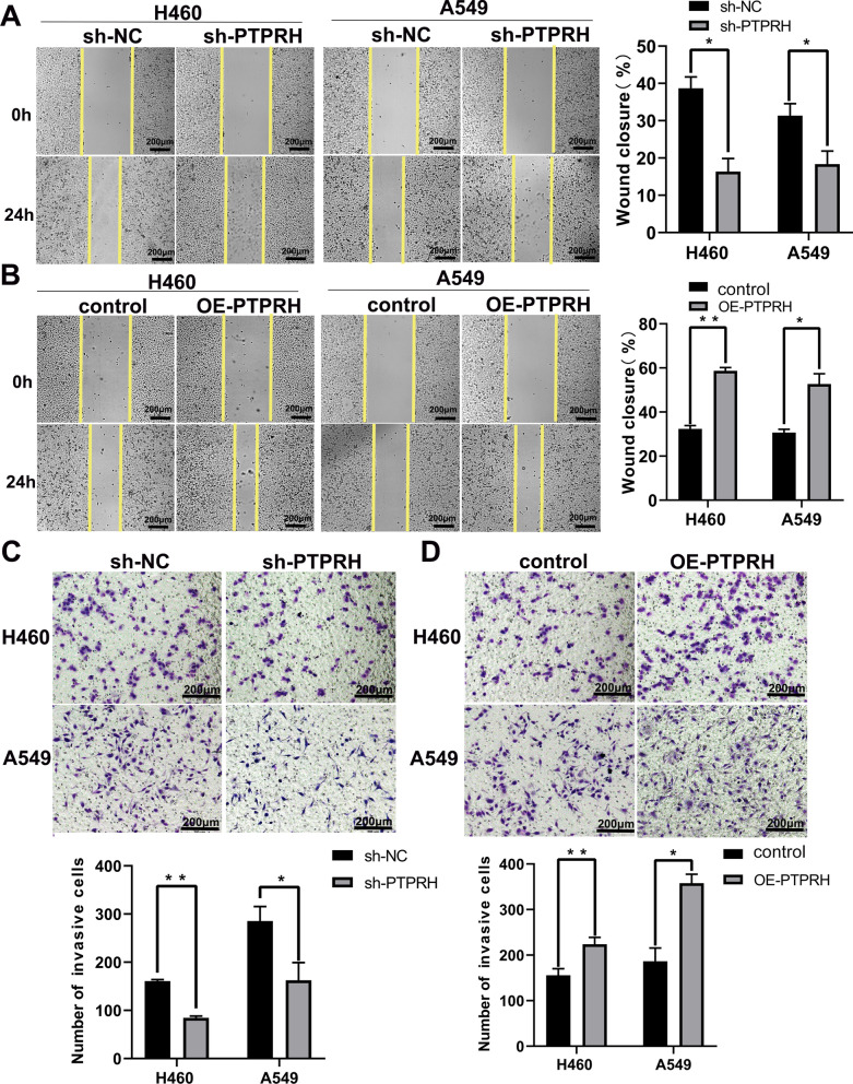
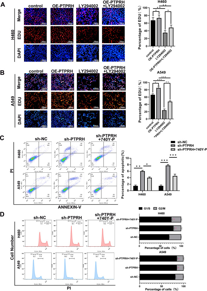
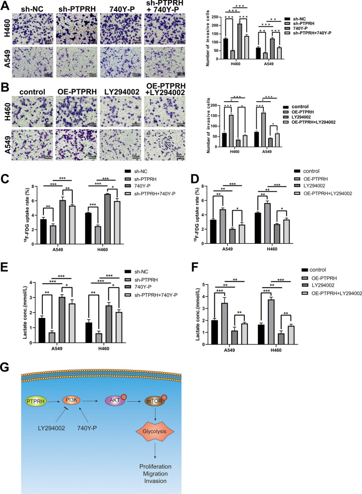
Methods: The expression of PTPRH in NSCLC cells was evaluated by IHC staining, qRT‒PCR and Western blotting. The effect of PTPRH on cell biological behavior was evaluated by colony assays, EdU experiments, Transwell assays, wound healing assays and flow cytometry. Changes in F-18-fluorodeoxyglucose (18F-FDG) uptake and glucose metabolite levels after altering PTPRH expression were detected via a gamma counter and lactic acid tests. The expression of glycolysis-related proteins in NSCLC cells was detected by Western blotting after altering PTPRH expression.

Results: The results showed that PTPRH was highly expressed in clinical patient tissue samples and closely related to tumor diameter and clinical stage. In addition, PTPRH expression was associated with glycometabolism indexes on 18F-FDG positron emission tomography/computed tomography (PET/CT) imaging, the expression level of Ki67 and the expression levels of glycolysis-related proteins. PTPRH altered cell behavior, inhibited apoptosis, and promoted 18F-FDG uptake, lactate production, and the expression of glycolysis-related proteins. In addition, PTPRH modulated the glycometabolism of NSCLC cells via the phosphatidylinositol-3-kinase (PI3K)/protein kinase B (AKT)/mammalian target of rapamycin (mTOR) signaling pathway, as assessed using LY294002 and 740Y-P (an inhibitor and agonist of PI3K, respectively). The same results were validated in vivo using a xenograft tumor model in nude mice. Protein expression levels of PTPRH, glycolysis-related proteins, p-PI3K/PI3K and p-AKT/AKT were measured by IHC staining using a subcutaneous xenograft model in nude mice.

Conclusions: In summary, we report that PTPRH promotes glycolysis, proliferation, migration, and invasion via the PI3K/AKT/mTOR signaling pathway in NSCLC and ultimately promotes tumor progression, which can be regulated by LY294002 and 740Y-P. These results suggest that PTPRH is a potential therapeutic target for NSCLC.

Keywords: Computed tomography; F-18-fluorodeoxyglucose; Glycolysis; Non-small cell lung cancer; Positron emission tomography; Protein tyrosine phosphatase H receptor.


Conflict of interest statement

The authors declare that they have no known competing financial interests or personal relationships that could have appeared to influence the work reported in this paper.

Figures

Fig. 1
Fig. 1
PTPRH is highly expressed in NSCLC and is closely related to poor prognosis. A, B One-way Cox analysis of 10 genes with prognostic value in two GEO-independent datasets (GSE31210 and GSE30219). C Genes with prognostic value from one-way Cox analysis of TCGA-LUAD data were identified by intersection with genes in the GEO dataset. D Analysis of differentially expressed genes in TCGA-LUAD, presented as volcano plots. EG Correlations of FAM83A, COL11A1, and PTPRH expression with survival prognosis. H Correlation analysis of PTPRH expression with glycolysis. I Correlation analysis of PTPRH expression with glycolysis in TCGA pancancer data. PTPRH: protein tyrosine phosphatase H receptor gene; NSCLC: non-small cell lung cancer; GEO: Gene Expression Omnibus; TCGA: The Cancer Genome Atlas; LUAD: lung adenocarcinoma
Fig. 2
Fig. 2
Correlation of PTPRH expression with 18F-fluorodeoxyglucose (18F-FDG) semiquantitative indicators, the expression of proliferation markers and the expression of glycolysis-related proteins. A, B Detection of PTPRH expression by immunohistochemistry (×400). C Correlation of PTPRH expression with 18F-FDG accumulation and the expression levels of Ki67. D In patients with high levels of PTPRH expression, immunohistochemical staining revealed high expression levels of GLUT1, HK2, PKM2, and LDHA (×400), whereas patients with low levels of PTPRH expression expressed low levels of GLUT1, HK2, PKM2 and LDHA (× 400). E The expression levels of PTPRH correlated positively with SUVmax, MTV, and TLG values and the expression of Ki67, GLUT1, HK2, PKM2, LDHA. 18F-FDG: 18F-fluorodeoxyglucose; GLUT1: glucose transporter type 1; HK2: hexokinase 2; PKM2: pyruvate kinase M2; LDHA: lactate dehydrogenase A; SUVmax: maximum standard uptake value
Fig. 3
Fig. 3
PTPRH promoted proliferation in NSCLC. A, B Detection of PTPRH expression using RT‒PCR and western blotting. C, D Expression of PTPRH after transfection by western blotting analysis and RT‒PCR. E The results of the colony formation assay.) F 3-(4,5-dimethylthiazol-2-yl)-2,5-diphenyltetrazolium bromide (MTT) assay. G, H 5-ethynyl-2′-deoxyuridine (EdU) assay show that PTPRH enhances the proliferation ability of NSCLC cells
Fig. 4
Fig. 4
PTPRH promoted migration and invasion in NSCLC. A, B The results of the scratch assay show that PTPRH promotes the migration ability of NSCLC cells. C, D The results of the Transwell assays show that PTPRH enhances the invasion ability of NSCLC cells
Fig. 5
Fig. 5
PTPRH inhibited cell cycle arrest and apoptosis. A, B Gene Set Enrichment Analysis (GSEA) of PTPRH. C, D Flow cytometry analysis of the apoptotic effect of PTPRH on NSCLC and statistical analysis. E, F Flow cytometry analysis of the effects of PTPRH expression on the cell cycle in NSCLC and statistical analysis. G Western blotting results showing the effects of short hairpin RNA-PTPRH (sh-PTPRH) treatment on cell cycle-related proteins. H Western blotting results showing the effects of sh-PTPRH treatment on the expression of apoptotic proteins
Fig. 6
Fig. 6
PTPRH enhanced glycolysis in vivo and in vitro. A Xenograft tumor sizes in the sh-PTPRH and sh-negative control (sh-NC) groups. B, C The volume and weight of tumors in the sh-PTPRH group were significantly decreased compared to those in the sh-NC group. D Representative hematoxylin–eosin (H&E) images of Ki67 and PTPRH staining in A549 xenografts. E, F Representative micro-PET images of mice in the sh-PTPRH and sh-NC groups. The closer the color of the tumor within the circle of mice is to red means the higher accumulation of 18F-FDG. The accumulation of 18F-FDG in the tumors of sh-NC-treated mice was significantly greater than that in the tumors of sh-PTPRH-treated mice. G Representative immunostained images of PTPRH, GLUT1, HK2, PKM2, and LDHA staining in A549 xenografts. H Cellular 18F‐FDG uptake was significantly decreased in the sh-PTPRH group and increased in the PTPRH-overexpressing (OE-PTPRH) group. I Lactate levels in the culture medium of the sh-PTPRH-treated group were significantly decreased, whereas they were increased in the OE-PTPRH-treated group. J Relative expression levels of glycolysis-related proteins in the sh-PTPRH and sh-NC groups by western blotting
Fig. 7
Fig. 7
PTPRH enhanced glycolysis via the phosphatidylinositol-3-kinase (PI3K)/Protein kinase B (AKT)/mammalian target of rapamycin (mTOR) signaling pathway. A GSEA to identify enriched signaling pathways for PTPRH. B Representative immunostaining images after staining of PI3K/AKT/mTOR signaling pathway-related proteins in A549 xenografts. C, D Relative expression levels of PI3K/AKT/mTOR signaling pathway-related proteins in the sh-PTPRH and sh-NC groups by western blotting. E Western blotting results showing the effects of sh-PTPRH and the PI3K activator 740Y-P alone and in combination on the expression levels of glycolysis-related proteins. F Western blotting results showing the effects of OE-PTPRH and the PI3K inhibitor LY294002 alone and in combination on the expression levels of glycolysis-related proteins
Fig. 8
Fig. 8
PTPRH promoted proliferation and invasion via PI3K/AKT/mTOR signaling pathway-mediated glycolysis. A, B EdU assay to verify the individual and combined effects of sh-PTPRH and the PI3K inhibitor LY294002 on the number of cells undergoing DNA replication. C, D Flow cytometry assays to detect the reversal of the proapoptotic effect of sh-PTPRH after treatment with the PI3K activator 740Y-P in H460 and A549 cells
Fig. 9
Fig. 9
PTPRH increased invasion, FDG uptake and lactate levels via PI3K/AKT/mTOR signaling pathway-mediated glycolysis. A, B Transwell assays to detect the individual and combined effects of sh-PTPRH and the PI3K activator 740Y-P and OE-PTPRH and the PI3K inhibitor LY294002 on the invasion ability of H460 and A549 cells. C, D 18F-FDG uptake assays to detect the individual and combined effects of sh-PTPRH and the PI3K activator 740Y-P and OE-PTPRH and the PI3K inhibitor LY294002 on the FDG uptake ability of H460 and A549 cells. E, F Cell metabolism assays to detect the individual and combined effects of sh-PTPRH and the PI3K activator 740Y-P and OE-PTPRH and the PI3K inhibitor LY294002 on the lactate levels of H460 and A549 cells. G A diagram showing the possible mechanism underlying the modulation of glycolysis by PTPRH

References

    1. Siegel RL, Miller KD, Fuchs HE, Jemal A. Cancer statistics, 2022. Cancer J Clin. 2022;72(1):7–33. doi: 10.3322/caac.21708. - DOI - PubMed
    1. Wood DE, Kazerooni EA, Baum SL, Eapen GA, Ettinger DS, Hou L, Jackman DM, Klippenstein D, Kumar R, Lackner RP, et al. Lung cancer screening, Version 3.2018, NCCN clinical practice guidelines in oncology. J Natl Compr Cancer Netw. 2018;16(4):412–441. doi: 10.6004/jnccn.2018.0020. - DOI - PMC - PubMed
    1. Kim S, Im JH, Kim WK, Choi YJ, Lee JY, Kim SK, Kim SJ, Kwon SW, Kang KW. Enhanced sensitivity of nonsmall cell lung cancer with acquired resistance to epidermal growth factor receptor-tyrosine kinase inhibitors to phenformin: the roles of a metabolic shift to oxidative phosphorylation and redox balance. Oxid Med Cell Longev. 2021;2021:5428364. doi: 10.1155/2021/5428364. - DOI - PMC - PubMed
    1. Warburg O. On respiratory impairment in cancer cells. Science. 1956;124(3215):269–270. doi: 10.1126/science.124.3215.269. - DOI - PubMed
    1. Liao X, Liu M, Wang R, Zhang J. Potentials of non-invasive (18)F-FDG PET/CT in immunotherapy prediction for non-small cell lung cancer. Front Genet. 2021;12:810011. doi: 10.3389/fgene.2021.810011. - DOI - PMC - PubMed

Publication types

MeSH terms

Substances