. 2023 Nov 16;21(1):430.
doi: 10.1186/s12951-023-02207-4.

Topical bismuth oxide-manganese composite nanospheres alleviate atopic dermatitis-like inflammation

Affiliations

Topical bismuth oxide-manganese composite nanospheres alleviate atopic dermatitis-like inflammation

Mengjie Li et al. J Nanobiotechnology. .

Retraction in

Abstract

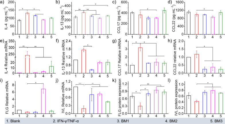
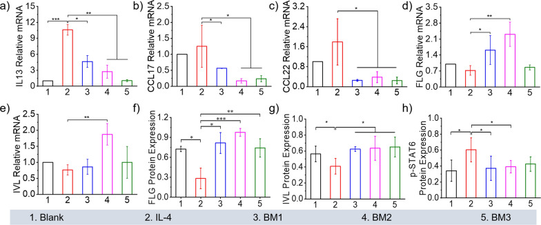
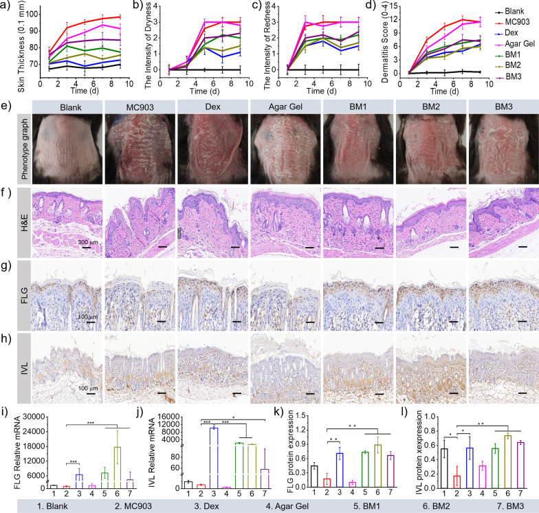
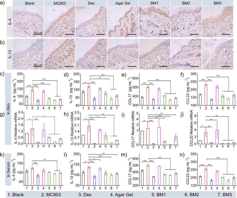
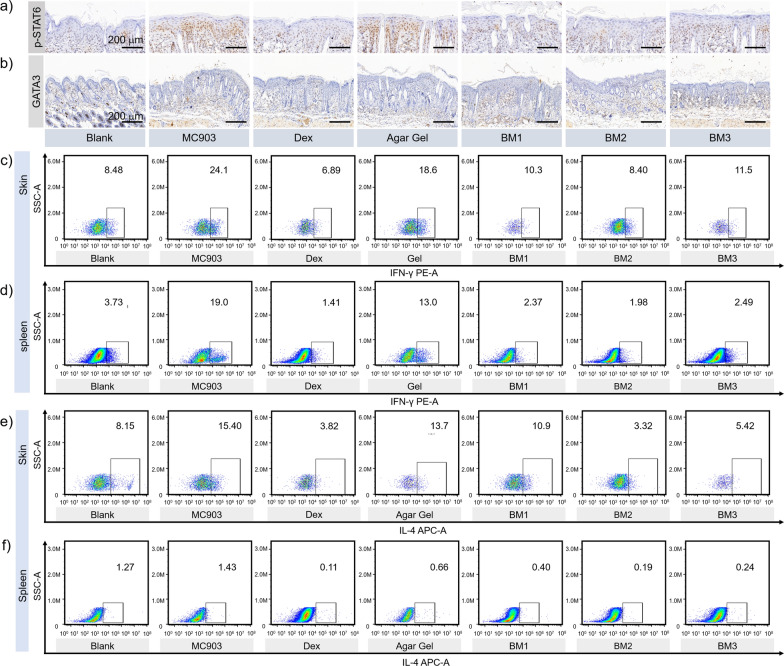
Atopic dermatitis (AD) is a common skin disease involving important immune mechanisms. There is an unmet need for a treatment for this condition. Herein, we focused on elucidating the role of Bi2-xMnxO3 nanospheres (BM) in alleviating skin inflammation in AD-like C57BL/6 mice. The BM was fabricated via sacrificial templates and its biosafety was systematically evaluated. The BM was applied topically to skin lesions of AD-like C57BL/6 mice. The phenotypic and histological changes in the skin were examined carefully. The responses of barrier proteins, inflammatory cytokines and cells to BM were evaluated in HaCaT cells and AD mouse models. The data demonstrated that BM treatment alleviated the AD phenotypes and decreased the level of inflammatory factors, while increasing the expression of the barrier proteins filaggrin/involucrin in the skin. BM effectively reduced the expression of phosphorylated STAT6, which in turn reduced the expression of GATA3, and further decreased the differentiation ratio of Th2 cells, thereby reducing the expression of IL-4. In conclusion, topical drug therapy with BM provides a safe and effective treatment modality for AD by reducing IL-4 and increasing barrier proteins.

Keywords: Atopic dermatitis; Bi2-xMnxO3 nanospheres; IL-4; STAT6; Skin inflammation.


Conflict of interest statement

The authors declare that they have no competing interests.

Figures

Fig. 1
Fig. 1
Schematic illustration of BM alleviating AD. Herein, BM were prepared via sacrificial Bi2S3 templates, which were loaded in agar gel as STAT phosphorylation regulators for treatment of MC903-induced AD-like mice. BM reduced the expression of IL-4, IL-13 and p-STAT6 in TNF-α/IFN-γ-induced HaCaT inflammatory cells and in the MC903-induced mouse AD model, thereby effectively improving the expression of the barrier protein FLG/IVL and promoting the recovery of damaged skin. Moreover, the Th1 and Th2 cell types in mouse skin and spleen were considerably decreased after BM administration in MC903-induced AD mice, indicating that the as-prepared BM inhibits the Th2 cell response in the AD mouse model
Fig. 2
Fig. 2
Characterization of BM2 and BM2 agar gel (a) SEM of BM nanospheres. b STEM of BM2 nanospheres and corresponding elemental mappings of Bi, Mn and O. Scale bars 500 nm. ce XPS spectrum of O 1 s peaks, Bi 4f, and Mn 2p. f SEM image and digital picture of the BM2 agar gel. g, h Rheological properties of the BM2 agar gel. i Elemental mappings of the BM2 agar gel containing Bi, Mn and O
Fig. 3
Fig. 3
The inflammation levels of HaCaT cells after TNF-α/IFN-γ stimulation and BM treatment. The levels of (a) IL-4, b IL-13, c CCL17, and (d) CCL22 in HaCaT cells analysed by ELISAs. The mRNA levels of the inflammatory factors IL-4, IL-13, CCL17 and CCL22 (eh) were analysed by qRT-PCR. Corresponding mRNA levels of (i) FLG and (j) IVL in HaCaT cells from each group. The levels of the proteins (k) FLG and (l) IVL in different groups were normalized against the protein expression of GAPDH, and the corresponding protein greyscale statistics were analysed. One-way ANOVA was used for data analysis in (ad) and (km), and the Kruskal–Wallis test was used for data analysis in (i, j). The results are expressed as the mean ± standard deviation (n = 5) (*P < 0.05, **P < 0.01, ***P < 0.001)
Fig. 4
Fig. 4
The levels of inflammatory factors and barrier proteins in HaCaT cells after IL-4 stimulation and BM treatment. The mRNA levels of (a) IL-13, (b) CCL17 and (c) CCL22 in HaCaT cells. Corresponding mRNA levels of (d) FLG, and (e) IVL in HaCaT cells from each group. (fh) Corresponding protein greyscale statistical analysis in HaCaT cells from each group normalized against protein expression of GAPDH. The results are expressed as the mean ± standard deviation. The Kruskal–Wallis test was used for data analysis in (ae), and one-way ANOVA was used for data analysis in (fh) (n = 5) (*P < 0.05, **P < 0.01, ***P < 0.001)
Fig. 5
Fig. 5
Skin penetration, cellular uptake and biosafety evaluation. a Penetration of NR-loaded BM agar gel (red) in AD-like lesion skin after topical administration by CLSM. Scale bar: 300 μm. b Cellular uptake evaluation of HaCaT cells after 4 h incubation with NR loaded BM at different concentrations (20, 40 and 60 μg mL−1). Scale bar: 50 μm. c, d Content of Mn retained in skin and major organs, including the liver, kidney, heart, spleen, and lung, calculated by ICP-MS. eh γ-GT, AST, UBN and CRE levels in mouse serum after 9 days of treatment with BM agar gel. i H&E staining images of the excised organs of AD-like mice at 9 days post treatment. The results are expressed as the mean ± standard deviation. One-way ANOVA was used for data analysis in (e), and the Kruskal–Wallis test was used for data analysis in (g, h) (n = 5) (* P < 0.05, ** P < 0.01)
Fig. 6
Fig. 6
Dermatitis score, the levels of inflammation factors and barrier proteins of mouse skin lesions during 9 days of the experiment (a) skin thickness, (b) dryness intensity, (c) redness intensity, (d) dermatitis score, (e) phenotype graph of mouse dorsum from each group. f H&E staining images of dorsal skin sections (magnification, × 200). g, h The expression of FLG and IVL determined by IHC analysis (magnification, × 100). i, j The mRNA expression of FLG and IVL in the skin from each group was analysed by real-time PCR. k, l Density levels of the FLG and IVL proteins in the dorsal skin of mice normalized against the protein expression of GAPDH. The results are expressed as the mean ± standard deviation. One-way ANOVA was used for data analysis in (i, j), and the Kruskal–Wallis test was used for data analysis in (k, l) (n = 5) (*P < 0.05, **P < 0.01, ***P < 0.001)
Fig. 7
Fig. 7
Inflammatory factor levels of IL-4, IL-13 CCL 17, and CCL 22 in mice after treatment. Protein expression of (a) IL-4 and (b) IL-13 by IHC analysis (magnification, × 200). cf Corresponding mRNA levels of IL-4, IL-13, CCL17 and CCL22 in skin from each group obtained by qRT-PCR. Relative concentrations of IL-4, IL-13, CCL17 and CCL22 analysed by ELISAs, (gj) in skin, (kn) in serum. The results are expressed as the mean ± standard deviation. One-way ANOVA was used for data analysis in (cf) and (kn), and the Kruskal–Wallis test was used for data analysis in (gj) (n = 5) (*P < 0.05, **P < 0.01, ***P < 0.001)
Fig. 8
Fig. 8
The effect of BM on Th2-mediated immune regulation in MC903-induced AD-like mice. The expression of (a) p-STAT6 and (b) GATA3 by IHC analysis (magnification, × 100). The proportion of CD4+/IFN-λ+ T-lymphocytes detected by flow cytometry originated from (c) skin and (d) spleen tissue. The proportion of CD4+/IL-4+ T cells detected by flow cytometry originating from skin (e) and spleen (f) tissue (n = 5)

References

    1. Guo Y, Li P, Tang J, Han X, Zou X, Xu G, Xu Z, Wei F, Liu Q, Wang M, et al. Prevalence of atopic dermatitis in chinese children aged 1–7 ys. Sci Rep. 2016;6:29751. doi: 10.1038/srep29751. - DOI - PMC - PubMed
    1. Langan SM, Irvine AD, Weidinger S. Atopic dermatitis. Lancet. 2020;396:345–360. doi: 10.1016/S0140-6736(20)31286-1. - DOI - PubMed
    1. Silverberg JI, Barbarot S, Gadkari A, Simpson EL, Weidinger S, Mina-Osorio P, Rossi AB, Brignoli L, Saba G, Guillemin I, et al. Atopic dermatitis in the pediatric population: a cross-sectional, international epidemiologic study. Ann Allergy Asthma Immunol. 2021;126:417–428.e412. doi: 10.1016/j.anai.2020.12.020. - DOI - PubMed
    1. Stander S. Atopic dermatitis. N Engl J Med. 2021;384:1136–1143. doi: 10.1056/NEJMra2023911. - DOI - PubMed
    1. Brunello L. Atopic dermatitis. Nat Rev Dis Primers. 2018;4:2. doi: 10.1038/s41572-018-0004-9. - DOI - PubMed

Publication types