

Unveiling a Rarity: A Case Report on Glomangiopericytoma in the nasal cavity
- PMID: 37974783
- PMCID: PMC10645710
- DOI: 10.1007/s12070-023-03975-z
Unveiling a Rarity: A Case Report on Glomangiopericytoma in the nasal cavity
Abstract
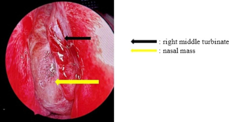
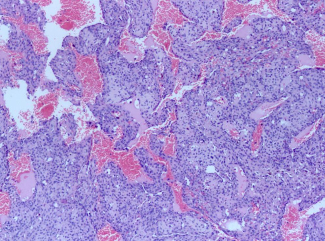
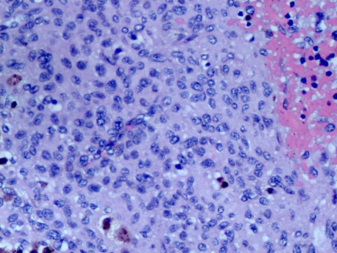
Sino-nasal glomangiopericytoma is a rare benign tumour comprising only about 0.5% of all sino-nasal tumours. Presenting as a bleeding nasal mass, it is among the myriad of differential diagnoses for the same. Clinical characterisation of mass becomes difficult; hence, histopathology and immunohistocytochemistry play an essential role in clenching the diagnosis. Optimal treatment includes complete tumour excision with endoscopic or open approaches with or without preoperative embolization and a long post-operative follow-up period. Here we report such a case treated with endoscopic approach.
Supplementary information: The online version contains supplementary material available at 10.1007/s12070-023-03975-z.
Keywords: Benign tumours of nose/ paranasal sinus; Glomangiopericytoma; Hemangiopericytoma; Spontaneous epistaxis; Vascular nasal mass.
© Association of Otolaryngologists of India 2023. Springer Nature or its licensor (e.g. a society or other partner) holds exclusive rights to this article under a publishing agreement with the author(s) or other rightsholder(s); author self-archiving of the accepted manuscript version of this article is solely governed by the terms of such publishing agreement and applicable law.
Conflict of interest statement
Conflicts of Interest/Competing InterestsNone to declare.
Figures
References
LinkOut - more resources
Full Text Sources