Review
. 2023 Sep 15;15(5):983-998.
doi: 10.1007/s12551-023-01129-7. eCollection 2023 Oct.

Phthalocyanine aggregates in the photodynamic therapy: dogmas, controversies, and future prospects

Affiliations
Review

Phthalocyanine aggregates in the photodynamic therapy: dogmas, controversies, and future prospects

Dmitry A Bunin et al. Biophys Rev. .

Abstract

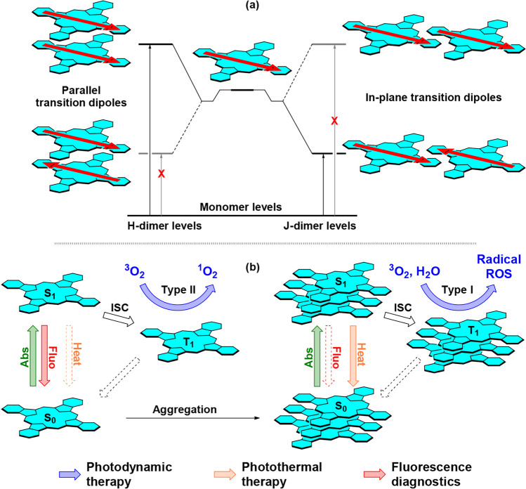
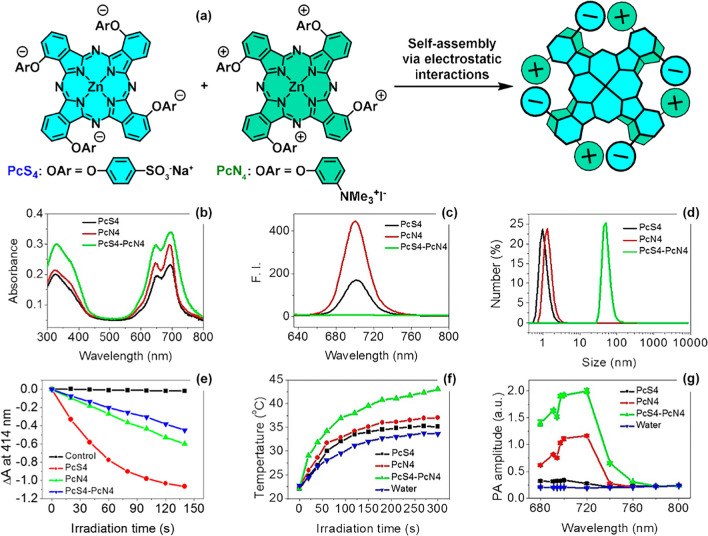
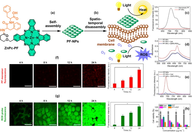
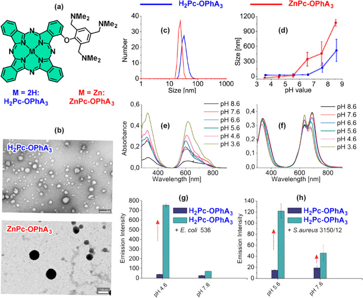
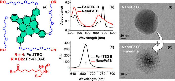
Photodynamic therapy (PDT), a rapidly developing method for the treatment of cancer and bacterial diseases, is based on the photosensitization of oxygen to generate reactive oxygen species (ROS) that destroy specific biological targets. Among the various photosensitizers, phthalocyanines (Pc) have attracted particular attention due to their excellent photophysical properties, most of which meet the therapeutic requirements. The statement that aggregation of Pc-based photosensitizers is undesirable because it suppresses ROS generation has become commonplace in PDT. In this review, we have collected and discussed a number of works whose results refute this well-established axiom and show that aggregated forms of phthalocyanines can still exhibit photodynamic activity, in some cases in synergy with the photothermal and optoacoustic effects. In addition, ROS generation can be induced by aggregates under the conditions of sonodynamic therapy.

Keywords: Nanocarrier; Nanomedicine; Photodynamic therapy; Photophysics; Reactive oxygen species; Self-assembly.


Conflict of interest statement

Conflict of interestThe authors declare no competing interests.

Figures

Fig. 1
Fig. 1
a Simplified diagram of energy levels in phthalocyanine monomers, H- and J-aggregates (dimers are considered for simplicity). The red arrows show orientations of molecular transition dipoles. The crossed-out arrows are forbidden transitions (Kasha et al. 1965). b Simplified Jablonski diagrams showing the formation and deactivation pathways of excited states of monomeric and aggregated Pcs
Fig. 2
Fig. 2
a Structures of the phthalocyanine SiPc-NH2 in cis- and trans-conformations. b Schematic representation of the formation of SiPcNano J-aggregates. c UV–vis spectra of monomeric SiPc-NH2 in DMF (red line) and as J-aggregate in water (black line). d Fluorescence images after irradiation of SiHa cells stained with DCFH-DA and incubated with SiPcNano. Green fluorescence is due to the oxidized form of DCFH-DA formed after its reaction with total ROS; the control is cells in PBS. e Fluorescence images of SiHa cells incubated with SiPcNano; control – cells in PBS. f Fluorescence images of SiHa cells incubated with SiPcNano and Cremofor EL surfactant. Used with permission of Royal Society of Chemistry, from Pan et al. (2020); permission conveyed through Copyright Clearance Center, Inc
Fig. 3
Fig. 3
a Zinc tetra-(6-hydroxyhexyloxy)-phthalocyaninate ZnPcHexOH and SEM images of aggregates ZnPcHexOH-NPs in water. UV–vis b and fluorescence c spectra of ZnPcHexOH (in DMSO, black lines) and aggregates (in water, red lines). d Fluorescence images of cells stained with Calcein AM and propidium iodide (PI) and incubated with aggregates before (left) and after irradiation (650 nm, 10 min). The disappearance of the green emission of Calcein AM indicates cell death, which is also indicated by the appearance of the red emission of propidium iodide, which does not penetrate into the cells but intercalates into the DNA of dead cells. e Generation of ROS mediated by aggregates in HeLa cells, detected by staining with DCFH-DA, the green emission indicates its oxidation. Reprinted from Wang et al. (2019), Copyright (2019), with permission from Elsevier
Fig. 4
Fig. 4
a Zinc tris-(dimethylaminomethyl)-phenoxyphthalocyaninate ZnPc-OPhA3 and its self-assembly into NanoPcA aggregates in aqueous solution. b Size distribution of NanoPcA aggregates found by DLS in water (black line) and in PBS buffer (red line); the inset shows a TEM image of a NanoPcA aggregate. c Cryo-TEM images of E. coli bacterial cells incubated with NanoPcA; red arrows indicate Pc nanoparticles. d Cryo-TEM images of E. coli bacterial cells incubated with NanoPcA after irradiation (655 nm, 10 min); yellow arrows indicate bacterial membrane damage. Copyright (2018) Wiley. Used with permission from Li et al. (2018a)
Fig. 5
Fig. 5
a Cationic zinc phthalocyaninates modified with and without octyl substituents ZnPcBinOct and ZnPcBin. Absorption spectra of intracellular contents after lysis of E. coli b and S. aureus c in DMSO; in both cases, higher ZnPcBin accumulation was observed in both bacterial cells. Copyright (2021) Wiley. Used with permission from Revuelta-Maza et al. (2021)
Fig. 6
Fig. 6
a Simplified scheme of coaggregation of sodium salt of tetra-(para-sulfophenoxy)-substituted zinc phthalocyaninate PsS4 and tetra-(trimethylaminophenoxy)-substituted zinc phthalocyaninate PcN4 with the formation of PsS4-PcN4 aggregates. UV–vis b, fluorescence c, and size distributions d of PsS4, PcN4 and PsS4-PcN4 in aqueous solution. e Monitoring of singlet oxygen trap — 1,3-diphenylizobensofuran, DPBF oxidation by PsS4, PcN4. and PsS4-PcN4 aggregate over time. f Temperature increase for PsS4 and PcN4 and the PsS4-PcN4 aggregate under irradiation (655 nm). g Photoacoustic signal amplitude for PsS4 and PcN4 and the PsS4-PcN4 aggregate at different wavelengths. Copyright (2020) Wiley. Used with permission from Li et al. (2020)
Fig. 7
Fig. 7
a Zinc phthalocyanine PcN4-BA functionalized with boronic acid residues, its self-assembly in aqueous solution and b interaction of PcN4-BA aggregates with glycans on the bacterial membrane surface. c TEM images of nanoparticles formed by PcN4-BA aggregates in aqueous solution. d E. coli bacterial cells incubated with PcN4-BA and e cryo-TEM images of E. coli with PcN4-BA. Red arrows indicate PcN4-BA nanoparticles. Reproduced from Ref. (Lee et al. 2020) with permission from the Royal Society of Chemistry
Fig. 8
Fig. 8
Zinc tetracarboxy-octafluoro- (F8(CO2H)4PcZn) and hexadecafluorophthalocyanates (F16PcZn) and their fluorescence spectra in HeLa cells. Reprinted from Oda et al. (2000), Copyright (2000), with permission from Elsevier
Fig. 9
Fig. 9
Zinc phthalocyaninates LysnPcZn conjugated with tri-, penta-, and heptalysine residues a, their absorption b, and fluorescence spectra c on the surface of the bacterial cell membrane and in the lysis buffer d. Used with permission of World Scientific Publishing Co., Inc., from Liu et al. (2018); permission conveyed through Copyright Clearance Center, Inc
Fig. 10
Fig. 10
Zinc phthalocyaninate conjugate with L-phenylalanine-L-phenylalanine dipeptide ZnPcPF, its self-assembly solution giving PF-NPs nanoparticles a and their disassembly b upon contact with the cell membrane; fluorescence spectra of monomeric ZnPcPF and PF-NPs nanoparticles c. UV–vis d and fluorescence e spectra of PF-NPs nanoparticles in water, in MCF-7 cell medium and in DSPE-PEG2000 lipid solution. f Fluorescence images of MCF-7 cells incubated with PF-NPs for 4, 8, 12, and 24 h and the intensity of red emission due to ZnPcPF phthalocyanine molecules leaving the PF-NPs nanoparticles. g Fluorescence images of MCF-7 cells incubated with PF-NPs for 4, 8, 12, and 24 h and with DCFH-DA with the intensity of emission. h Cell survival in PDT experiments, PTT experiments and in the combined mode. Copyright (2019) Wiley. Used with permission from Li et al. (2019)
Fig. 11
Fig. 11
a Structures of phthalocyanines H2Pc-OPhA3 and ZnPc-OPhA3. b TEM images of H2Pc-OPhA3 and ZnPc-OPhA3 aggregates in water. Dependence of the size of H2Pc-OPhA3c and ZnPc-OPhA3d aggregates on the pH of the aqueous solution. UV–vis of H2Pc-OPhA3e and ZnPc-OPhA3f at different pH of aqueous solution. Fluorescence intensity changes upon incubation with E. coli g and S. aureus h at different pH for H2Pc-OPhA3. Adapted with permission from Galstyan et al. (2020). Copyright 2020 American Chemical Society
Fig. 12
Fig. 12
Tetraethylene glycol–substituted zinc phthalocyaninate Pc-4TEG and its biotinylated analogue Pc-4TEG-B a, absorption b, and fluorescence spectra c in DMSO and in water, where these Pcs form aggregates. The aggregate formed by Pc-4TEG-B d and in the presence of avidin after incubation for 24 h e undergoes partial disassembly as evidenced by TEM. Adapted with permission from Li et al. (2017). Copyright 2017 American Chemical Society

References

    1. Bächle F, Maichle-Mössmer C, Ziegler T. Helical self-assembly of optically active glycoconjugated phthalocyanine J -aggregates. ChemPlusChem. 2019;84:1081–1093. doi: 10.1002/cplu.201900381. - DOI - PubMed
    1. Bandera Y, Burdette MK, Shetzline JA, et al. Synthesis of water soluble axially disubstituted silicon (IV) phthalocyanines with alkyne & azide functionality. Dye Pigment. 2016;125:72–79. doi: 10.1016/j.dyepig.2015.10.007. - DOI
    1. Bettini S, Valli L, Giancane G. Applications of photoinduced phenomena in supramolecularly arranged phthalocyanine derivatives: a perspective. Molecules. 2020;25:3742. doi: 10.3390/molecules25163742. - DOI - PMC - PubMed
    1. Bialas D, Kirchner E, Röhr MIS, Würthner F. Perspectives in dye chemistry: a rational approach toward functional materials by understanding the aggregate state. J Am Chem Soc. 2021;143:4500–4518. doi: 10.1021/jacs.0c13245. - DOI - PubMed
    1. Borzęcka W, Domiński A, Kowalczuk M (2021) Recent progress in phthalocyanine-polymeric nanoparticle delivery systems for cancer photodynamic therapy. Nanomaterials 11. 10.3390/nano11092426 - PMC - PubMed

LinkOut - more resources