

The emerging role of Piezo1 channels in skeletal muscle physiology
- PMID: 37975010
- PMCID: PMC10643716
- DOI: 10.1007/s12551-023-01154-6
The emerging role of Piezo1 channels in skeletal muscle physiology
Abstract
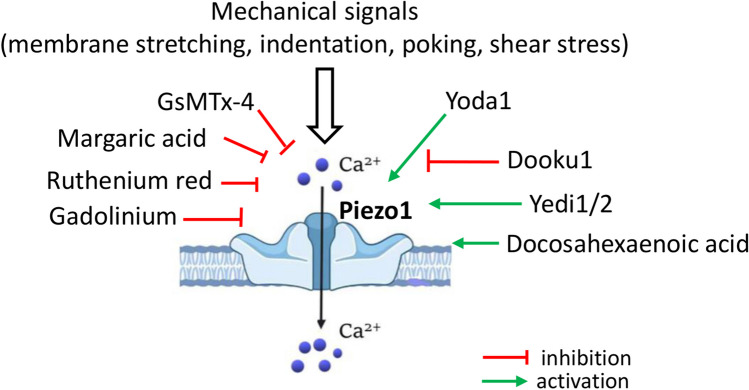
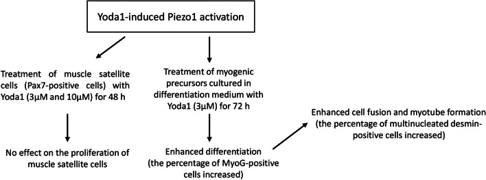
Piezo1 channels are mechanically activated (MA) cation channels that are involved in sensing of various mechanical perturbations, such as membrane stretch and shear stress, and play a crucial role in cell mechanotransduction. In response to mechanical stimuli, these channels open up and allow cations to travel into the cell and induce biochemical reactions that can change the cell's metabolism and function. Skeletal muscle cells/fibers inherently depend upon mechanical cues in the form of fluid shear stress and contractions (physical exercise). For example, an exposure of skeletal muscles to chronic mechanical loading leads to increased anabolism and fiber hypertrophy, while prolonged mechanical unloading results in muscle atrophy. MA Piezo1 channels have recently emerged as key mechanosensors that are capable of linking mechanical signals and intramuscular signaling in skeletal muscle cells/fibers. This review will summarize the emerging role of Piezo1 channels in the development and regeneration of skeletal muscle tissue as well as in the regulation of skeletal muscle atrophy. In addition, an overview of potential Piezo1-related signaling pathways underlying anabolic and catabolic processes will be provided. A better understanding of Piezo1's role in skeletal muscle mechanotransduction may represent an important basis for the development of therapeutic strategies for maintaining muscle functions under disuse conditions and in some disease states.
Keywords: Intracellular signaling; Mechanotransduction; Myogenesis; Piezo1; Skeletal muscle.
© International Union for Pure and Applied Biophysics (IUPAB) and Springer-Verlag GmbH Germany, part of Springer Nature 2023. Springer Nature or its licensor (e.g. a society or other partner) holds exclusive rights to this article under a publishing agreement with the author(s) or other rightsholder(s); author self-archiving of the accepted manuscript version of this article is solely governed by the terms of such publishing agreement and applicable law.
Conflict of interest statement
Competing InterestsThe author declares no competing interests.
Figures


References
-
- Blythe NM, Muraki K, Ludlow MJ, Stylianidis V, Gilbert HTJ, Evans EL, Cuthbertson K, Foster R, Swift J, Li J, Drinkhill MJ, van Nieuwenhoven FA, Porter KE, Beech DJ, Turner NA. Mechanically activated Piezo1 channels of cardiac fibroblasts stimulate p38 mitogen-activated protein kinase activity and interleukin-6 secretion. J Biol Chem. 2019;294(46):17395–17408. doi: 10.1074/jbc.RA119.009167. - DOI - PMC - PubMed
Publication types
LinkOut - more resources
Full Text Sources
