Review
. 2023 Nov-Dec;13(6):1074-1090.
doi: 10.1016/j.jceh.2023.05.010. Epub 2023 May 25.

My 40-Year Encounter with ERCP: A Saga of Service, Syndromes, and Solutions

Affiliations
Review

My 40-Year Encounter with ERCP: A Saga of Service, Syndromes, and Solutions

Mohammad S Khuroo. J Clin Exp Hepatol. 2023 Nov-Dec.

Abstract

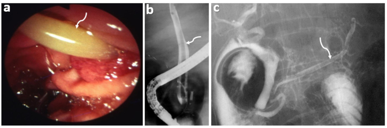
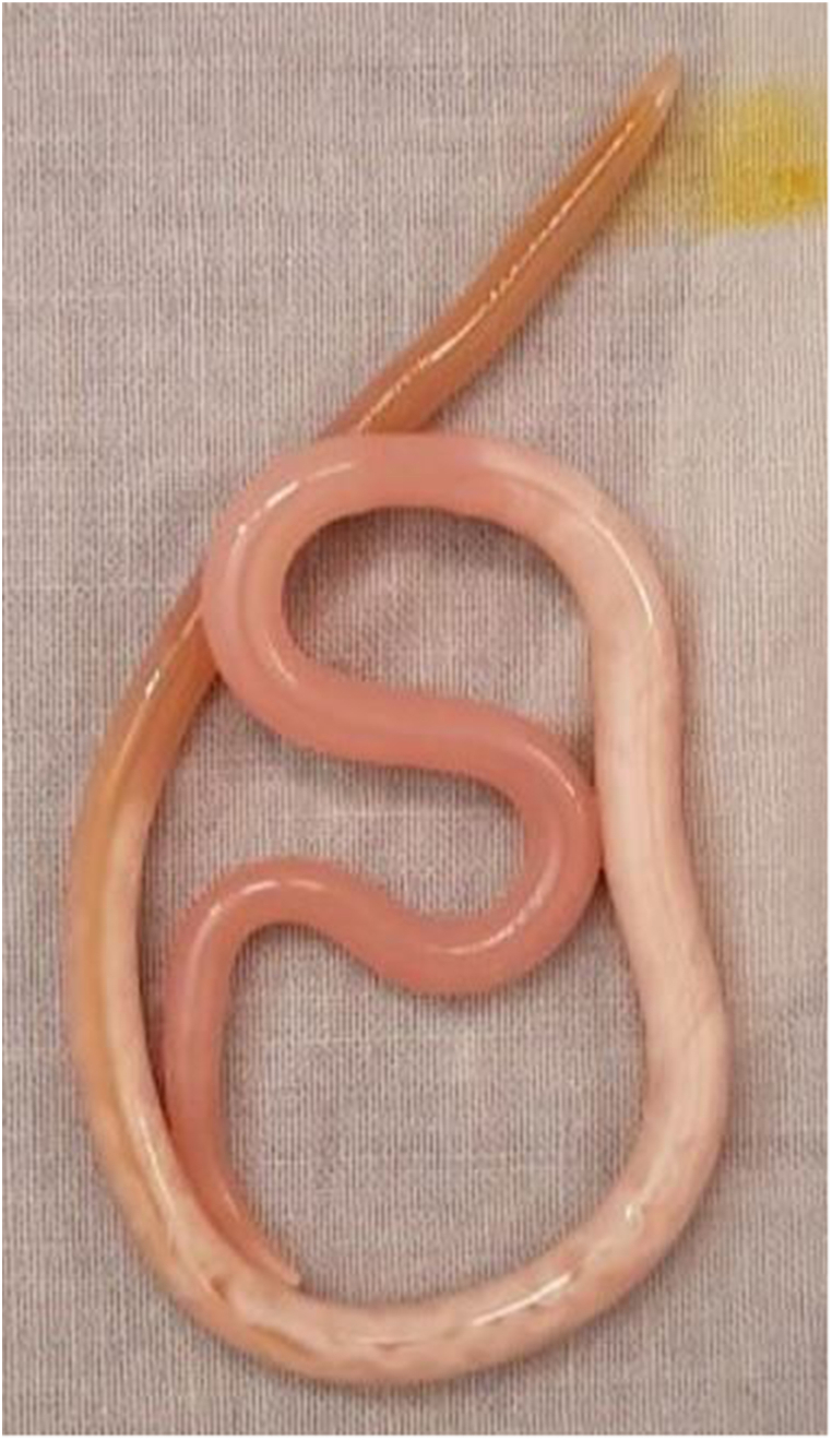
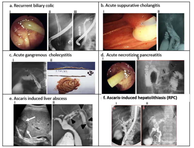
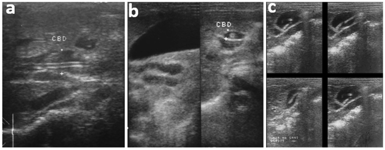
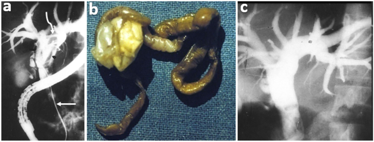
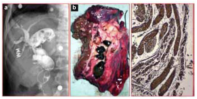
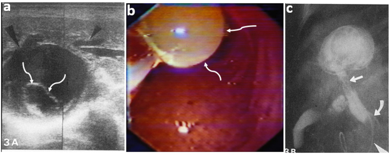
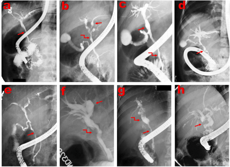
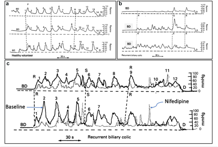
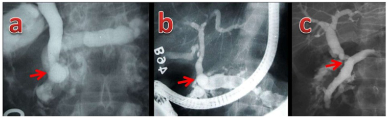
Endoscopic retrograde cholangiopancreatography (ERCP) has been a significant development in gastrointestinal endoscopy. I did my first ERCP at SKIMS on December 5, 1982, and over the last 40 years, I have performed 10,100 ERCP procedures, including 600 Sphincter of Oddi manometries (SOM), and 3200 therapeutic ERCPs. We were confronted with many clinical challenges that needed answers by applying ERCP as a primary diagnostic tool. These studies gave birth to and/or recognition of several clinical syndromes. The hepatobiliary and pancreatic ascariasis (HBPA) as a clinical disease was recognized in 1985. The nematode, Ascaris lumbricoides, was the most common cause of hepatobiliary and pancreatic diseases in Kashmir, and its impact on healthcare, clinical profile, management policies, and control measures was identified. Kashmir was recognized as an endemic zone for recurrent pyogenic cholangitis (RPC), which constituted 12.5% of all biliary diseases. RPC in this population was found essentially to be an aftermath of HBPA. A subset of patients with hepatic hydatidosis with rupture into the biliary tract was recognized at ERCP and primarily treated by endotherapy. Cholangiographic abnormalities in children with portal cavernoma evolved into the recognition of portal biliopathy. Extensive studies of the sphincter of Oddi manometry in patients with unexplained biliary and/or pancreatic pain following cholecystectomy identified the entity of the sphincter of Oddi dyskinesia (SOD). In a cross-over trial, Nifedipine, compared with a placebo, showed a significant clinical response in 20 of 28 such patients. ERCP studies done in patients with tropical calcific pancreatitis showed an anomalous union of bile and pancreatic ducts. Forty of the 220 patients with liver transplantation had biliary complications namely biliary leaks, bile duct strictures, SOD, and recurrence of underlying primary biliary cholangitis. Biliary complications caused considerable morbidity and mortality in patients with liver transplantation.

Keywords: ERCP; Portal biliopathy; ascariasis; recurrent pyogenic cholangitis; sphincter of Oddi dyskinesia.


Figures

Figure 1
Figure 1
Definition: Hepatobiliary and pancreatic ascariasis (HBPA). Panel a, b, and c. The disease is caused by the nematode, Ascaris lumbricoides, seen in the duodenum and invading the ampulla of Vater (panel a, arrow); and entering into the biliary tract (panel b, arrow) and pancreatic ducts (panel c, arrow). [Adopted from Khuroo et al.19].
Figure 2
Figure 2
Photograph of the nematode, Ascaris lumbricoides, extracted out of the bile duct from an elderly male with cholangitis at the first ERCP done at SKIMS on December 5, 1982. This was mistaken for a snake extracted out of the belly by the conference delegates watching this session and most of them ran out of the endoscopy lab.
Figure 3
Figure 3
Ascaris Snapshots. Clinical syndromes of HBPA. Panels a–f. a. Recurrent biliary colic. i. Duodenoscopy: Ascaris in the ampulla of Vater with tail in the duodenum. ii. ERCP: worm in the bile duct. iii. ERCP after worm extraction. Following worm extraction, the patient had rapid relief of biliary colic. b. Acute suppurative cholangitis. i. Duodenoscopy: pus exuding from the papilla. ii. ERCP: multiple worms palisading the bile duct. 37 worms were extracted from the duct. c. Acute gangrenous cholecystitis. i. ERCP: worm in the bile duct which had entered the cystic duct and blocked it with a distended gallbladder. ii. Urgent open cholecystectomy specimen: a gangrenous gallbladder with 2 worms, and a few small stones. d. Acute necrotizing pancreatitis. i. Duodenoscopy: worm in the ampulla of Vater. ii. ERCP: the worm in the pancreatic duct. e. Ascaris-induced liver abscess. i. ultrasound liver: a large hypoechoic lesion right lobe liver. ii. ERCP: multiple worms in the hepatic ducts. f. Ascaris-induced hepatolithiasis. i. ERCP 5 years before present admission: worms in the bile ducts. ii. ERCP on present admission: cholangiographic features of RPC with stones and sludge packing the hepatic ducts. A naso-biliary drain has been placed to treat pyogenic cholangitis. [Adopted from Khuroo et al.19,27,54].
Figure 4
Figure 4
Sonographic appearances in HBPA. a. Dilated CBD containing an echogenic strip with a central longitudinal anechoic tube, representing the alimentary canal of a worm. b. A composite of 2 images showing distended GB (left) and a small nonshadowing echogenic disk in CBD. c. Multiple GB images each containing a nonshadowing curved echogenic structure. The worm was highly motile giving varied appearances on the sonograms. [Adopted from Khuroo et al.27].
Figure 5
Figure 5
Endotherapy in HBPA. a. ERCP shows gross biliary dilation with a worm (curved arrow) in the ducts. An endoscopic basket (straight arrow) has been passed into the bile duct to engage the worm. b. Dead macerated worm mass with sludge removed from the duct. c. Repeat cholangiogram after worm extraction. [Adopted from Khuroo et al.19].
Figure 6
Figure 6
Recurrent pyogenic cholangitis. a. ERCP: gross dilation of common and left hepatic ductal system, with stones and sludge, b. Resected left lobe liver: multiple brown pigment stones are placed in the ducts, c. Histology brown pigment stone (Masson's trichome ×100): multiple linear Ascaris fragments with ova and encrusted mineral deposits. [Adopted from Khuroo et al.54].
Figure 7
Figure 7
Biliary hydatidosis. a. Sonogram: ruptured cyst communicating with dilated hepatic ducts (small arrowhead) with loss of continuity of the cyst wall adjacent to the dilated hepatic ducts (large arrowhead). b. Duodenoscopy: large daughter cyst projecting out of the ampullary orifice. c. ERCP shows a dilated main bile duct with a leaf-like filling defect possibly due to ruptured membranes or ruptured daughter vesicles (arrow), and a round filling defect due to an intact daughter vesicle at the lower end of the common bile duct {curved arrow). A cyst cavity filled with contrast medium through the right hepatic duct and a nasobiliary drainage catheter in situ (arrowhead) is seen. [Adopted from Khuroo et al.58].
Figure 8
Figure 8
Portal biliopathy snapshots. 8 ERCPs plates with various cholangiographic abnormalities. a. Extrinsic impression on common bile duct (arrow). b. Ectasia of left hepatic duct (arrow) and caliber irregularity of common hepatic duct (curved arrow). c. Ectasia of the common hepatic duct (arrow) and caliber irregularity of the common bile duct (curved arrow). d. Angulation (arrow) of a common bile duct. e. Large smooth impression (arrow) on the common bile duct. f. Angulation of common hepatic duct (curved arrow) and gross ectasia of intrahepatic ducts (arrow); g. Long smooth stricture of the common bile duct (arrow) with upstream dilatation (curved arrow); h. dilated bile ducts with multiple filling defects (arrow). {Adopted from Khuroo et al.65].
Figure 9
Figure 9
Sphincter of Oddi manometry tracing. Panel a, b, and c. a. Healthy volunteer. The pressure tracing shows SO normal pressures: BD (16 mmHg), SO basal (30 mmHg), SO phasic (37 mmHg), frequency phasic waves (4.0/min), duration phasic waves (6.0 s) and sequence phasic waves (antegrade 56%; retrograde 35% and simultaneous 9%). b. Sphincter of Oddi Dyskinesia. The patient had post-cholecystectomy right upper quadrant pain with elevated alkaline phosphatase. The tracing showed elevated ductal & basal pressures and abnormal retrograde contractions. BD (25 mm Hg), SO basal (38.4 mm Hg), SO phasic (28.4 mm Hg), frequency (7.1/min), duration (5.0/s), and Sequence phasic waves (antegrade 0%; simultaneous 25%; retrograde 75%). c. Sphincter of Oddi Dyskinesia. Pressure profile before (solid line tracing) and after 10 mg of sublingual nifedipine (broken line tracing). Nifedipine caused an appreciable fall in SO basal and phasic pressure with no effect on frequency, duration, and sequence of phasic waves. The pressures before and after nifedipine were CBD 18 and 8 mmHg; SO basal 38 & 22 mmHg; SO phasic 54 & 31 mmHg; frequency 4 & 5/min; duration 8 & 6 s; sequence phasic wave (antegrade 9 & 12%; retrograde 58 & 50%, & simultaneous 33 & 38%). [Adopted from Khuroo et al.70]. BD = bile duct, D = duodenum, Numbers define the corresponding phasic waves recorded by 2/3 lumen catheters and were used to check the sequence of phasic waves, P = progressive phasic wave, S = simultaneous phasic wave, R = retrograde phasic wave.
Figure 10
Figure 10
Tropical calcific pancreatitis. Anomalous pancreatobiliary ductal union. Panel a, b, and c. a. ERCP: grossly dilated pancreatic duct, filling defects in the side branches, dilated bile duct joining pancreatic neck region (arrow) to form an anomalous pancreaticobiliary ductal union of the B-P type. b. ERCP: grossly dilated pancreatic duct, multiple large filling defects, and the bile duct joining the body region (arrow) to form an anomalous pancreaticobiliary ductal union of the B-P type. c. ERCP: grossly dilated pancreatic duct, filling defects in the side branches and the bile duct joining it in the distal body region (arrow) to form a very long anomalous pancreaticobiliary ductal union of the B-P type. [Adopted from Khuroo et al.76].

Similar articles

Cited by

References

    1. Kozarek R.A. The past, present, and future of endoscopic retrograde cholangiopancreatography. Gastroenterol Hepatol (N Y) Oct 2017;13:620–622. - PMC - PubMed
    1. McCune W.S., Shorb P.E., Moscovitz H. Endoscopic cannulation of the ampulla of vater: a preliminary report. Ann Surg. 1968;167:752–756. doi: 10.1097/00000658-196805000-00013. - DOI - PMC - PubMed
    1. Oi I., Kobayashi S., Kondo T. Endoscopic pancreatocholangiography. Endoscopy. 1970;2:103–106.
    1. Ogoshi K., Tobita Y., Hara Y. Endoscopic observation of the duodenum and pancreatocholedochography, using duodenal fibrescope under direct vision. Gastroent Endosc (Tokyo) 1970;12:83–86. (Translation)
    1. Cotton P.B., Blumgart L.H., Davies G.T., et al. Cannulation of papilla of Vater via fiber-duodenoscope: assessment of retrograde cholangiopancreatography in 60 patients. Lancet. 1972;299:53–58. doi: 10.1016/S0140-6736(72)90059-1. - DOI - PubMed

LinkOut - more resources