. 2023 Nov 15;9(46):eadi5326.
doi: 10.1126/sciadv.adi5326. Epub 2023 Nov 17.

Zona incerta dopamine neurons encode motivational vigor in food seeking

Affiliations

Zona incerta dopamine neurons encode motivational vigor in food seeking

Qiying Ye et al. Sci Adv. .

Abstract

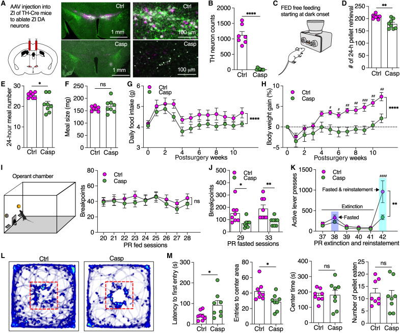
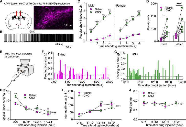
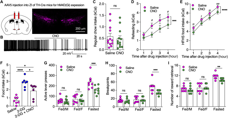
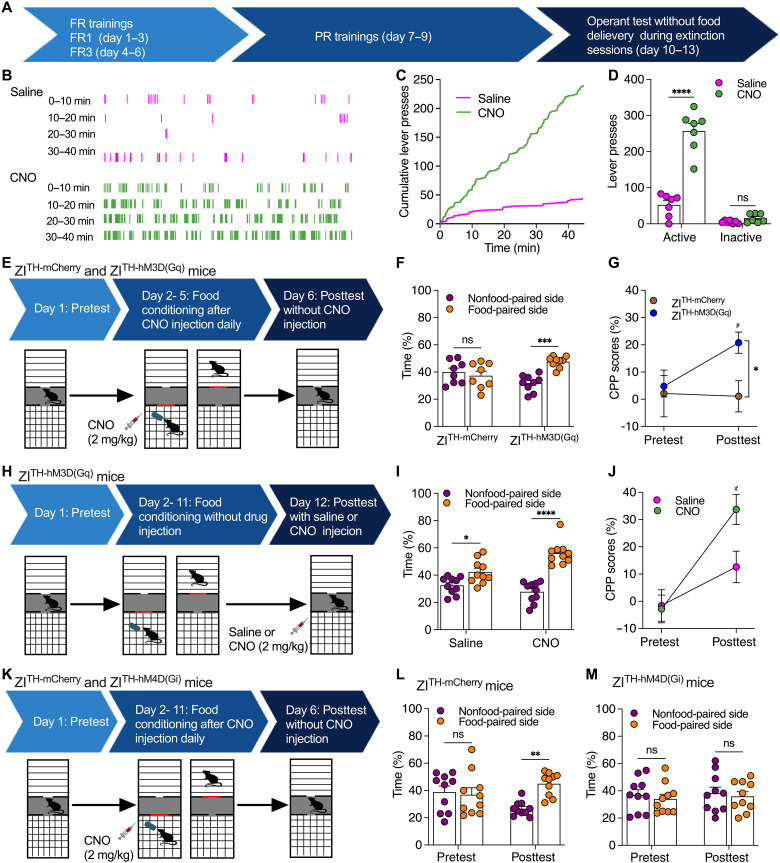
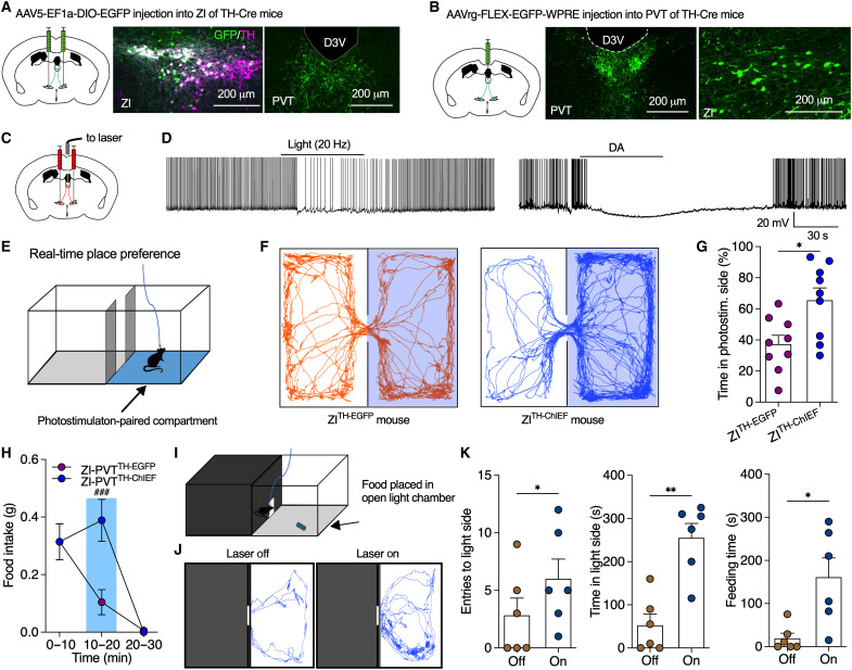
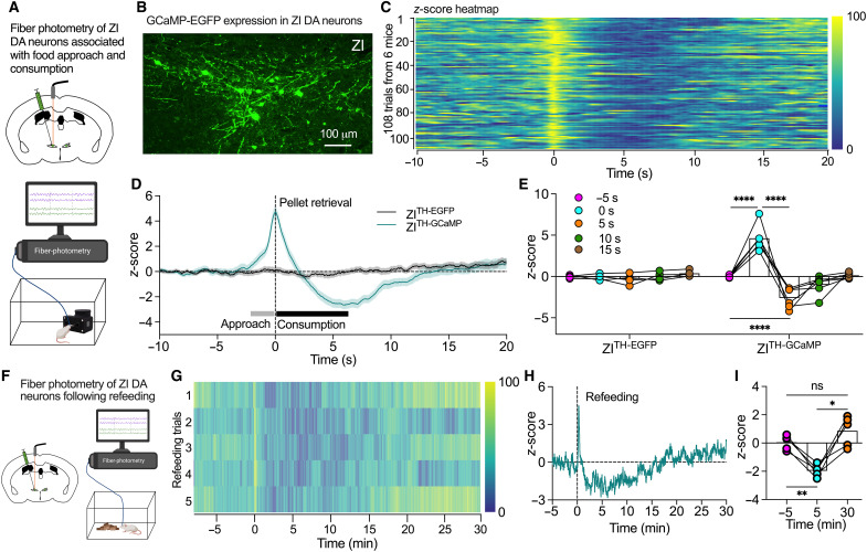
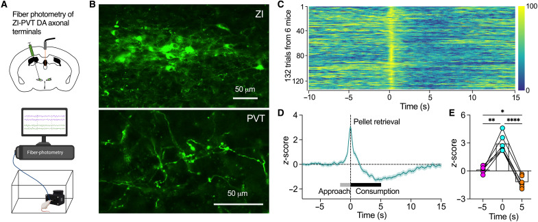
Energy deprivation triggers food seeking to ensure homeostatic consumption, but the neural coding of motivational vigor in food seeking during physical hunger remains unknown. Here, we report that ablation of dopamine (DA) neurons in zona incerta (ZI) but not ventral tegmental area potently impaired food seeking after fasting. ZI DA neurons and their projections to paraventricular thalamus (PVT) were quickly activated for food approach but inhibited during food consumption. Chemogenetic manipulation of ZI DA neurons bidirectionally regulated feeding motivation to control meal frequency but not meal size for food intake. Activation of ZI DA neurons promoted, but silencing of these neurons blocked, contextual memory associate with food reward. In addition, selective activation of ZI DA projections to PVT promoted food seeking for food consumption and transited positive-valence signals. Together, these findings reveal that ZI DA neurons encode motivational vigor in food seeking for food consumption through their projections to PVT.


Figures

Fig. 1.
Fig. 1.. Selective ablation of ZI DA neurons impaired motivational vigor in fasting-triggered food seeking.
(A) AAV was injected into bilateral ZI of TH-Cre mice to induce mCherry (n = 8 mice) or mCherry plus caspase expression (n = 8 mice) in ZI DA neurons. (B) TH-immunoreactive ZI neurons (green) were ablated by virus-induced caspase/mCherry (magenta) expression. (C to F) Meal pattern analysis using FED free feeding shows total number of pellet retrieval, meal numbers, and averaged meal size of 24 hours. (G) Daily food intake for both control and caspase mice for 11 weeks following virus injection. (H) Body weight gain for both control and caspase mice. (I) Breakpoint reached by fed mice during operant PR sessions of 45 min. (J) Breakpoints reached by 24-hour fasted mice. (K) Active lever presses during PR extinction and reinstatement sessions. (L) Real-time activity of mice in open-field chambers with food pellets placed in the center. (M) The latency to the first entry of the center area, the total entries to center area, the total time in the center area, and the number of food pellets consumed by mice in open-field chambers with food pellets placed in the center. Unaired t test for (B), (D), (E), (F), and (M), two-way ANOVA with post hoc Bonferroni test for (G) to (K).*P < 0.05, **P < 0.01, ***P < 0.001, ****P < 0.0001, and ####P < 0.0001; ns, no significance.
Fig. 2.
Fig. 2.. Chemogenetic activation of ZI DA neurons promoted motivational vigor in food seeking to regulate food consumption.
(A) AAV was injected to bilateral ZI of TH-Cre mice to express excitatory hM3D(Gq) in ZI DA neurons. (B) A representative trace from slice recording shows that CNO (3 μM) excited an hM3D(Gq)-positive ZI DA neuron. (C) Regular food consumption during light cycle in both male (n = 12) and female (n = 8) TH-Cre mice with hM3D(Gq) expression in ZI DA neurons following IP injection of saline or CNO (2.0 mg/kg). (D) Breakpoints reached by both fed (n = 9) and partially fasted mice (70% of their daily food intake, n = 9) during PR sessions following IP injections of saline or CNO (2.0 mg/kg). (E to G) Real-time feeding bout sizes of a female ZITH-hM3D(Gq) mouse following IP injection of saline and CNO (2.0 mg/kg). FED devices at free-feeding modes were used for collecting real-time food intake in home cages. (H to J) Bar graphs showing meal numbers, intermeal intervals, and meal sizes of 6 hours. n = 9 female mice each group. Three-way ANOVA with post hoc Bonferroni test for (C), two-way repeated-measures (RM) ANOVA with post hoc Bonferroni test [(D) and (H) to (J)].
Fig. 3.
Fig. 3.. Chemogenetic inhibition of ZI DA neurons decreased motivated food seeking and consumption.
(A) AAV was injected to bilateral ZI of TH-Cre mice to express inhibitory hM4D(Gi) in ZI DA neurons. (B) A representative trace shows that CNO (3.0 μM) inhibited a ZITH-hM4D(Gi) neuron. (C) Regular food intake of fed mice (n = 12) over 4 hours following saline and CNO injection. (D) Refeeding after 24 hours fasting following saline and CNO injection. (E) HFHS food intake following saline and CNO injection. (F) Food intake over 4 hours following injection of saline, 2-DG (200 mg/kg), or 2-DG plus CNO (2.0 mg/kg). n = 7 each group. (G to I) Bar graphs showing active lever presses, breakpoints, and number of rewards earned in both fed and fasted mice (70% of their daily food intake overnight) during operant PR tests of 45 min following saline or CNO (2.0 mg/kg) injection. Paired t test for (C), two-way RM ANOVA with post hoc Bonferroni test [(D), (E), and (G) to (I)], one-way ANOVA with post hoc Bonferroni test (F).
Fig. 4.
Fig. 4.. ZI DA neurons regulate both acquisition and expression of contextual food memory.
(A) Timeline for operant training and tests. (B) Real-time lever presses during operant tests without reward delivery. (C) Cumulative lever presses during operant tests without reward delivery. (D) Presses of the food-paired active levers and the nonfood-paired inactive levers during operant tests without reward delivery. n = 7 mice each group. (E) Timeline for testing food CPP acquisition in both ZITH-mCherry (n = 8) or ZITH-hM3D(Gq) (n = 9) mice. (F) Percentage of time that mice spent in both nonfood-paired and food-paired sides during postconditioning sessions. (G) CPP scores during both preconditioning and postconditioning tests. (H) Timeline for testing food CPP expression. ZITH-hM3D(Gq) mice received 10-day place conditioning with HFHS food and pre- and postconditioning place preference tests. (I) Percentage of time that mice spent in both nonfood-paired and food-paired sides during postconditioning sessions following saline (n = 10 mice) or CNO (2.0 mg/kg, n = 10 mice) injection. (J) CPP scores in mice during both preconditioning and postconditioning tests. (K) Timeline for testing food CPP acquisition in both ZITH-mCherry (n = 10) or ZITH-hM4D(Gi) (n = 10) mice. (L) Percentage of time that control ZITH-mCherry mice spent in both nonfood-paired and food-paired sides during pre- and postconditioning tests. (M) Percentage of time that ZITH-hM4D(Gi)-mCherry mice spent in both nonfood-paired and food-paired sides during pre- and postconditioning tests. Two-way RM ANOVA with post hoc Bonferroni test for (D), (G), (J), (L), and (M). Two-way ANOVA with post hoc Bonferroni test for (F) and (I).
Fig. 5.
Fig. 5.. Optogenetic activation of ZI-PVT DA projections produced positive valence to promote food seeking for consumption.
(A) AAV was injected into bilateral ZI of TH-Cre mice to induce EGFP expression in ZI DA neurons that sent EGFP-positive axons to PVT. (B) Retrograde AAV was injected into PVT of TH-Cre mice to trace ZI DA neurons that projected to PVT. (C) AAV was injected into bilateral ZI of TH-Cre mice to induce ChIEF or EGFP expression in ZI DA neurons. (D) Both photostimulation and DA (30 μM) inhibited PVT neurons surrounded by ChIEF-positive ZI DA axons. (E) A two-compartment chamber was used for real-time place preference test with one compartment paired with photostimulation (20 Hz). The laser was only turned on when mice entered the photostimulation-paired side. (F) Real-time motion of a control ZITH-EGFP and a ZITH-ChIEF mouse in the two-compartment chamber. (G) Percentage of time that mice spent in photostimulation-paired compartment. n = 9 mice each group. (H) HFHS food intake of mice before, during, and after PVT photostimulation. n = 7 mice each group. (I) A light/dark box with entrance connecting the two sides for food intake test over 10 min when food pellets were placed in the light side. (J) Real-time activity tracking in light side of the chambers during 10-min food intake test with or without photostimulation (20 Hz) of DAZI-PVT pathway. (K) Total entries to light side, cumulative time in light side, and cumulative feeding time during 10-min food intake. n = 6 mice per group. Unpaired t test for (G), two-way ANOVA with post hoc Bonferroni test for (H), and paired t test for (K).
Fig. 6.
Fig. 6.. ZI DA neurons responded to food approach and consumption.
(A) AAV was injected into ZI of TH-Cre mice to express GCaMP or EGFP in ZI DA neurons for fiber-photometry imaging of ZI DA neurons associated with food approach and consumption. (B) GCaMP7f was expressed in ZI DA neurons of TH-Cre mice. (C) Heatmap of normalized z-scores of ZI DA neuron activity from 108 feeding trials of ZITH-GCaMP mice (n = 6). (D) z-scores of ZI DA neurons aligned to the time of pellet retrieval from 85 feeding trials of control ZITH-EGFP mice and 108 trials of ZITH-GCaMP mice. (E) Increase in z-scores for food approach and decease during food consumption compared to 5 s before food retrieval in ZITH-GCaMP (n = 6) but not ZITH-EGFP mice (n = 5). Two-way RM ANOVA with post hoc Bonferroni test. (F) A diagram showing that AAV was injected into ZI of TH-Cre mice to express GCaMP in ZI DA neurons for fiber-photometry tests following refeeding. (G) Heatmaps of normalized z-scores of ZI DA neuron activity from refeeding trials of five fasted ZITH-GCaMP mice. (H) z-scores of ZI DA neurons aligned to the time of food delivery for refeeding from five fasted ZITH-GCaMP mice. (I) Bar graph with data plots showing the averages of z-score at the different time points before and after refeeding (n = 5 mice). One-way RM ANOVA with post hoc Bonferroni test.
Fig. 7.
Fig. 7.. ZI DA axonal terminals in the PVT were activated for food approach but inhibited during food consumption.
(A) AAV-hSynapsin1-FLEx-axon-GCaMP6s was injected into ZI of TH-Cre mice for fiber-photometry imaging of ZI DA axon terminals in PVT. (B) Representative images show GCaMP6s was expressed in both ZI DA somas (top) and axonal terminals in the PVT (bottom). (C) Heatmap of normalized z-scores of ZI-PVT DA axonal terminals from 132 feeding trials of six ZITH-GCaMP mice. (D) z-scores of ZI-PVT DA axonal terminals aligned to the time of pellet retrieval from 132 trials of six ZITH-GCaMP mice. (E) Bar graph with data plots showing the averages of z-score at the time of −5, 0, and 5 s (n = 6 mice). One-way RM ANOVA with post hoc Bonferroni test.

Update of

Similar articles

Cited by

References

    1. Q. Y. Zhou, R. D. Palmiter, Dopamine-deficient mice are severely hypoactive, adipsic, and aphagic. Cell 83, 1197–1209 (1995). - PubMed
    1. L. Boekhoudt, T. J. M. Roelofs, J. W. de Jong, A. E. de Leeuw, M. C. M. Luijendijk, I. G. Wolterink-Donselaar, G. van der Plasse, R. A. H. Adan, Does activation of midbrain dopamine neurons promote or reduce feeding? Int. J. Obes. (Lond) 41, 1131–1140 (2017). - PubMed
    1. R. A. M. Marino, R. A. McDevitt, S. C. Gantz, H. Shen, M. Pignatelli, W. Xin, R. A. Wise, A. Bonci, Control of food approach and eating by a GABAergic projection from lateral hypothalamus to dorsal pons. Proc. Natl. Acad. Sci. U.S.A. 117, 8611–8615 (2020). - PMC - PubMed
    1. R. D. Palmiter, Is dopamine a physiologically relevant mediator of feeding behavior? Trends Neurosci. 30, 375–381 (2007). - PubMed
    1. X. Zhang, A. N. van den Pol, Hypothalamic arcuate nucleus tyrosine hydroxylase neurons play orexigenic role in energy homeostasis. Nat. Neurosci. 19, 1341–1347 (2016). - PMC - PubMed

LinkOut - more resources