. 2024 Jan;49(1):312-319.
doi: 10.1007/s00261-023-04097-4. Epub 2023 Nov 17.

Iodine density mapping for the diagnosis of acute bowel ischemia using fast kV-switching dual-energy CT

Affiliations

Iodine density mapping for the diagnosis of acute bowel ischemia using fast kV-switching dual-energy CT

Jack Junchi Xu et al. Abdom Radiol (NY). 2024 Jan.

Abstract

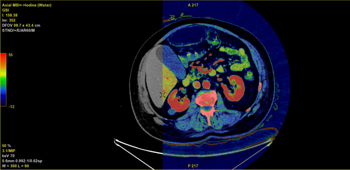
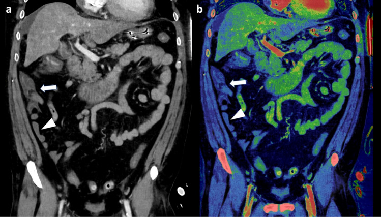
Purpose: To evaluate the diagnostic capabilities of a supplementary color ramped iodine density map compared to virtual monoenergetic images (VMIs) at 74 keV in the diagnosis of acute bowel ischemia (ABI).

Methods: Data for this study were prospectively gathered and retrospectively evaluated. Patients referred to the Department of Diagnostic Radiology between October 2020 and August 2022 on the suspicion of ABI and underwent surgery < 12 h following fast kV-switching venous phase abdominal dual-energy CT (DECT) were consecutively included. Images were evaluated by two board-certified radiologists and two radiology residents. First round included only 74 keV VMIs resembling conventional 120 kVp images, and the second round included a supplementary iodine density map. Readers were asked to register presence of ABI as well as their confidence in their diagnosis based on a 5-point Likert scale. Sensitivity, specificity, positive predictive value (PPV), and negative predictive value (NPV) were calculated for each observer with the surgical findings as the gold-standard. McNemar's and Wilcoxon signed-rank test were used to compare registrations and diagnostic confidence across assessment rounds.

Results: A total of 29 patients resulting in 31 DECT scans were included. Fourteen cases of ischemic/necrotic bowel were reported following surgery. Sensitivity and NPV were decreased with the use of supplementary iodine map images compared to 120 kVp-like images without supplementary iodine map images for three of four observers (round 1 range: 71.4-92.9% and 78.0-94.8%; round 2 range: 57.1-78.6% and 70.1-83.3%, respectively), while specificity and PPV were increased for three of four observers (round 1 range: 64.7-94.1% and 67.4-93.1%; round 2 range: 88.2-94.1% and 73.8-91.1%, respectively). However, no significant difference in ABI diagnosis or diagnostic confidence was found (p-value range: 0.07-1.00 and 0.23-0.58, respectively).

Conclusion: No significant difference for the diagnosis of ABI was found using supplementary iodine mapping. Our study may suggest a trend of increased specificity and decreased sensitivity, hence, the use of supplementary iodine mapping should be carefully considered.

Keywords: Bowel ischemia; Computed tomography; Dual-energy CT.


Conflict of interest statement

The authors J.J.X. and P.S.U. have received consulting fees from GE healthcare for presenting at GE Healthcare related webinars. The other authors of this manuscript declare no relationships with any companies whose products or services may be related to the subject matter of the article.

Figures

Fig. 1
Fig. 1
Axial image of the abdomen with an iodine overlay (colored map) on top of a virtual non-contrast image (underlying gray scale image). The color ramp ranging from − 12 to 55 (shown to the far left) represents the varying iodine concentrations in μgI/cm3
Fig. 2
Fig. 2
Flowchart of the patient inclusion process
Fig. 3
Fig. 3
A patient with surgically confirmed ischemic/necrotic caecum, ascending colon, and proximal portion of the transverse colon. Three of four observers evaluated the colon to be ischemic (arrow) in the 120 kVp-like images (a) during the first evaluation round. However, during the second evaluation round, with the iodine map (c) and underlying virtual unenhanced (VUE) image (b), only one of four observers deemed the bowel to be ischemic. In c the ischemic section of the colon may be interpreted as perfused (arrow) when compared to other vital bowel segments (arrowhead). Upon closer inspection of the VUE images, it is noticeable that the ischemic bowel maintains some bowel wall attenuation (arrow) compared to the non-ischemic bowel (arrowhead) suggesting a paired effect. Additionally, pseudo hyperenhancement due to an adjacent air distended bowel segment, may also contribute to the enhancement seen on the iodine map
Fig. 4
Fig. 4
A patient with surgically confirmed small bowel ischemia due to closed loop bowel obstruction (arrow). Despite some attenuation in the bowel wall seen on the conventional 120 kVp-like image (a), one resident observer suspected ischemia and became more confident using iodine map (b). Additionally, one radiologist only reported ischemia using the iodine map
Fig. 5
Fig. 5
A case for which all observers suspected bowel ischemia located to the ileum (arrowhead), caecum and ascending colon (arrow). A confidence rating of three was given by three observers while the last observer rated four in the 120 kVp-like images (a). Following evaluation with iodine mapping (b), the three observers who initially gave a confidence rating of three now confidently diagnosed ischemia with an increased confidence rating of five, while the last observer maintained a confidence rating of four. However, in this case the surgical report found no signs of ischemia

Similar articles

Cited by

References

    1. Patel A, Kaleya RN, Sammartano RJ. Pathophysiology of Mesenteric Ischemia. Surgical Clinics of North America. 1992;72:31–41. doi: 10.1016/S0039-6109(16)45626-4. - DOI - PubMed
    1. Davarpanah AH, Ghamari Khameneh A, Khosravi B, et al (2021) Many faces of acute bowel ischemia: overview of radiologic staging (page 1-23). Insights Imaging 12. - PMC - PubMed
    1. Bala M, Kashuk J, Moore EE, et al. Acute mesenteric ischemia: guidelines of the World Society of Emergency Surgery. World Journal of Emergency Surgery. 2017;12:38. doi: 10.1186/s13017-017-0150-5. - DOI - PMC - PubMed
    1. Jan M. Diagnostic accuracy of multidetector CT in acute mesenteric ischemia: Systematic review and meta-analysis. Radiology. 2010;256:93–101. doi: 10.1148/radiol.10091938. - DOI - PubMed
    1. Luther B, Mamopoulos A, Lehmann C, Klar E. The Ongoing Challenge of Acute Mesenteric Ischemia. Visc Med. 2018;34:215–221. doi: 10.1159/000490318. - DOI - PMC - PubMed

Publication types

LinkOut - more resources