. 2023 Nov 17;14(1):7452.
doi: 10.1038/s41467-023-43062-z.

Mutation of key signaling regulators of cerebrovascular development in vein of Galen malformations

Shujuan Zhao #  1   2 Kedous Y Mekbib #  2   3 Martijn A van der Ent #  4 Garrett Allington #  2   5 Andrew Prendergast  6 Jocelyn E Chau  7 Hannah Smith  2   3 John Shohfi  2   3 Jack Ocken  3 Daniel Duran  8 Charuta G Furey  3   9   10 Le Thi Hao  2 Phan Q Duy  11 Benjamin C Reeves  3 Junhui Zhang  12 Carol Nelson-Williams  12 Di Chen  4 Boyang Li  13 Timothy Nottoli  14 Suxia Bai  14 Myron Rolle  2 Xue Zeng  7   15 Weilai Dong  12   15 Po-Ying Fu  1 Yung-Chun Wang  1 Shrikant Mane  12 Paulina Piwowarczyk  16 Katie Pricola Fehnel  16 Alfred Pokmeng See  16 Bermans J Iskandar  17 Beverly Aagaard-Kienitz  17   18 Quentin J Moyer  2 Evan Dennis  2 Emre Kiziltug  2 Adam J Kundishora  3 Tyrone DeSpenza Jr  3 Ana B W Greenberg  2 Seblewengel M Kidanemariam  19 Andrew T Hale  20 James M Johnston  20 Eric M Jackson  21 Phillip B Storm  22   23 Shih-Shan Lang  22   23 William E Butler  2 Bob S Carter  2 Paul Chapman  2 Christopher J Stapleton  2 Aman B Patel  2 Georges Rodesch  24   25 Stanislas Smajda  25 Alejandro Berenstein  26 Tanyeri Barak  3 E Zeynep Erson-Omay  3 Hongyu Zhao  12   13 Andres Moreno-De-Luca  27 Mark R Proctor  16 Edward R Smith  16 Darren B Orbach  16   28 Seth L Alper  29 Stefania Nicoli  12   30   31 Titus J Boggon  7   30 Richard P Lifton  15 Murat Gunel  3 Philip D King  32 Sheng Chih Jin  33   34 Kristopher T Kahle  35   36   37   38
Affiliations

Mutation of key signaling regulators of cerebrovascular development in vein of Galen malformations

Shujuan Zhao et al. Nat Commun. .

Abstract

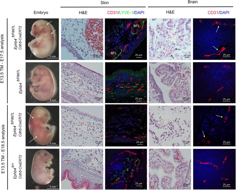
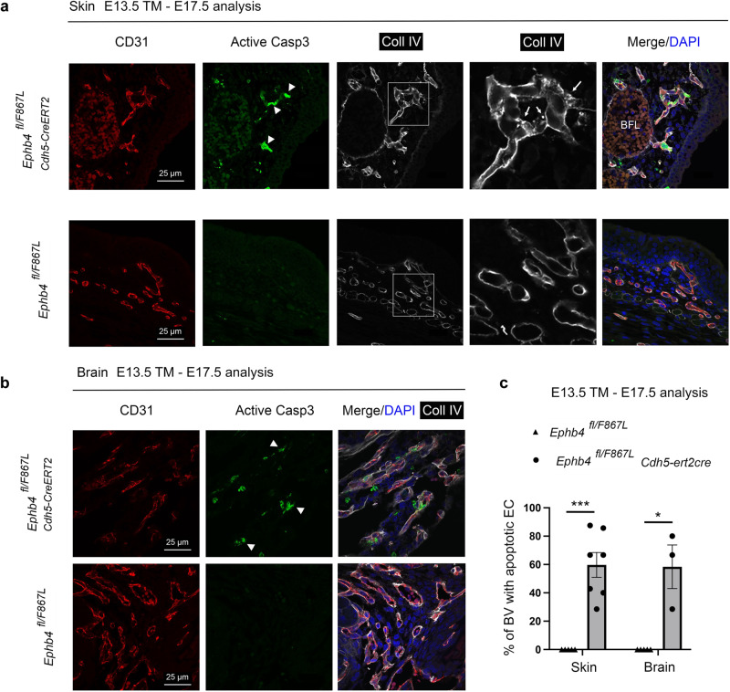
To elucidate the pathogenesis of vein of Galen malformations (VOGMs), the most common and most severe of congenital brain arteriovenous malformations, we performed an integrated analysis of 310 VOGM proband-family exomes and 336,326 human cerebrovasculature single-cell transcriptomes. We found the Ras suppressor p120 RasGAP (RASA1) harbored a genome-wide significant burden of loss-of-function de novo variants (2042.5-fold, p = 4.79 x 10⁻⁷). Rare, damaging transmitted variants were enriched in Ephrin receptor-B4 (EPHB4) (17.5-fold, p = 1.22 x 10⁻⁵), which cooperates with p120 RasGAP to regulate vascular development. Additional probands had damaging variants in ACVRL1, NOTCH1, ITGB1, and PTPN11. ACVRL1 variants were also identified in a multi-generational VOGM pedigree. Integrative genomic analysis defined developing endothelial cells as a likely spatio-temporal locus of VOGM pathophysiology. Mice expressing a VOGM-specific EPHB4 kinase-domain missense variant (Phe867Leu) exhibited disrupted developmental angiogenesis and impaired hierarchical development of arterial-capillary-venous networks, but only in the presence of a "second-hit" allele. These results illuminate human arterio-venous development and VOGM pathobiology and have implications for patients and their families.


Conflict of interest statement

The authors declare no competing interests.

Figures

Fig. 1
Fig. 1. VOGM-associated variants in RASA1 and EPHB4.
a In the normal anatomy shown on a lateral projection subtracted angiogram of a 4-year-old boy (left), the deep veins drain into the straight sinus (white star). The normal deep venous anatomy involves the internal cerebral veins (white arrow) and the basal vein of Rosenthal (white arrowhead). VOGM is shown on a lateral projection subtracted angiogram of a 3 day-old boy (right), where the falcon sinus (black star) is commonly observed as the draining vein and involves prominent supply from splenial vessels deriving from the anterior cerebral artery (black arrows), as well as posterior choroidal arteries (black arrowhead). The normal drainage of the torcular typically involves the bilateral transverse and sigmoid sinuses (white dashed line). In the abnormal venous anatomy, VOGM typically recruit dilated occipital sinuses (black dashed line). b Quantile-quantile plot of observed versus expected p-values (one-sided Poisson test, not adjusted) for rare damaging (D-mis + LoF) DNVs with MAF ≤ 4 x 10−4 in the Exome Aggregation Consortium database for all genes. The exome-wide significant cutoff was 8.6 × 10−7 (0.05 / (3 × 19,347)). D-mis are missense variants predicted to be deleterious per MetaSVM or MPC ≤ 2. LoF, loss-of-function variants. DNV, de novo variant. MAF, minor allele frequency. c RASA1 and EPHB4 functional domains (green rectangles) with the location of VOGM variants and phylogenetic conservation of wild-type amino acids (red text) at each mutated position. 5 LoF variants were found in RASA1, including 2 DNVs (p. R427X and p. V527Mfs*16), and 3 transmitted variants (p. H743fs*24, p. R709X and p. Y872*). Transmitted damaging variants in EPHB4 including 2 variants in the Fibronectin type III domain (p. E432Gfs*7 and p. A509G), and 3 D-mis variants (p. K650N, p. R838W, p. F867L) in the kinase domain. d Quantile-quantile plots of observed versus expected p-values (one-sided Fisher’s exact test, not adjusted) for rare damaging (D-mis + LoF) variants with MAF ≤ 5 x 10−5 in the Genome Aggregation database (gnomAD) from case-control burden test. The genome-wide significant cutoff was 2.6 × 10−6 (0.05 of 19,347).
Fig. 2
Fig. 2. Preserved protein stability with compromised tyrosine kinase activity in EPHB4 kinase domain variants.
a Steady-state abundance of EPHB4 D-mis mutants. Cos-7 cells were transiently transfected with c-myc-tagged WT or EPHB4 D-mis mutants together with an eGFP-encoding vector. Numbers at top indicate percentage of GFP+ cells determined by flow cytometry (see Supplementary Fig. 3). Cells were lysed and EPHB4 abundance determined by Western blot for c-myc (left) or EPHB4 (right). Blots were probed for tubulin or beta actin (ActB) respectively to demonstrate equivalent protein loading. Numbers below indicate normalized abundance of EPHB4 D-mis mutants relative to WT EPHB4. Each experiment was repeated two times with similar results. b Stability of EPHB4 D-mis mutants. Cos-7 cells transiently transfected with c-myc-tagged WT or EPHB4 D-mis mutants were treated with cycloheximide (CHX) for the indicated times before lysis and determination of EPHB4 and ActB abundance by Western blot. Numbers indicate normalized EPHB4 abundance relative to CHX-untreated cells for each time point. Shown are the results of a single experiment. c Kinase activity of EPHB4 D-mis mutants. EPHB4 was immunoprecipitated from Cos-7 cells transiently transfected with c-myc-tagged WT or EPHB4 D-mis mutants. EPHB4 kinase activity was determined by Western blot of immunoprecipitates with an anti-phosphotyrosine antibody (pTyr). Numbers indicate normalized pTyr content of EPHB4 D-mis mutants relative to WT EPHB4. Shown are the results of one experiment of two repeats.
Fig. 3
Fig. 3. VOGM-associated variants in ACVRL1 and other Mendelian vascular disease genes.
a Top 10 GO molecular function, GO biological process, and Reactome pathway enrichment terms. The y-axis depicts GO term and Reactome pathway ID numbers. The x-axis depicts -log (p-value) and the dotted line represents the α = 0.05 significance threshold (one-sided Fisher’s exact test, Bonferroni multiple-testing adjusted). The GO term and Reactome pathway term name overline their respective bars. b Interactome of mutated Ras signaling genes in VOGM. Genes with damaging Ras signaling variants (WP4223 in Wiki pathway analysis) and EPHB4, NOTCH1, ACVRL1, and ITGB1 (27 genes in total) were inputted into String (https://string-db.org/cgi/about.pl) and mapped onto a single STRING interactome that includes a higher than expected number of interactions (PPI enrichment p-value < 1.0 x 10-16, one-sided hypergeometric test, Bonferroni multiple-testing adjusted). All 6 VOGM risk genes (highlighted by red box) contribute significantly to the PPI enrichment of this network. c Multi-generational VOGM family in KVOGM-91. Two transmitted D-mis variants (p. Cys344Tyr and p. Arg484Gln) are located in the kinase domain of ALK1 (ACVRL1) shown in ribbon structure (PDB ID: 3MY0). Cys344 and Arg484 are indicated with yellow spheres. A small molecule inhibitor (purple) is bound in the catalytic cleft. HHT, hereditary hemorrhagic telangiectasia.
Fig. 4
Fig. 4. Depleting VOGM candidate genes in zebrafish lead to aneurysm-like phenotypes.
a Schematic showing layout of cranial arteries (red) and veins (blue) in 48 hpf zebrafish wt and mutant embryos. We attempted to identify phenotypes in any of the following vessels: primordial hindbrain channel (PHBC), basilar artery (BA), posterior communicating segment (PCS), and dorsal aorta (DA). Also pictured: duct of Cuvier (DoC). Generally, we observed enlarged PHBC and PCS vessels. b Cranial vasculature indicated by Tg(kdr:gfp) and dextran microangiography of 48 hpf wt and ephb4a/b-depleted zebrafish embryo. Scale bar: 100 µm. Cranial vasculature is indicated by Tg(kdr:gfp), dextran microangiogram, and merged channels of the wt and ephb4a/b-deleted embryo. Ventrally restricted confocal substacks emphasizing PCS (dashed lines) are shown. Ventrally restricted confocal substack which emphasizing PCS (dashed lines) shows enlarged PCS in ephb4a/b-depleted zebrafish. Altogether, 46 independent larvae were imaged from 15 independent experiments. c Cranial vasculature indicated by Tg(kdr:gfp) and dextran microangiography of 48 hpf embryos following acvrl1 gRNA targeting, rescue, and false rescue with C344Y variant. Scale bar: 150 µm. Cranial vasculature is indicated by Tg(kdr:gfp) and dextran microangiogram (merged). Ventrally restricted confocal substacks emphasizing PCS (lower row) are shown. This confocal substack shows enlarged PCS in targeted embryos which can be rescued with wt mRNA but not C344Y mRNA. Note that PHBC is also enlarged in targeted embryos. d Quantification of PCS results (two-sided Wilcoxon signed-rank test, not adjusted). we also injected scrambled acvrl1 gRNA as a further negative control. *** ≤ 0.001, ** ≤ 0.01, * ≤  0.05. n = 66 independent larvae taken from 14 independent experiments. Error bars are ± standard error of the mean.
Fig. 5
Fig. 5. VOGM genes converge in a VEGFR-Ras signaling network in fetal cerebral endothelial cells.
a Enrichment in VOGM gene modules of the fetal human cortex, compared to other disease genes. Numbers displayed exceed the Bonferroni-corrected statistical significance threshold tested by one-sided Fisher’s exact test and are -log10(p-value). Height: human height; MMD: moyamoya disease; CCM: cavernous malformation; AVM_VOGM: arteriovenous malformation and vein of Galen aneurysmal malformation; VOGM: high-confidence vein of Galen aneurysmal malformation gene set; pVOGM: probable VOGM gene set; AVM: arteriovenous malformation (see Methods for gene set details). b Temporal dynamics of modules enriched with VOGM genes. Peak expression of “Midnight Blue” module is at post-conception week (PCW) 37. Both the “Black” and “Green-yellow” modules exhibited peak gene expression early in development at PCW 9-17. “Light Cyan” module is expressed much later at postnatal age 10–12 months. c Gene Ontology (GO) biological processes and WikiPathways of midnight blue module converge on Focal Adhesion-PI3K-Akt-mTOR Signaling Pathway, Positive Regulation of Vascular Development, and Regulation of Cell Migration (one-sided Fisher’s exact test, Bonferroni multiple-testing adjusted). The significance threshold is denoted by the vertical dashed line. Top enriched terms have bolded text and purple bars. d Cell-type enrichment of VOGM and other disease genes in the fetal human cortex. Numbers displayed exceed the Bonferroni-corrected statistical significance threshold tested by one-sided Fisher’s exact test and are -log10(p-value). Different cell types are noted on the x-axis, see text for details. e Cell-type enrichment of VOGM genes in the developing human cerebrovasculature. Numbers displayed exceed the Bonferroni-corrected statistical significance threshold tested by one-sided Fisher’s exact test and are -log10(p-value). Different cell types are noted on the x-axis (see text for details). f Enrichment of disease genes in fetal human cortex modules. Numbers displayed exceed the Bonferroni-corrected statistical significance threshold tested by one-sided Fisher’s exact test and are -log10(p-value). g GO molecular function and WikiPathways analyses of enriched module 11 and 12 (one-sided Fisher’s exact test, Bonferroni multiple-testing adjusted). The significance threshold is denoted by the vertical dashed line. Enriched terms of interest have bolded text and purple bars.
Fig. 6
Fig. 6. VOGM-specific EPHB4 variant disrupts developmental angiogenesis.
Ephb4+/F867L mice were intercrossed. a Number of live born Ephb4+/+ wild-type (WT), Ephb4+/F867L heterozygote (Het), and Ephb4F867L/F867L homozygote (Hom) mice. **p = 0.0046; Chi-squared test (two-sided). b Images of littermate embryos of the indicated genotypes at E10.5. Note abnormal vascularization of the yolk sac, reduced size and distended pericardial sac (arrow) of homozygous Ephb4F867L/F867L embryos. c Mean +/- 1 SEM of embryo length at E10.5 (WT, n = 10; Het, n = 18; Hom, n = 8). ****P < 0.0001; ns, not significant; Student’s two-sample two-sided t-test. d E10.5 yolk sacs were stained with anti-CD31 antibodies to identify vasculature. Images at right are higher magnifications of the boxed areas of images at left. Note hierarchical vascular networks in yolk sacs of wild-type and heterozygous embryos and primitive vascular plexuses in yolk sacs of homozygous Ephb4F867L/F867L embryos that present as classical honeycomb-like structures at higher magnification. e CD31 antibody staining of E9.5 embryos of the indicated genotypes. For each embryo, the right image is a higher magnification of the boxed area in the left image. Arrows show trunk angiogenesis toward the midline in wild-type and heterozygous embryos but not homozygous Ephb4F867L/F867L embryos. Images in (d, e) are representative of at least 2 embryos of each genotype.
Fig. 7
Fig. 7. Vascular-specific expression of EPHB4 F867L alone during mid to late gestation results in blood and lymphatic vascular abnormalities.
Embryos of the indicated genotypes were administered tamoxifen (TM) at E13.5 and harvested at E17.5 or E18.5. At left are images of whole embryos (n = 3 each genotype). Note evidence of blood-filled lymphatics (BFL) in skin of E17.5 Ephb4fl/F867L Cdh5-CreERT2 embryos (arrows) but not in control littermate Ephb4fl/F867L embryos. Staining of skin sections with H&E and anti-CD31, and anti-LYVE-1 antibodies revealed dilated blood-filled lymphatics in Ephb4fl/F867L Cdh5-CreERT2 embryos. Note generalized hemorrhage in skin of E18.5 Ephb4fl/F867L Cdh5-CreERT2 embryos (arrows) but not in control littermate Ephb4fl/+ Cdh5-CreERT2 embryos, confirmed by staining of tissue sections with H&E. Staining of E18.5 tissue sections with anti-CD31 anti-LYVE-1 antibodies revealed near complete absence of lymphatic vessels in skin of E18.5 Ephb4fl/F867L Cdh5-CreERT2 embryos compared to E17.5 and E18.5 controls. Images of H&E-stained brain sections are of the cerebellar/choroid plexus region. Note evidence of hemorrhage (extravascular erythrocytes) in E17.5 Ephb4fl/F867L Cdh5-CreERT2 embryos (arrows) but not in controls. The E18.5 brain of Ephb4fl/F867L Cdh5-CreERT2 embryos shows extensive loss of cellularity and total absence of erythrocytes. CD31 staining of brain sections shows disorganized vascular structures in Ephb4fl/F867L Cdh5-CreERT2 embryos compared to controls in the region of the choroid plexus and cerebellum.
Fig. 8
Fig. 8. Apoptotic death of skin and brain endothelial cells in embryos induced to express EPHB4 F867L alone within the vasculature during mid-to late gestation.
Embryos of indicated genotypes (n = 3) were administered tamoxifen at E13.5 and harvested at E17.5. Sections of skin (a) and brain (b) were stained with antibodies against CD31, active caspase-3, and collagen IV (Coll IV). Note active caspase-3 positive endothelial cells in blood vessels of skin and brain of Ephb4fl/F867L Cdh5-CreERT2 embryos (arrowheads) but not in littermate Ephb4fl/F867L controls. Note also intracellular accumulation of collagen IV in skin vascular endothelial cells of Ephb4fl/F867L Cdh5-CreERT2 embryos (arrows) but not in controls. c Mean +/-1 SEM of % of vessels with at least one apoptotic endothelial cell per field of the choroidal plexus/cerebellum region (Ephb4 fl/F867L skin, n = 5; Ephb4 fl/F867L Cdh5-ert2cre skin, n = 7; Ephb4 fl/F867L brain, n = 5; Ephb4 fl/F867L Cdh5-ert2cre brain, n = 3 independent fields). **p =  0.0025; *p = 0.0179; two-sided Mann–Whitney test.

References

    1. Adams RH, Eichmann A. Axon guidance molecules in vascular patterning. Cold Spring Harb. Perspect. Biol. 2010;2:a001875. doi: 10.1101/cshperspect.a001875. - DOI - PMC - PubMed
    1. Fish JE, Wythe JD. The molecular regulation of arteriovenous specification and maintenance. Dev. Dyn. 2015;244:391–409. doi: 10.1002/dvdy.24252. - DOI - PubMed
    1. Wang HU, Chen ZF, Anderson DJ. Molecular distinction and angiogenic interaction between embryonic arteries and veins revealed by ephrin-B2 and its receptor Eph-B4. Cell. 1998;93:741–753. doi: 10.1016/S0092-8674(00)81436-1. - DOI - PubMed
    1. Sabin FR. Preliminary note on the differentiation of angioblasts and the method by which they produce blood-vessels, blood-plasma and red blood-cells as seen in the living chick. 1917. J. Hematother. Stem Cell Res. 2002;11:5–7. doi: 10.1089/152581602753448496. - DOI - PubMed
    1. Eichmann A, et al. Vascular development: from precursor cells to branched arterial and venous networks. Int J. Dev. Biol. 2005;49:259–267. doi: 10.1387/ijdb.041941ae. - DOI - PubMed

Publication types

Substances