. 2023 Nov 17;21(1):821.
doi: 10.1186/s12967-023-04712-4.

A role of TRIM59 in pulmonary hypertension: modulating the protein ubiquitylation modification

Affiliations

A role of TRIM59 in pulmonary hypertension: modulating the protein ubiquitylation modification

Yingli Liu et al. J Transl Med. .

Abstract

Background: Pulmonary hypertension (PH), an infrequent disease, is characterized by excessive pulmonary vascular remodeling and proliferation of pulmonary artery smooth muscle cells (PASMCs). However, its underlying molecular mechanisms remain unclear. Uncovering its molecular mechanisms will be beneficial to the treatment of PH.

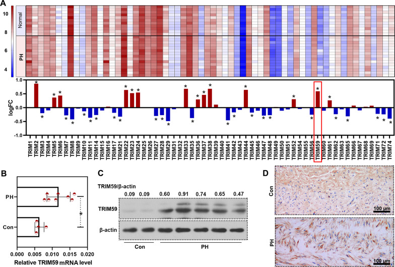
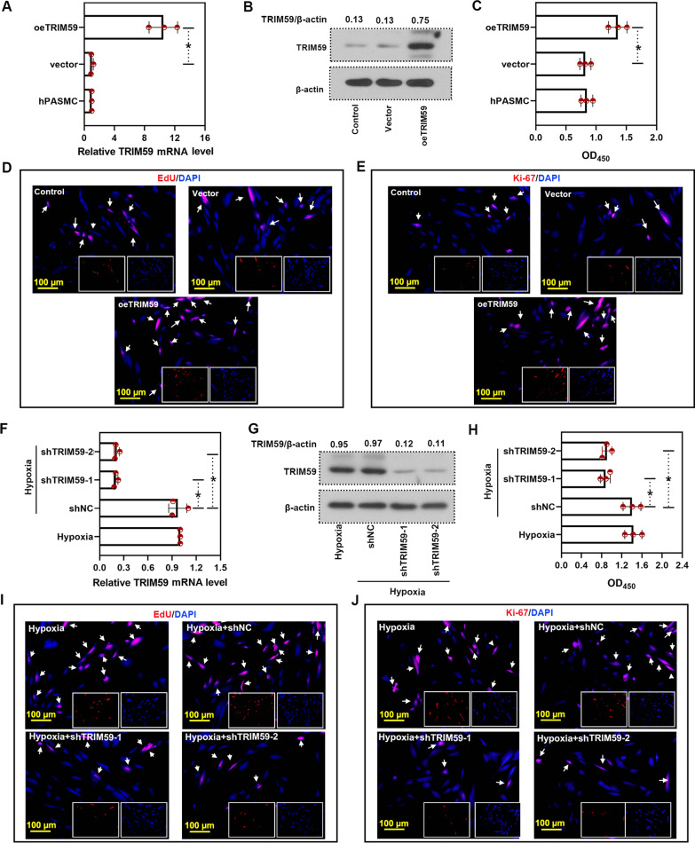
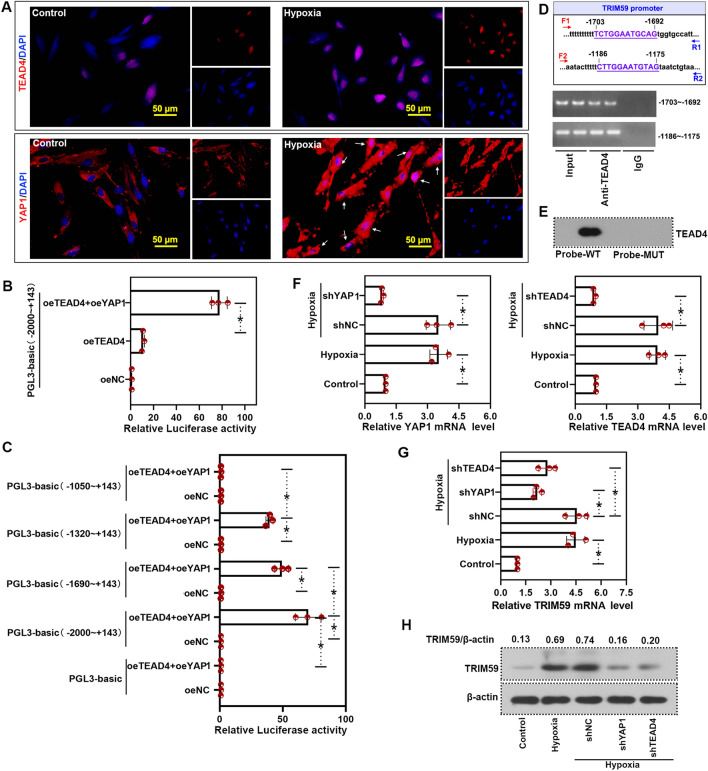
Methods: Differently expressed genes (DEGs) in the lung tissues of PH patients were analyzed with a GEO dataset GSE113439. From these DEGs, we focused on TRIM59 which was highly expressed in PH patients. Subsequently, the expression of TRIM59 in the pulmonary arteries of PH patients, lung tissues of PH rat model and PASMCs cultured in a hypoxic condition was verified by quantitative real-time PCR (qPCR), western blot and immunohistochemistry. Furthermore, the role of TRIM59 in PAMSC proliferation and pathological changes in PH rats was assessed via gain-of-function and loss-of-function experiments. In addition, the transcriptional regulation of YAP1/TEAD4 on TRIM59 was confirmed by qPCR, western blot, luciferase reporter assay, ChIP and DNA pull-down. In order to uncover the underlying mechanisms of TRIM59, a protein ubiquitomics and a CoIP- HPLC-MS/MS were companied to identify the direct targets of TRIM59.

Results: TRIM59 was highly expressed in the pulmonary arteries of PH patients and lung tissues of PH rats. Over-expression of TRIM59 accelerated the proliferation of PASMCs, while TRIM59 silencing resulted in the opposite results. Moreover, TRIM59 silencing mitigated the injuries in heart and lung and attenuated pulmonary vascular remodeling during PH. In addition, its transcription was positively regulated by YAP1/TEAD4. Then we further explored the underlying mechanisms of TRIM59 and found that TRIM59 overexpression resulted in an altered ubiquitylation of proteins. Accompanied with the results of CoIP- HPLC-MS/MS, 34 proteins were identified as the direct targets of TRIM59.

Conclusion: TRIM59 was highly expressed in PH patients and promoted the proliferation of PASMCs and pulmonary vascular remodeling, thus contributing to the pathogenesis of PH. It is indicated that TRIM59 may become a potential target for PH treatment.

Keywords: Pulmonary artery smooth muscle cells; Pulmonary hypertension; TRIM59; Ubiquitylation modification; YAP1/TEAD4.


Conflict of interest statement

The authors declare that they have no competing interests.

Figures

Fig. 1
Fig. 1
TRIM59 was highly expressed in PH patients. A Levels of TRIM family members in the lung tissues of PH patients according to data from GSE113439. B qPCR was performed to determine the expression of TRIM59 in the pulmonary arteries from PH patients and controls. C The protein level of TRIM59 in the pulmonary arteries of PH patients was detected by western blot. D Immunohistochemistry was conducted to detect TRIM59 in the pulmonary arteries of PH patients. Amplification: 200 ×. *p < 0.05
Fig. 2
Fig. 2
TRIM59 was highly expressed in the lung tissues of rat model and cell model of PH. A A rat model of PH was established via a single subcutaneous injection of Sugen-5416 (20 mg/kg) and fed in a hypoxic condition. B The right ventricular systolic pressure was measured via right heart catheterization. C Immunohistochemistry staining was performed to detect the expression and distribution of TRIM59. Amplification: 100 × and 400 ×. D The expression of TRIM59 in the lung tissues of PH rats was determined by qPCR. E Western blot was performed to detect the TRIM59 expression in the lung tissues of PH rats. F Double immunofluorescence staining was performed to detect the TRIM59 expression and distribution in lung tissues. Amplification: 100 × . G Human PASMCs were cultured in hypoxic condition for 24 h. H The level of TRIM59 was detected by western blot. I qPCR was conducted to determine the expression of TRIM59 in PASMCs under a hypoxic condition. *p < 0.05
Fig. 3
Fig. 3
TRIM59 accelerated the proliferation of PASMCs. A After TRIM59 over-expression, the mRNA level of TRIM59 was determined by qPCR. B Western blot was performed to detect the protein level of TRIM59 after TRIM59 over-expression. C After TRIM59 over-expression, the cell viability of PASMCs with TRIM59 over-expression was evaluated by CCK-8 assay. D The proliferation of PASMCs was assessed by EdU incorporation assay. Magnification: × 200. White arrows indicated EdU-positive cells. E Imunofluorescence staining with Ki-67 antibody was conducted after TRIM59 over-expression. Magnification: × 200. White arrows indicated Ki-67-positive cells. F PASMCs with TRIM59 silencing were treated under a hypoxic condition, then the mRNA level of TRIM59 was determined by qPCR. G The level of TRIM59 was determined by western blot after indicated treatment. H CCK-8 assay was conducted in cells with indicated treatment. I EdU incorporation assay was performed to assess the proliferation of PASMC after indicated treatment. Magnification: × 200. White arrows indicated EdU-positive cells. J Ki-67 inofluorescence staining was conducted after TRIM59 over-expression. Magnification: × 200. White arrows indicated Ki-67-positive cells. *p < 0.05
Fig. 4
Fig. 4
TRIM59 silencing remitted PH. A SuHx rat model of PH was established following infection with AAV2 carrying TRIM59 shRNA, then the mRNA level of TRIM59 was determined by qPCR. B Western blot was conducted to detect the TRIM59 protein level after TRIM59 silencing in PH rats. C After TRIM59 silencing, the right ventricular systolic pressure was measured. D HE staining was conducted to evaluate the histopathological changes of heart. Magnification: × 200. E HE staining was performed to assess the histopathological changes of lung. Magnification: × 200. F Immunohistochemical staining was performed to detect the level of α-SMA. Magnification: × 200. G Imunofluorescence staining with TRIM59 and α-SMA was performed. Magnification: 100 ×, 400 ×. H The level of α-SMA and Ki-67 was determined by imunofluorescence staining. Magnification: 400 ×. * p < 0.05
Fig. 5
Fig. 5
TRIM59 was transcriptionally regulated by YAP1/TEAD4. A Immunofluorescence staining was performed to detect YAP1 or TEAD4 expression and distribution during hypoxia. Amplification: 400 ×. B The promoter sequence of TRIM59 was inserted into pGL3-basic plasmid. Then the luciferase reporter assay was performed to determine the effect of YAP1 and TEAD4 on the transcription of TRIM59. C Differently truncated fragments of TRIM59 promoter were inserted into pGL3-basic plasmid. Then the luciferase reporter assay was performed to determine the effect of YAP1 and TEAD4 on the transcription of TRIM59. D ChIP assay was conducted to verify the bind between TEAD4 and TRIM59 promoter. IgG served as the negative control. E The binding of TRIM59 promoter and TEAD4 was confirmed by DNA pull down assay. F In order to explore the effect of YAP1 and TEAD4 on the transcription of TRIM59, PASMCs were infected with adenovirus carrying YAP1 shRNA or TEAD4 shRNA. Then the levels of YAP1 and TEAD4 were measured by qPCR. G After infection, the mRNA level of TRIM59 was determined by qPCR. H Western blot was performed to detect the protein level of TRIM59. *p < 0.05
Fig. 6
Fig. 6
TRIM59 modulated protein ubiquitylation. A In PASMCs with TRIM59 over-expression, the level of flag (indicating TRIM59) and total ubiquitylation was detected by western blot. B The TRIM59 overexpressed PASMCs were lysed and subjected to protein ubiquitomics. C Heatmap of differently ubiquitylated proteins in PASMCs with TRIM59 over-expression. D The ubiquitylation-upregulated and ubiquitylation-downregulated proteins were subjected to GO analysis. E The differently ubiquitylated proteins were subjected to KEGG analysis
Fig. 7
Fig. 7
Binding proteins of TRIM59. A CoIP was performed with flag antibody (indicating TRIM59) to detect the proteins binding to TRIM59. B CoIP binding proteins were subjected to GO analysis. C CoIP binding proteins were subjected to KEGG analysis
Fig. 8
Fig. 8
Direct targets of TRIM59. A Venn diagram of CoIP binding proteins and up- ubiquitylated proteins. B Direct targets of TRIM59 identified by protein ubiquitomics and CoIP-LC–MS/MS. C TRIM59 direct targets were subjected to GO analysis. D TRIM59 direct targets were subjected to KEGG analysis

References

    1. Humbert M, Guignabert C, Bonnet S, Dorfmuller P, Klinger JR, Nicolls MR, Olschewski AJ, Pullamsetti SS, Schermuly RT, Stenmark KR, Rabinovitch M. Pathology and pathobiology of pulmonary hypertension: state of the art and research perspectives. Eur Respir J. 2019;53:1801887. doi: 10.1183/13993003.01887-2018. - DOI - PMC - PubMed
    1. Rubin LJ. Therapy of pulmonary hypertension: the evolution from vasodilators to antiproliferative agents. Am J Respir Crit Care Med. 2002;166:1308–1309. doi: 10.1164/rccm.2208008. - DOI - PubMed
    1. Montani D, Chaumais MC, Guignabert C, Gunther S, Girerd B, Jais X, Algalarrondo V, Price LC, Savale L, Sitbon O, et al. Targeted therapies in pulmonary arterial hypertension. Pharmacol Ther. 2014;141:172–191. doi: 10.1016/j.pharmthera.2013.10.002. - DOI - PubMed
    1. Dilasser F, Rio M, Rose L, Tesse A, Guignabert C, Loirand G, Sauzeau V. Smooth muscle Rac1 contributes to pulmonary hypertension. Br J Pharmacol. 2022;179:3418–3429. doi: 10.1111/bph.15805. - DOI - PMC - PubMed
    1. Zhang L, Wang Y, Wu G, Rao L, Wei Y, Yue H, Yuan T, Yang P, Xiong F, Zhang S, et al. Blockade of JAK2 protects mice against hypoxia-induced pulmonary arterial hypertension by repressing pulmonary arterial smooth muscle cell proliferation. Cell Prolif. 2020;53:e12742. doi: 10.1111/cpr.12742. - DOI - PMC - PubMed

Publication types

MeSH terms