. 2023 Nov 17;20(1):267.
doi: 10.1186/s12974-023-02938-z.

Pericytes are protective in experimental pneumococcal meningitis through regulating leukocyte infiltration and blood-brain barrier function

Affiliations

Pericytes are protective in experimental pneumococcal meningitis through regulating leukocyte infiltration and blood-brain barrier function

Nina C Teske et al. J Neuroinflammation. .

Abstract

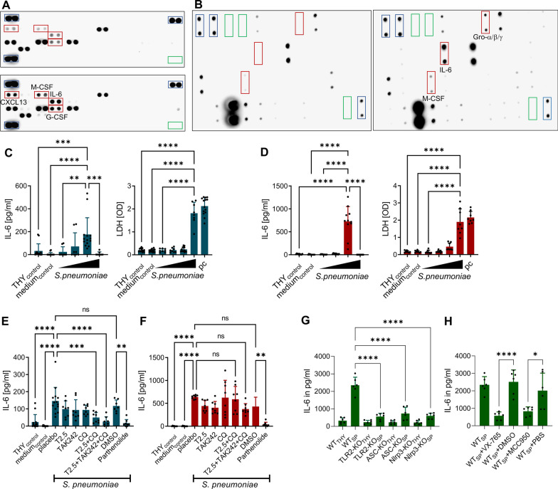
Background: Brain pericytes participate in the regulation of cerebral blood flow and the maintenance of blood-brain barrier integrity. Because of their perivascular localization, their receptor repertoire, and their potential ability to respond to inflammatory and infectious stimuli by producing various cytokines and chemokines, these cells are also thought to play an active role in the immune response to brain infections. This assumption is mainly supported by in vitro studies, investigations in in vivo disease models are largely missing. Here, we analysed the role of brain pericytes in pneumococcal meningitis, in vitro and in vivo in two animal models of pneumococcal meningitis.

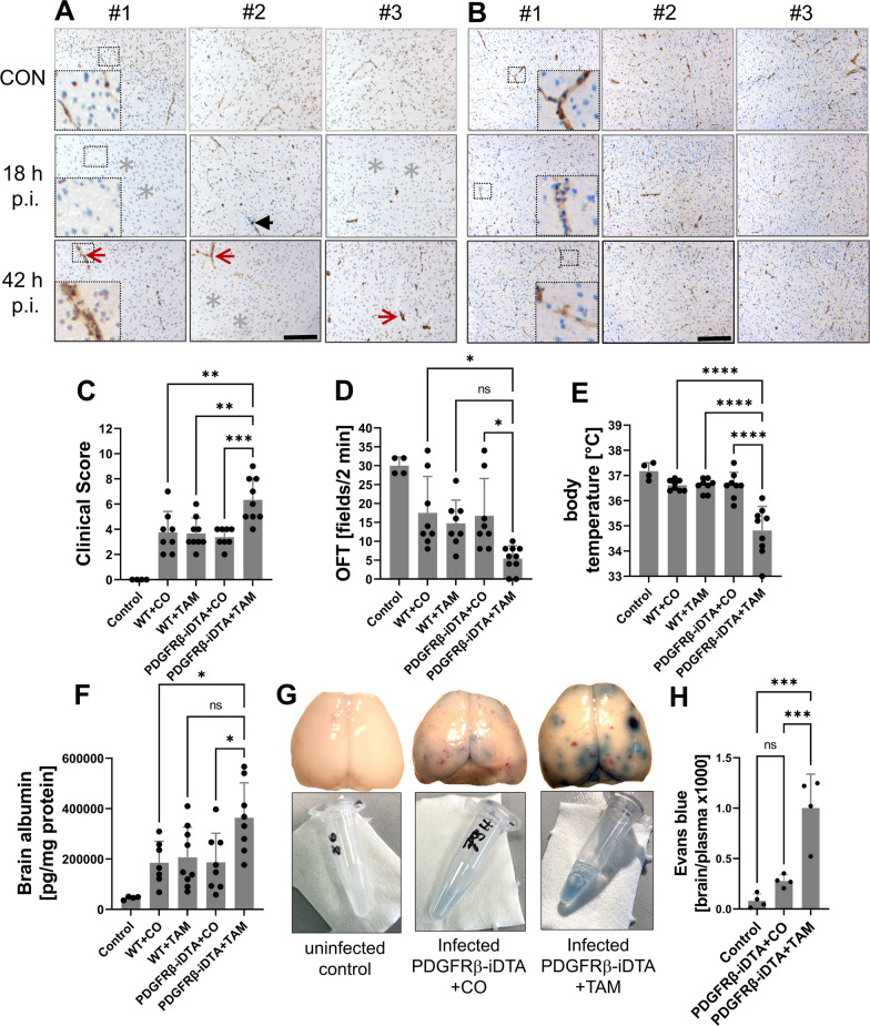
Methods: Primary murine and human pericytes were stimulated with increasing concentrations of different serotypes of Streptococcus pneumoniae in the presence or absence of Toll-like receptor inhibitors and their cell viability and cytokine production were monitored. To gain insight into the role of pericytes in brain infection in vivo, we performed studies in a zebrafish embryo model of pneumococcal meningitis in which pericytes were pharmacologically depleted. Furthermore, we analyzed the impact of genetically induced pericyte ablation on disease progression, intracranial complications, and brain inflammation in an adult mouse model of this disease.

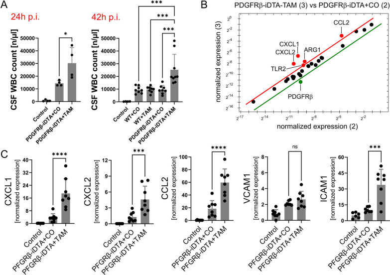
Results: Both murine and human pericytes reacted to pneumococcal exposure with the release of selected cytokines. This cytokine release is pneumolysin-dependent, TLR-dependent in murine (but not human) pericytes and can be significantly increased by macrophage-derived IL-1b. Pharmacological depletion of pericytes in zebrafish embryos resulted in increased cerebral edema and mortality due to pneumococcal meningitis. Correspondingly, in an adult mouse meningitis model, a more pronounced blood-brain barrier disruption and leukocyte infiltration, resulting in an unfavorable disease course, was observed following genetic pericyte ablation. The degree of leukocyte infiltration positively correlated with an upregulation of chemokine expression in the brains of pericyte-depleted mice.

Conclusions: Our findings show that pericytes play a protective role in pneumococcal meningitis by impeding leukocyte migration and preventing blood-brain barrier breaching. Thus, preserving the integrity of the pericyte population has the potential as a new therapeutic strategy in pneumococcal meningitis.

Keywords: Blood–brain barrier; Pericytes; Pneumococcal meningitis; Streptococcus pneumoniae.


Conflict of interest statement

The authors declare that they have no competing interests.

Figures

Fig. 1
Fig. 1
Human and murine brain pericytes release selected cytokines upon S. pneumoniae challenge. Protein array analyses of cell-culture supernatants obtained from murine (A) and human (B) primary brain pericytes stimulated with S. pneumoniae (MOI = 40) or its vehicle for 6 h. The differentially expressed proteins are outlined with red rectangles, the positive controls with blue rectangles and the negative controls with green rectangles. IL-6 concentrations (determined by ELSA) in cell-culture supernatants of murine (blue bars; C) and human (red bars; D) primary brain pericytes 6 h after exposure to increasing concentrations of antibiotic-lysed serotype 2 S. pneumoniae (MOI = 2.5, 10, 40, 160). THY (= Todd–Hewitt broth supplemented with 0.2% yeast extract, used for culturing S. pneumoniae) and cell-culture medium served as negative controls. Effect of various anti-TLR antagonists (T2.5 = neutralizing antir-TLR2 antibody; TAK242 = a TLR4 antagonist; CQ = chloroquine = an endosomal TLR antagonist) and the NF-κB inhibitor parthenolide on S. pneumoniae (MOI = 40)-induced IL-6 release from murine (E) and human (F) primary brain pericytes. Response of human brain pericytes to conditioned media (green bars) from wild-type (WT), TLR2-deficient, ASC-deficient, and Nlrp3-deficient THP-1 cells stimulated with either THY or S. pneumoniae (MOI = 80; G). Response of human brain pericytes to conditioned media from S. pneumoniae-stimulated WT THP-1 treated either with the caspase-1 inhibitor VX-765, the Nlrp3 inhibitor MCC950, or its vehicles (DMSO or PBS, H). Data are given as individual values as well as means ± SD. *P < 0.05, **P < 0.01, ***P < 0.001, ****P < 0.0001, using ANOVA with Tukey’s multiple comparisons test
Fig. 2
Fig. 2
Confocal microscopy images from brains of TgBAC(pdgfrb:EGFP/Tg(fli 1 a:Myr-mCherry) transgenic zebrafish embryos that have had vehicle (uninfected controls, AC) or S. pneumoniae (meningitis, DF) injected into their hindbrain ventricles (three representative examples each). The embryos express eGFP in mural cells (namely, pericytes) and mCherry in endothelial cells. White bold arrows: compared to controls, infected embryos showed a marked reduction in mCherry-positive cells indicating meningitis-associated endothelial cell destruction (DF). Thin double-headed arrows: a strong decrease in eGFP-positive cells was seen in some infected embryos, suggesting pericyte loss (E, F). Thin arrows: in others, there was predominantly a loss of contact between eGFP-positive and mCherry-positive cells (D, E). The scale bar indicates 100 μm length. (A–F). Effect of pharmacological pericyte depletion on the survival of wild-type (on the left) and TgBAC(pdgfrb:EGFP/Tg(flila:Myr-mCherry; on the right side) zebrafish embryos (G, H). Wild-type embryos were injected with 1900 cfu of live S. pneumoniae (= meningitis; or phenol red solution = controls; G); TgBAC(pdgfrb:EGFP/Tg(flila:Myr-mCherry embryos received 2200 cfu of live S. pneumoniae (= meningitis; or phenol red solution = controls, H). Embryos were treated either with DMSO as a placebo or the PDGFRβ inhibitor AG1296. Data are given as means ± SEM. Histopathological analysis of non-infected and Streptococcus pneumonia-infected wild-type zebrafish embryos treated either with DMSO as placebo (IL) or the PDGFRβ inhibitor AG1296 (MP) at 32 h post-injection (hpi). White arrows in M: embryos treated with AG1296 showed multiple cystic abnormalities in their eyes, compared to placebo-treated embryos (I). After S. pneumoniae infection, placebo- and AG1296-treated embroys showed signs of meningitis with presence of polymorphonuclear leukocytes in the ventricles (black asterisks) (K, O). White arrow heads in O: the brain parenchyma of AG1296-treated, infected embryos showed distinct edema, while the parenchyma of placebo-treated, infected embryos remained compact (K). Black arrows in L, P: the perivascular space was broadened in AG1296-treated embryos, compared to placebo-treated embryos
Fig. 3
Fig. 3
Regional changes in the immunoreactivity for PDGFRβ (A) and CD13 (B) in murine parietal cortices during PM. Brain sections were obtained from healthy controls (upper images) as well as infected mice at 18 and 42 h (h) after intracisternal application of S. pneumoniae (p.i. = post-infection; 3 mice per group, #1, #2, and #3; middle and lower images, respectively). The sections were stained either with anti-murine PDGFRβ or anti-murine CD13 antibodies and counterstained with hematoxylin–eosin. The scale bars on the lower middle images indicates 100 µm in length. PDGFRβ immunoreactivity appeared regionally disrupted (grey asterisks) and was missing at some vascular segments (black arrow), whereas in other vascular segments, PDGFRβ staining appears unchanged or even enhanced (red arrows). The determination of the disease phenotype 42 h after infection (CE) showed a clear worsening of clinical symptoms in tamoxifen (TAM)-treated PDGFRB::creER2-iDTA mice (PDGFRβ-iDTA) which were largely missing pericytes, compared to corn oil (CO)-treated PDGFRB::creER2-iDTA mice (controls), as evidenced by significantly increased clinical score values (E), more restricted motor activities in the open-field test (OFT) (D), and lower body temperatures (E). The worsening of disease was associated with increased brain edema formation, as evidenced by increased brain albumin concentrations 42 h after infection (F) and enhanced Evans blue extravasation 24 h after infection (G, H) in TAM-treated PDGFRB::creER2-iDTA mice compared to controls. G Representative brain images taken immediately after perfusion and removal from one control mouse, one TAM- and one CO-treated PDGFRB::creER2-iDTA mouse (upper images) as well as the associated Evans blue extracts. Data are given as individual values as well as means ± SD. *P < 0.05, **P < 0.01, ***P < 0.001, ****P < 0.0001, using ANOVA with Tukey’s multiple comparisons test
Fig. 4
Fig. 4
Cerebrospinal (CSF) white blood cell (WBC) counts at 24 and 42 h post-infection in TAM-treated PDGFRB::creER2-iDTA mice (PDGFRβ-iDTA) that are largely missing pericytes compared to control groups (A). Prime™ PCR array analysis of cDNA obtained from brains of infected transgenic mice revealed higher expression levels of CXCL1, CXCL2, and CCL2 (and also of TLR2 and ARG1), but lower PDGFRβ expression following pericyte depletion (B). The array results were confirmed by supplemental RT-PCR analyses (C), which showed increased brain expression of CXCL1, CXCL2, and CCL2 in TAM-treated PDGFRB::creER2-iDTA mice. Data are given as individual values as well as means ± SD. *P < 0.05, **P < 0.01, ***P < 0.001, ****P < 0.0001, using ANOVA with Tukey’s multiple comparisons test

References

    1. Van de Beek D, Brouwer M, Hasbun R, Koedel U, Whitney CG, Wijdicks E. Community-acquired bacterial meningitis. Nat Rev Dis Primers. 2016;2:16074. doi: 10.1038/nrdp.2016.74. - DOI - PubMed
    1. Van de Beek D, Brouwer MC, Koedel U, Wall EC. Community-acquired bacterial meningitis. Lancet. 2021;398:1171–1183. doi: 10.1016/S0140-6736(21)00883-7. - DOI - PubMed
    1. Weber JR, Tuomanen EI. Cellular damage in bacterial meningitis: an interplay of bacterial and host driven toxicity. J Neuroimmunol. 2007;184:45–52. doi: 10.1016/j.jneuroim.2006.11.016. - DOI - PubMed
    1. Zanluqui NG, McGavern DB. Bacterial meningitis hits an immunosuppressive nerve. Nature. 2023;615:396. doi: 10.1038/d41586-023-00540-0. - DOI - PMC - PubMed
    1. Pinho-Ribeiro FA, Deng L, Neel DV, Erdogan O, Basu H, Yang D, Choi S, Walker AJ, Carneiro-Nascimento S, He K, Wu G, Stevens B, Doran KS, Levy D, Chiu IM. Bacteria hijack a meningeal neuroimmune axis to facilitate brain invasion. Nature. 2023;615:472. doi: 10.1038/s41586-023-05753-x. - DOI - PMC - PubMed

LinkOut - more resources