. 2024 May;20(5):1032-1053.
doi: 10.1080/15548627.2023.2281156. Epub 2023 Nov 18.

Autophagy activates EGR1 via MAPK/ERK to induce FGF2 in renal tubular cells for fibroblast activation and fibrosis during maladaptive kidney repair

Affiliations

Autophagy activates EGR1 via MAPK/ERK to induce FGF2 in renal tubular cells for fibroblast activation and fibrosis during maladaptive kidney repair

Man J Livingston et al. Autophagy. 2024 May.

Abstract

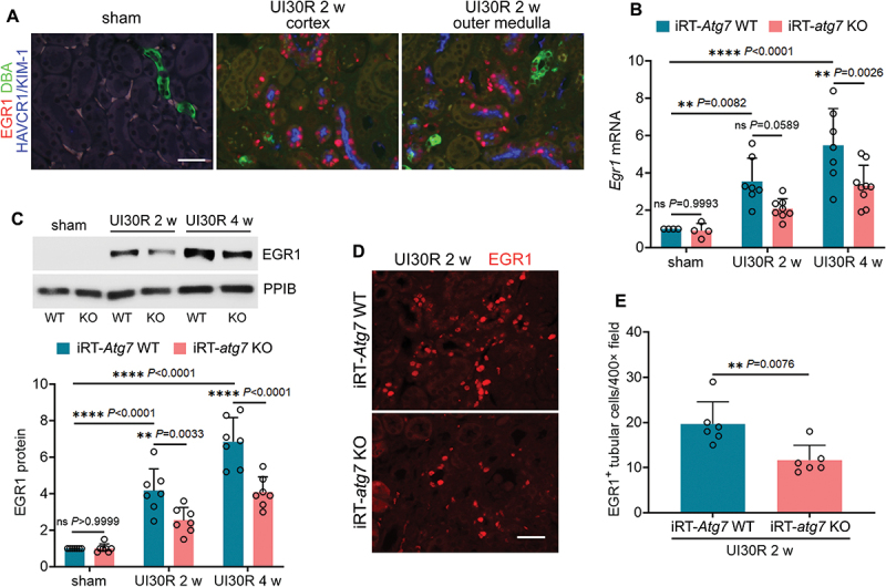
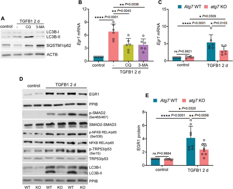
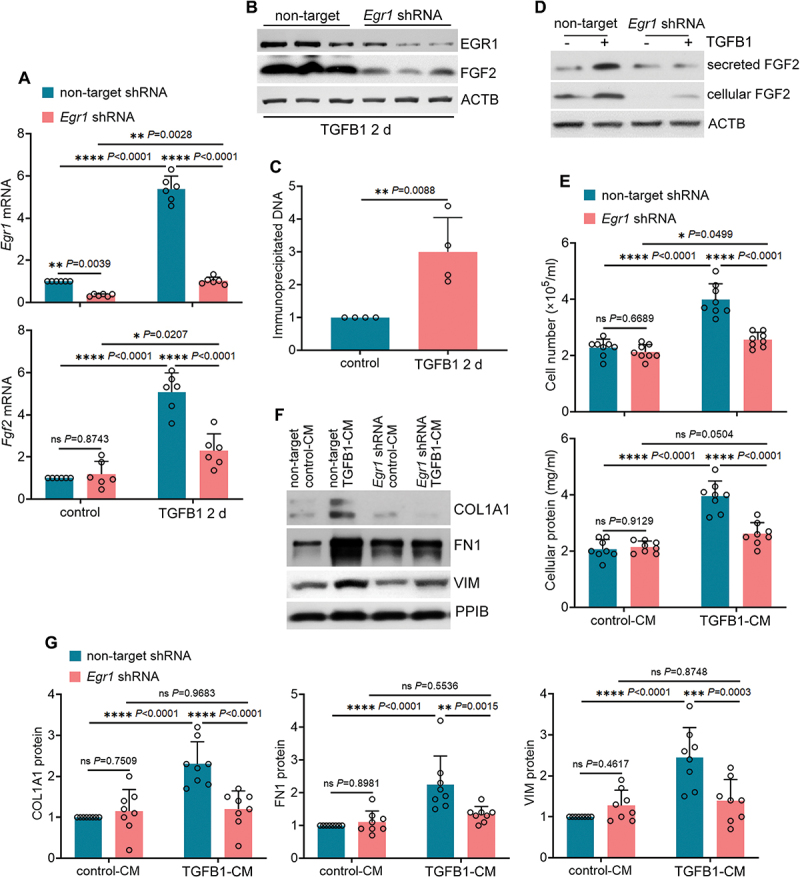
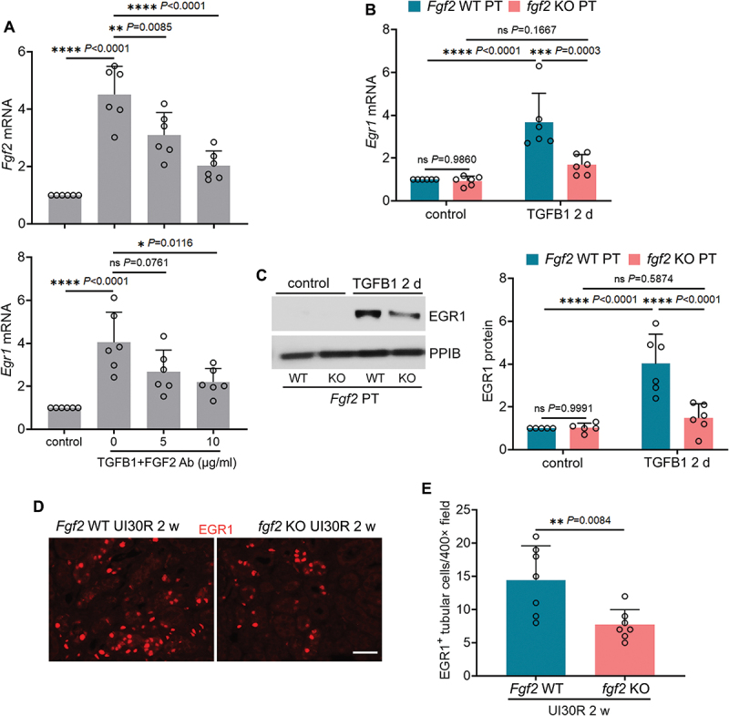
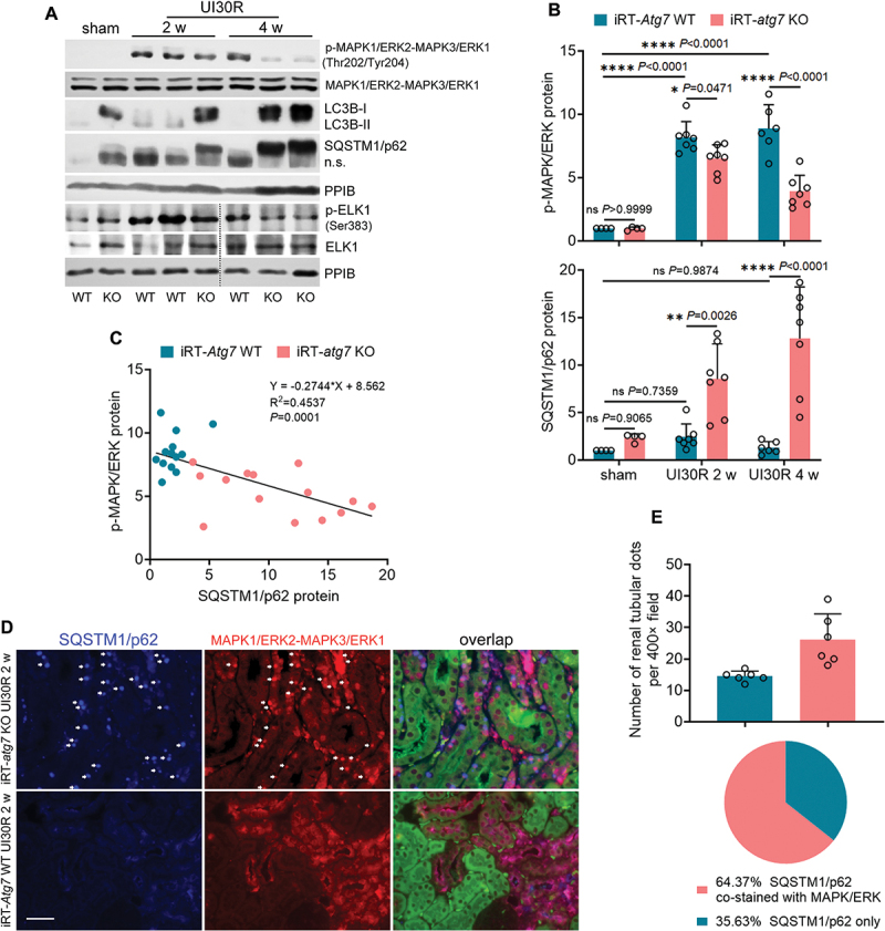
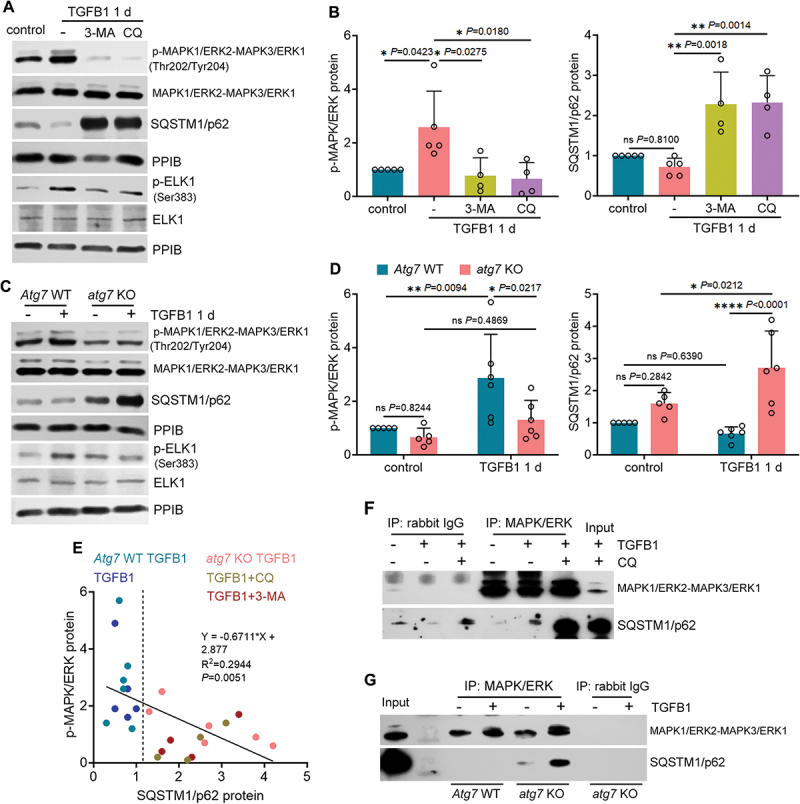
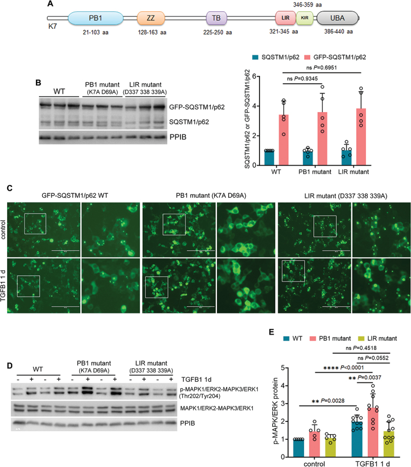
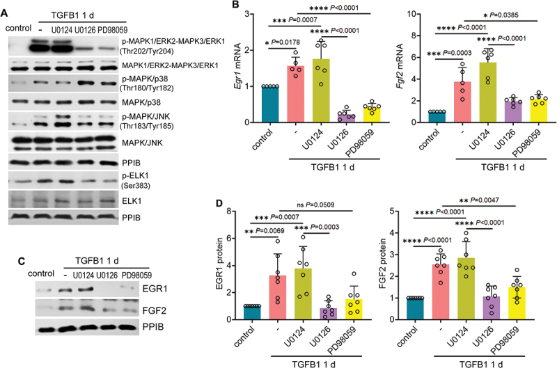
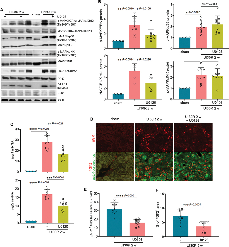
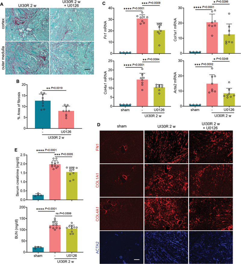
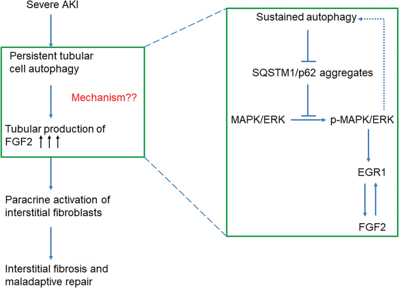
Macroautophagy/autophagy contributes to maladaptive kidney repair by inducing pro-fibrotic factors such as FGF2 (fibroblast growth factor 2), but the underlying mechanism remains elusive. Here, we show that EGR1 (early growth response 1) was induced in injured proximal tubules after ischemic acute kidney injury (AKI) and this induction was suppressed by autophagy deficiency in inducible, renal tubule-specific atg7 (autophagy related 7) knockout (iRT-atg7 KO) mice. In cultured proximal tubular cells, TGFB1 (transforming growth factor beta 1) induced EGR1 and this induction was also autophagy dependent. Egr1 knockdown in tubular cells reduced FGF2 expression during TGFB1 treatment, leading to less FGF2 secretion and decreased paracrine effects on fibroblasts. ChIP assay detected an increased binding of EGR1 to the Fgf2 gene promoter in TGFB1-treated tubular cells. Both Fgf2 and Egr1 transcription was inhibited by FGF2 neutralizing antibody, suggesting a positive feedback for EGR1-mediated FGF2 autoregulation. This feedback was confirmed using fgf2-deficient tubular cells and fgf2-deficient mice. Upstream of EGR1, autophagy deficiency in mice suppressed MAPK/ERK (mitogen-activated protein kinase) activation in post-ischemic renal tubules. This inhibition correlated with SQSTM1/p62 (sequestosome 1) aggregation and its sequestration of MAPK/ERK. SQSTM1/p62 interacted with MAPK/ERK and blocked its activation during TGFB1 treatment in autophagy-deficient tubular cells. Inhibition of MAPK/ERK suppressed EGR1 and FGF2 expression in maladaptive tubules, leading to the amelioration of renal fibrosis and improvement of renal function. These results suggest that autophagy activates MAPK/ERK in renal tubular cells, which induces EGR1 to transactivate FGF2. FGF2 is then secreted into the interstitium to stimulate fibroblasts for fibrogenesis.Abbreviation: 3-MA: 3-methyladenine; ACTA2/α-SMA: actin alpha 2, smooth muscle, aorta; ACTB/β-actin: actin, beta; AKI: acute kidney injury; aa: amino acid; ATG/Atg: autophagy related; BUN: blood urea nitrogen; ChIP: chromatin immunoprecipitation; CKD: chronic kidney disease; CM: conditioned medium; COL1A1: collagen, type I, alpha 1; COL4A1: collagen, type IV, alpha 1; CQ: chloroquine; DBA: dolichos biflorus agglutinin; EGR1: early growth response 1; ELK1: ELK1, member of ETS oncogene family; FGF2: fibroblast growth factor 2; FN1: fibronectin 1; GAPDH: glyceraldehyde-3-phosphate dehydrogenase; HAVCR1/KIM-1: hepatitis A virus cellular receptor 1; IP: immunoprecipitation; LIR: LC3-interacting region; MAP1LC3B/LC3B: microtubule-associated protein 1 light chain 3 beta; MAP2K/MEK: mitogen-activated protein kinase kinase; MAPK: mitogen-activated protein kinase; NFKB: nuclear factor kappa B; PB1: Phox and Bem1; PFT: pifithrin α; PPIB/cyclophilin B: peptidylprolyl isomerase B; RT-qPCR: real time-quantitative PCR; SQSTM1/p62: sequestosome 1; TGFB1/TGF-β1: transforming growth factor beta 1; VIM: vimentin.

Keywords: Interstitial fibrosis; SQSTM1/p62; ischemic acute kidney injury; kidney repair; tubulo-interstitial communication.


Conflict of interest statement

No potential conflict of interest was reported by the author(s).

Figures

Figure 1.
Figure 1.
Autophagy deficiency inhibits EGR1 in renal tubular cells during maladaptive kidney repair after ischemic AKI in mice. (A) C57BL/6 mice underwent sham operation (n = 3) or 30-min unilateral renal ischemia followed by reperfusion for 2 weeks (n = 3). Left kidneys were harvested for co-immunofluorescence of EGR1 (red) and HAVCR1/KIM-1 (blue). Fluorescein-DBA (green) was used as a marker for renal distal tubules. Scale bar: 20 µm. (B-E) WT and iRT-atg7 KO mice underwent sham operation or 30-min unilateral renal ischemia followed by reperfusion for up to 4 weeks. Left kidneys were harvested. (B) RT-qPCR of Egr1 mRNA (sham: n = 4; UI30R 2 w: WT n = 7, KO n = 8; UI30R 4 w: WT n = 7, KO n = 9). (C) EGR1 immunoblot and densitometry (n = 7 for each group). PPIB/cyclophilin B was used as an internal loading control. (D) EGR1 immunofluorescence. Scale bar: 20 µm. (E) quantification of EGR1-positive tubular cells (n = 6 for each group). Data in (B), (C) and (E) are presented as mean ± SEM. For statistics, two-way ANOVA with multiple comparisons was used for (B) and (C). 2-tailed, unpaired Student t-test was used for (E).
Figure 2.
Figure 2.
Autophagy deficiency inhibits EGR1 in cultured mouse renal proximal tubular cells during TGFB1 treatment. (A and B) subconfluent BUMPT cells were exposed to 5 ng/ml TGFB1 in serum-free DMEM for 2 days alone or with 5 µM CQ or 1 mM 3-MA. Control cells were kept in serum-free medium without TGFB1. Cells were collected for immunoblot of LC3B and SQSTM1/p62 (A) and RT-qPCR of Egr1 mRNA (B) (n = 6 experiments). (C-E) subconfluent WT and atg7 KO cells were exposed to 5 ng/ml TGFB1 in serum-free DMEM for 2 days. Control cells were kept in serum-free medium without TGFB1. (C) RT-qPCR of Egr1 mRNA (n = 6 experiments). (D) immunoblot of EGR1, p-SMAD2 (Ser465/467), SMAD2-SMAD3, p-NFKB RELA/p65 (Ser536), NFKB RELA/p65, p-TRP53/p53 (Ser15), TRP53/p53 and LC3B. (E) densitometry of EGR1 immunoblot (n = 6 experiments). Data in (B), (C) and (E) are presented as mean ± SEM. For statistics, one-way ANOVA with multiple comparisons was used for (B). Two-way ANOVA with multiple comparisons was used for (C) and (E).
Figure 3.
Figure 3.
EGR1 transactivates FGF2 in TGFB1-treated mouse proximal tubular cells for subsequent paracrine activation of renal fibroblasts. (A and B) subconfluent, non-target shRNA-transfected or Egr1 shRNA-transfected BUMPT cells were exposed to 5 ng/ml TGFB1 in serum-free DMEM for 2 days. Control cells were kept in serum-free medium without TGFB1. Cells were collected for RT-qPCR of Egr1 and Fgf2 mRNA (A) and immunoblot of EGR1 and FGF2 (B) (n = 6 experiments). (C) subconfluent BUMPT cells were exposed to 5 ng/ml TGFB1 in serum-free DMEM for 2 days. Control cells were kept in serum-free medium without TGFB1. Cells were collected for ChIP assay of EGR1 binding to Fgf2 promoter sequence (n = 4 experiments). (D) subconfluent, non-target shRNA-transfected or Egr1 shRNA-transfected BUMPT cells were exposed to 5 ng/ml TGFB1 in serum-free DMEM for 2 days. Control cells were kept in serum-free medium without TGFB1. The old culture media for both TGFB1-treated and control cells were replaced by fresh media free of TGFB1 at the end of day 2, incubated with the cells for an additional day, and then collected as tubular cell-CM for immunoblot of secreted FGF2 (n = 3 experiments). Cell lysates were also collected in the same experiments for immunoblot of cellular FGF2. ACTB/β-actin was used as an internal loading control. (E-G) subconfluent NRK-49F fibroblasts were incubated with CMs from BUMPT cells (non-target shRNA control-CM, non-target shRNA TGFB1-CM, Egr1 shRNA control-CM, and Egr1 shRNA TGFB1-CM) for 2 days. The fibroblasts were then collected for cell number counting and cellular protein measurement (E), immunoblot of COL1A1, FN1 and VIM (F) and densitometry (G) (n = 7 experiments). Data in (A), (C), (E) and (G) are presented as mean ± SEM. For statistics, 2-tailed, unpaired Student t-test was used for (C). Two-way ANOVA with multiple comparisons was used for (A), (E) and (G).
Figure 4.
Figure 4.
EGR1 and FGF2 form a positive feedback for FGF2 autoregulation in renal proximal tubular cells during TGFB1 treatment. (A) subconfluent BUMPT cells were exposed to 5 ng/ml TGFB1 in serum-free DMEM for 3 days in the presence of FGF2 neutralizing antibody at 5 and 10 µg/ml or mouse IgG (indicated as FGF2 ab of 0 µg/ml) as negative control. Control cells were kept in serum-free medium without TGFB1. Cells were collected for RT-qPCR of Fgf2 and Egr1 mRNA (n = 6 experiments). (B and C) subconfluent isolated primary proximal tubular cells from fgf2 KO mice (fgf2 KO PT) and their WT littermates (Fgf2 WT PT) were exposed to 5 ng/ml TGFB1 in serum-free DMEM for 2 days. Control cells were kept in serum-free medium without TGFB1. Cells were collected for RT-qPCR of Egr1 mRNA (B) and immunoblot of EGR1 (C) (n = 6 experiments). (D) fgf2 KO mice (n = 7) and their WT littermates (n = 7) underwent 30-min unilateral renal ischemia followed by reperfusion for 2 weeks. Left kidneys were collected for EGR1 immunofluorescence. Scale bar: 20 µm. (E) quantification of the numbers of EGR1-positive tubular cells. Data in (A), (B), (C) and (E) are presented as mean ± SEM. For statistics, one-way ANOVA with multiple comparisons was used for (A). Two-way ANOVA with multiple comparisons was used for (B) and (C). 2-tailed, unpaired Student t-test was used for (E).
Figure 5.
Figure 5.
Autophagy deficiency inhibits MAPK1/ERK2-MAPK3/ERK1 activation in maladaptive renal tubules and this inhibition correlates with SQSTM1/p62 aggregation and sequestration of MAPK1/ERK2-MAPK3/ERK1 in iRT-atg7 KO mice. WT and iRT-atg7 KO mice underwent sham operation or 30-min unilateral renal ischemia followed by reperfusion for up to 4 weeks. Left kidneys were harvested. (A) immunoblot of p-MAPK1/ERK2-MAPK3/ERK1 (Thr202/Tyr204), MAPK1/ERK2-MAPK3/ERK1, LC3B, SQSTM1/p62, p-ELK1 (Ser383) and ELK1. (B) densitometry of p-MAPK1/ERK2-MAPK3/ERK1 (Thr202/Tyr204) and SQSTM1/p62 immunoblot (sham: n = 4; UI30R 2 w: WT n = 7, KO n = 7; UI30R 4 w: WT n = 6, KO n = 7). (C) correlation analysis of protein fold changes between p-MAPK1/ERK2-MAPK3/ERK1 (Thr202/Tyr204) and SQSTM1/p62. (D) co-immunofluorescence of MAPK1/ERK2-MAPK3/ERK1 (red) and SQSTM1/p62 (blue). Scale bar: 20 µm. (E) quantification of SQSTM1/p62 aggregates with or without MAPK1/ERK2-MAPK3/ERK1 co-staining in post-ischemic iRT-atg7 KO mice (n = 6). Data in (B) and (E) presented as mean ± SEM. For statistics, two-way ANOVA with multiple comparisons was used for (B). Pearson correlation analysis followed by simple linear regression was used for (C). 2-tailed, unpaired Student t-test and fraction of total analysis were used for (E).
Figure 6.
Figure 6.
SQSTM1/p62 interacts with MAPK1/ERK2-MAPK3/ERK1 to block its activation by TGFB1 in autophagy-deficient mouse proximal tubular cells. (A and B) subconfluent BUMPT cells were exposed to 5 ng/ml TGFB1 in serum-free DMEM for 1 day alone or with 20 µM CQ or 5 mM 3-MA. Control cells were kept in serum-free medium without TGFB1. Cells were collected for immunoblot of p-MAPK1/ERK2-MAPK3/ERK1 (Thr202/Tyr204), MAPK1/ERK2-MAPK3/ERK1, SQSTM1/p62, p-ELK1 (Ser383) and ELK1 (n = 5 experiments). (C and D) subconfluent WT and atg7 KO cells were exposed to 5 ng/ml TGFB1 in serum-free DMEM for 1 day. Control cells were kept in serum-free medium without TGFB1. Cells were collected for immunoblot of p-MAPK1/ERK2-MAPK3/ERK1 (Thr202/Tyr204), MAPK1/ERK2-MAPK3/ERK1, SQSTM1/p62, p-ELK1 (Ser383) and ELK1 (n = 5 experiments). (E) correlation analysis of protein fold changes between p-MAPK1/ERK2-MAPK3/ERK1 (Thr202/Tyr204) and SQSTM1/p62. (F) co-IP of MAPK1/ERK2-MAPK3/ERK1 and SQSTM1/p62 in BUMPT cells (n = 3 experiments). (G) co-IP of MAPK1/ERK2-MAPK3/ERK1 and SQSTM1/p62 in WT and atg7 KO cells (n = 3 experiments). Data in (B) and (D) are presented as mean ± SEM. For statistics, one-way ANOVA with multiple comparisons was used for (B). Two-way ANOVA with multiple comparisons was used for (D). Pearson correlation analysis followed by simple linear regression was used for (E).
Figure 7.
Figure 7.
Inhibition of SQSTM1/p62 aggregation further enhances MAPK1/ERK2-MAPK3/ERK1 phosphorylation in TGFB1-treated mouse proximal tubular cells. (A) structural domains in SQSTM1/p62 protein. (B) BUMPT cells stably expressing GFP-SQSTM1/p62 WT, PB1 mutant (K7A D69A), or LIR mutant (D337 338 339A) were collected for immunoblot analysis (n = 5 experiments). (C-E) BUMPT cells stably expressing GFP-SQSTM1/p62 WT, PB1 mutant (K7A D69A), or LIR mutant (D337 338 339A) were incubated for 1 day with or without 5 ng/ml TGFB1 in serum-free DMEM for morphological and immunoblot analyses. (C) Representative fluorescence images showing the formation of GFP-SQSTM1/p62 aggregates. Scale bar: 200 µm. The boxed areas were enlarged and placed on the right side. (D) immunoblots of p-MAPK1/ERK2-MAPK3/ERK1 (Thr202/Tyr204) and MAPK1/ERK2-MAPK3/ERK1. (E) densitometry of p-MAPK1/ERK2-MAPK3/ERK1 (Thr202/Tyr204) immunoblot (n = 5 experiments). Data in (B) and (E) are presented as mean ± SEM and two-way ANOVA with multiple comparisons was used for statistics.
Figure 8.
Figure 8.
Inhibition of MAPK1/ERK2-MAPK3/ERK1 suppresses EGR1 activation and FGF2 production in TGFB1-treated mouse proximal tubular cells. Subconfluent BUMPT cells were exposed to 5 ng/ml TGFB1 in serum-free DMEM for 1 day alone or with negative control inhibitor (10 µM U0124) or with MAPK1/ERK2-MAPK3/ERK1 inhibitors (10 µM U0126, 50 µM PD98059). Control cells were kept in serum-free medium without TGFB1. (A) immunoblot of p-MAPK1/ERK2-MAPK3/ERK1 (Thr202/Tyr204), MAPK1/ERK2-MAPK3/ERK1, p-MAPK/p38 (Thr180/Tyr182), MAPK/p38, p-MAPK/JNK (Thr183/Tyr185), MAPK/JNK, p-ELK1 (Ser383) and ELK1 (n = 3 experiments). (B) RT-qPCR of Fgf2 and Egr1 mRNA (n = 5 experiments). (C and D) immunoblot and densitometry of EGR1 and FGF2 (n = 7 experiments). Data in (B) and (D) are presented as mean ± SEM. For statistics, one-way ANOVA with multiple comparisons was used for both (B) and (D).
Figure 9.
Figure 9.
Inhibition of MAPK1/ERK2-MAPK3/ERK1 suppresses EGR1 and FGF2 in renal tubules during maladaptive kidney repair after ischemic AKI in mice. C57BL/6 mice underwent sham operation (n = 4) or 30-min unilateral renal ischemia followed by reperfusion alone (n = 11) or with U0126 (n = 10) (20 mg/kg, i.P., every other day injection, starting from day 2 of reperfusion) for 2 weeks. Left kidneys were harvested. (A) immunoblot of p-MAPK1/ERK2-MAPK3/ERK1 (Thr202/Tyr204), MAPK1/ERK2-MAPK3/ERK1, p-MAPK/p38 (Thr180/Tyr182), MAPK/p38, p-MAPK/JNK (Thr183/Tyr185), MAPK/JNK, HAVCR1/KIM-1, p-ELK1 (Ser383) and ELK1. (B) densitometry of p-MAPK1/ERK2-MAPK3/ERK1 (Thr202/Tyr204), p-MAPK/p38 (Thr180/Tyr182), p-MAPK/JNK (Thr183/Tyr185) and HAVCR1/KIM-1 immunoblot. (C) RT-qPCR of Egr1 and Fgf2 mRNA. (D) immunofluorescence of EGR1 and FGF2. Scale bar: 20 µm. (E) quantification of the numbers of EGR1-positive tubular cells. (F) quantification of FGF2-positive staining areas. Data in (B), (C), (E) and (F) are presented as mean ± SEM. For statistics, one-way ANOVA with multiple comparisons was used for (B) and (C). 2-tailed, unpaired Student t-test was used for (E) and (F).
Figure 10.
Figure 10.
Inhibition of MAPK1/ERK2-MAPK3/ERK1 ameliorates interstitial fibrosis and improves renal function in post-ischemic mice. C57BL/6 mice were subjected to the treatments as described in Figure 9. (A) Sirius red/Fast Green staining. Scale bar: 100 µm. (B) quantification of collagen-stained areas. (C) RT-qPCR of Fn1, Col1a1, Col4a1 and Acta2/α-sma mRNA. (D) immunofluorescence of FN1, COL1A1, COL4A1 and ACTA2/α-SMA. Scale bar: 100 µm. (E) 2 weeks after unilateral ischemia-reperfusion, right kidneys were removed, and blood samples were collected 24 h after right nephrectomy for measurements of BUN and serum creatinine. Data in (B), (C) and (E) are presented as mean ± SEM. For statistics, 2-tailed, unpaired Student t-test was used for (B). One-way ANOVA with multiple comparisons was used for (C) and (E).
Figure 11.
Figure 11.
Graphic summary highlighting how tubular cell autophagy promotes interstitial fibrosis for maladaptive kidney repair through the SQSTM1/p62-MAPK/ERK-EGR1-FGF2 signaling cascade.

References

    1. Ronco C, Bellomo R, Kellum JA.. Acute kidney injury. Lancet. 2019;394(10212):1949–1964. doi: 10.1016/S0140-6736(19)32563-2 - DOI - PubMed
    1. See EJ, Jayasinghe K, Glassford N, et al. Long-term risk of adverse outcomes after acute kidney injury: a systematic review and meta-analysis of cohort studies using consensus definitions of exposure. Kidney Int. 2019;95(1):160–172. doi: 10.1016/j.kint.2018.08.036 - DOI - PubMed
    1. Zuk A, Bonventre JV. Recent advances in acute kidney injury and its consequences and impact on chronic kidney disease. Curr Opin Nephrol Hypertens. 2019;28(4):397–405. doi: 10.1097/MNH.0000000000000504 - DOI - PMC - PubMed
    1. Yu SM, Bonventre JV. Acute kidney injury and maladaptive tubular repair leading to renal fibrosis. Curr Opin Nephrol Hypertens. 2020;29(3):310–318. doi: 10.1097/MNH.0000000000000605 - DOI - PMC - PubMed
    1. Ferenbach DA, Bonventre JV. Mechanisms of maladaptive repair after AKI leading to accelerated kidney ageing and CKD. Nat Rev Nephrol. 2015;11(5):264–276. doi: 10.1038/nrneph.2015.3 - DOI - PMC - PubMed

Publication types

MeSH terms

Substances