. 2024 Jan:238:109723.
doi: 10.1016/j.exer.2023.109723. Epub 2023 Nov 16.

Characterization of neural damage and neuroinflammation in Pax6 small-eye mice

Affiliations

Characterization of neural damage and neuroinflammation in Pax6 small-eye mice

James D Cole et al. Exp Eye Res. 2024 Jan.

Abstract

Aniridia is a panocular condition characterized by a partial or complete loss of the iris. It manifests various developmental deficits in both the anterior and posterior segments of the eye, leading to a progressive vision loss. The homeobox gene PAX6 plays an important role in ocular development and mutations of PAX6 have been the main causative factors for aniridia. In this study, we assessed how Pax6-haploinsufficiency affects retinal morphology and vision of Pax6Sey mice using in vivo and ex vivo metrics. We used mice of C57BL/6 and 129S1/Svlmj genetic backgrounds to examine the variable severity of symptoms as reflected in human aniridia patients. Elevated intraocular pressure (IOP) was observed in Pax6Sey mice starting from post-natal day 20 (P20). Correspondingly, visual acuity showed a steady age-dependent decline in Pax6Sey mice, though these phenotypes were less severe in the 129S1/Svlmj mice. Local retinal damage with layer disorganization was assessed at P30 and P80 in the Pax6Sey mice. Interestingly, we also observed a greater number of activated Iba1+ microglia and GFAP + astrocytes in the Pax6Sey mice than in littermate controls, suggesting a possible neuroinflammatory response to Pax6 deficiencies.


Conflict of interest statement

Declaration of competing interest None.

Figures
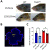
Figure 1.
Figure 1.. Pax1. Pax6Sey phenotype results in smaller retinas.
<p/>A) Pax1Sey in both 129S1/Svlmj and C57BL/6 exhibit small-eye phenotypes. Yellow dashed lines highlight the ocular anatomy. B) Example of whole-mounted retina stained with DAPI showing the contour shape for retinal area quantification (S: superior, T: temporal, I: inferior, N: nasal). C) Pax6Sey mice in both backgrounds show significantly smaller whole-mount area compared to their WT counterparts. 129S1/Svlmj is labeled as 129S and C57BL/6 as C57; same for all figures below. *: P < 0.05 in one-way ANOVA post-hoc Tukey’s test.
Figure 2.
Figure 2.. Intraocular pressure (IOP) increases, and visual acuity decreases significantly with age in Pax6Sey mice.
A) Pax6Sey C57BL/6 mice show a significant increase of IOP by P30; Pax6Sey 129S1/Svlmj mice show a significant IOP increase by P60. B) Visual acuity was significantly decreased in Pax6Sey of both C57BL/6 and 129S1/Svlmj backgrounds at P20. C) Plots of acuity versus IOP. *: P < 0.05, ***: P < 0.001; ****: P < 0.0001 in one-way ANOVA post-hoc Tukey’s test.
Figure 3.
Figure 3.. Abnormal layer organization in Pax6Sey mouse retinas.
A) Layer disorganization was present in Pax6Sey mice of both genetic backgrounds. P90 full sagittal retina sections are shown as well as high magnification of normal regions and hotspots, the localized damages. B) Quantification of total retinal thickness of the hotspots in WT, Pax6Sey normal area and Pax6Sey hotspots (hs). *: P < 0.05 in one-way ANOVA post-hoc Tukey’s test. Sample numbers are shown within the bars.
Figure 4.
Figure 4.. Retinal subpopulations show varied densities in Pax6Sey mice.
Confocal imaging of retinal cell-type markers, Rbpms, ChAT, and AP-2 in C57BL/6 (A) and 129S1/Svlmj (B) P90 retina sections. The somas labeled Rbpms, ChAT, and AP-2 showed varied distributions in Pax6Sey mice.
Figure 5.
Figure 5.. C57BL/6 Pax6Sey mice exhibit increased density of activated (Iba1+) microglial cells.
A) Confocal imaging of a whole-mounted P80 retina immuno-stained with Iba1 (red) and DAPI (blue). S: superior, T: temporal, I: inferior, N: nasal. B) Enlarged images of Iba1 staining and quantification of Iba1+ staining at P25 (dark red) and P80 (light red) in C57BL/6 background (W = wildtype, P = Pax6Sey). C) Enlarged images of Iba1 staining and quantification of Iba1+ staining at P25 (dark red) and P80 (light red) in 129S1/Svlmj background (W = wildtype, P = Pax6Sey). Each point represents one eye for each mouse *: P < 0.05, ***: P < 0.001 in one-way ANOVA post-hoc Tukey’s test.
Figure 6.
Figure 6.. C57BL/6 Pax6Sey mice also exhibit an increased density of astrocytes.
A) Confocal imaging of whole-mounted P80 retina immuno-stained with GFAP (green), a marker for astrocytes in the superficial layer of the retina. S: superior, T: temporal, I: inferior, N: nasal. B) Two magnified images that have been stained with GFAP (green) and DAPI (blue) with an area of 30,000 μm2 and have been used for cell counting. C) Quantification of cells with GFAP+ staining above the GCL at P25 (dark blue) and P80 (light blue) (W = wildtype, P = Pax6Sey). Each point represents one eye for each mouse. **: P < 0.01 in one-way ANOVA post-hoc Tukey’s test.

Similar articles

Cited by

References

    1. Axton R, Hanson I, Danes S, Sellar G, van Heyningen V, Prosser J, 1997. The incidence of PAX6 mutation in patients with simple aniridia: an evaluation of mutation detection in 12 cases. J Med Genet 34, 279–286. - PMC - PubMed
    1. Beckmann L, Cai Z, Cole J, Miller DA, Liu M, Grannonico M, Zhang X, Ryu HJ, Netland PA, Liu X, Zhang HF, 2021. In vivo imaging of the inner retinal layer structure in mice after eye-opening using visible-light optical coherence tomography. Exp Eye Res 211, 108756. 10.1016/j.exer.2021.108756 - DOI - PMC - PubMed
    1. Bhan A, Browning AC, Shah S, Hamilton R, Dave D, Dua HS, 2002. Effect of corneal thickness on intraocular pressure measurements with the pneumotonometer, Goldmann applanation tonometer, and Tono-Pen. Invest Ophthalmol Vis Sci 43, 1389–1392. - PubMed
    1. Bobilev AM, McDougal ME, Taylor WL, Geisert EE, Netland PA, Lauderdale JD, 2016. Assessment of PAX6 alleles in 66 families with aniridia. Clin Genet 89, 669–677. 10.1111/cge.12708 - DOI - PMC - PubMed
    1. Bosco A, Steele MR, Vetter ML, 2011. Early microglia activation in a mouse model of chronic glaucoma. J Comp Neurol 519, 599–620. 10.1002/cne.22516 - DOI - PMC - PubMed

Publication types