. 2023 Nov 18;9(1):419.
doi: 10.1038/s41420-023-01712-7.

Iron derived from NCOA4-mediated ferritinophagy causes cellular senescence via the cGAS-STING pathway

Affiliations

Iron derived from NCOA4-mediated ferritinophagy causes cellular senescence via the cGAS-STING pathway

Hong-Ying Li et al. Cell Death Discov. .

Abstract

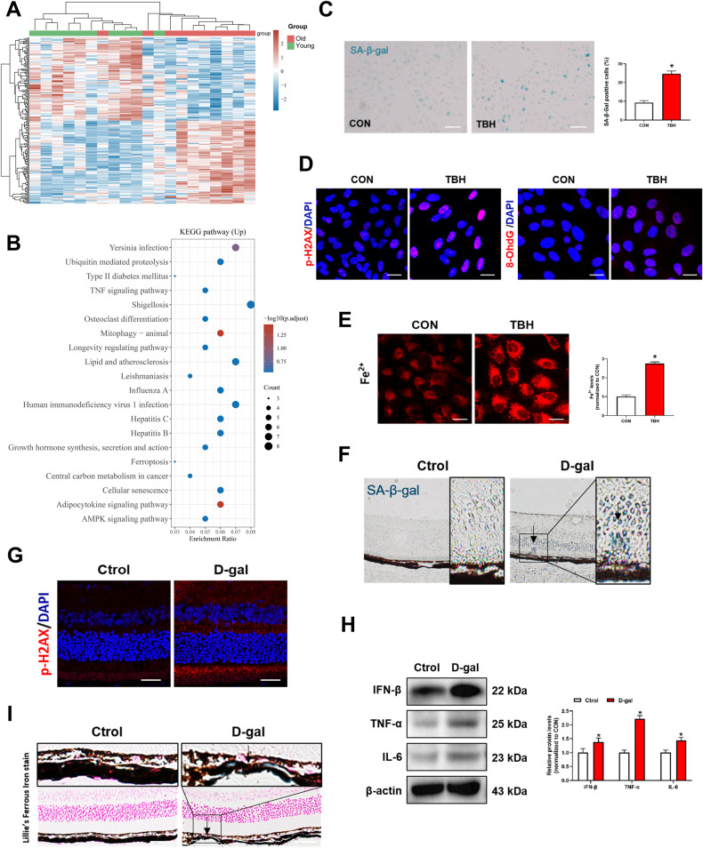
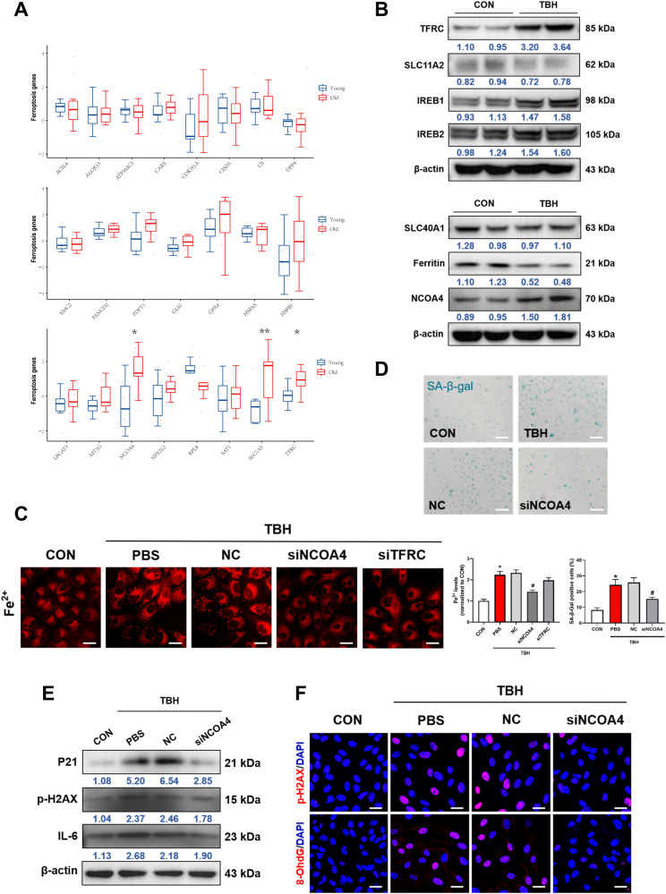
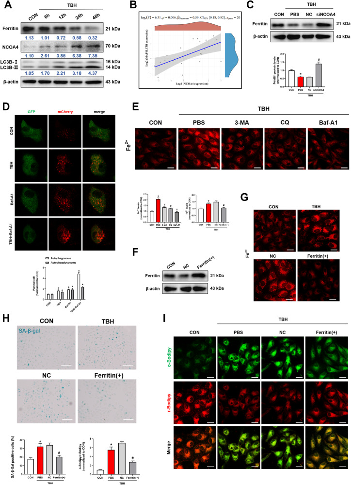
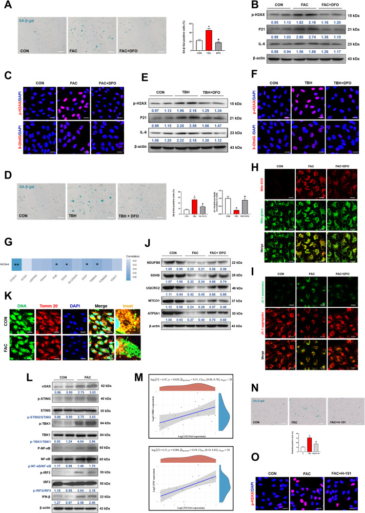
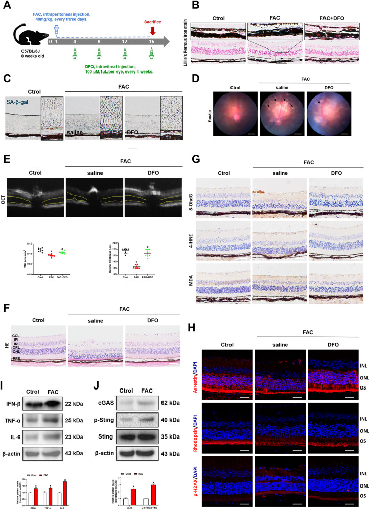
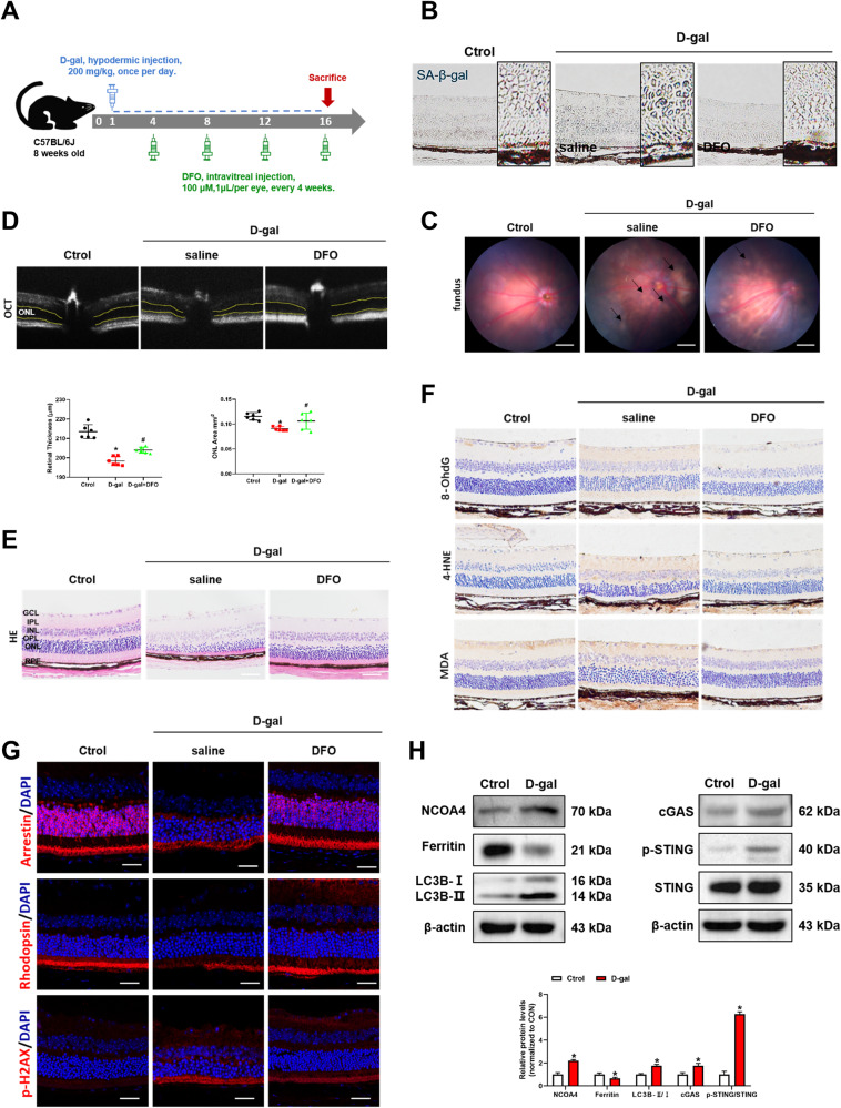
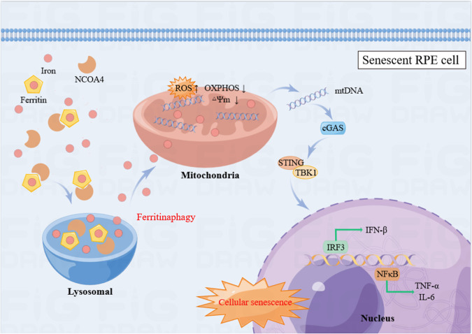
Cellular senescence is a hallmark of aging and has been linked to age-related diseases. Age-related macular degeneration (AMD), the most common aging-related retinal disease, is prospectively associated with retinal pigment epithelial (RPE) senescence. However, the mechanism of RPE cell senescence remains unknown. In this study, tert-butyl hydroperoxide (TBH)-induced ARPE-19 cells and D-galactose-treated C57 mice were used to examine the cause of elevated iron in RPE cell senescence. Ferric ammonium citrate (FAC)-treated ARPE-19 cells and C57 mice were used to elucidated the mechanism of iron overload-induced RPE cell senescence. Molecular biology techniques for the assessment of iron metabolism, cellular senescence, autophagy, and mitochondrial function in vivo and in vitro. We found that iron level was increased during the senescence process. Ferritin, a major iron storage protein, is negatively correlated with intracellular iron levels and cell senescence. NCOA4, a cargo receptor for ferritinophagy, mediates degradation of ferritin and contributes to iron accumulation. Besides, we found that iron overload leads to mitochondrial dysfunction. As a result, mitochondrial DNA (mtDNA) is released from damaged mitochondria to cytoplasm. Cytoplasm mtDNA activates the cGAS-STING pathway and promotes inflammatory senescence-associated secretory phenotype (SASP) and cell senescence. Meanwhile, iron chelator Deferoxamine (DFO) significantly rescues RPE senescence and retinopathy induced by FAC or D-gal in mice. Taken together, these findings imply that iron derived from NCOA4-mediated ferritinophagy causes cellular senescence via the cGAS-STING pathway. Inhibiting iron accumulation may represent a promising therapeutic approach for age-related diseases such as AMD.


Conflict of interest statement

The authors declare no competing interests.

Figures

Fig. 1
Fig. 1. Iron is elevated during aging.
A Heat map displays the changes of differential gene expression in two groups. Data were derived from Gene Expression Omnibus (GEO) data sets GSE58137. B KEGG pathway analysis of upregulated genes. ARPE-19 cells were treated with TBH (100 μM) for 48 h to induce cell senescence models. C Images of SA-β-Gal staining of ARPE-19 cells (n = 3). Scale bars, 100 μm. D Immunofluorescence staining of p-H2AX and 8-OhdG. Scale bars, 25 μm. E Intracellular Fe2+ levels were detected by the FerroOrange probe (1 μM, 30 min) (n = 3). Scale bars, 25 μm. F SA-β-Gal staining of mouse retina. Scale bars, 100 μm. G Immunofluorescence staining of p-H2AX in mouse retina. Scale bars, 25 μm. H The protein content of IFN-β, TNF-α and IL-6 in mouse retina was assessed by Western blot (n = 3). Mean ± SD. I lillie’s Ferrous Iron stain showing Fe2+ deposition in mouse retina. Blue depositions represent the accumulation of Fe2+. Scale bars, 100 μm. Data are shown as Mean ± SD. *p < 0.05 vs. control group.
Fig. 2
Fig. 2. NCOA4 mediates the elevation of iron in senescent ARPE-19 cells.
A The analysis of expression levels of 24 ferroptosis-associated genes from GEO datasets (GEO58137). B The expression of iron metabolism-related proteins was detected by Western blot (n = 3). C Intracellular Fe2+ was detected after the knockdown of NCOA4 by the FerroOrange probe (1 μM, 30 min) (n = 3). Scale bars, 25 μm. D The expression of SA-β-Gal staining positive cells were ameliorated after knockdown of NCOA4 (n = 3). Scale bars, 100 μm. E The expression of P21, p-H2AX and IL-6 were ameliorated after knockdown of NCOA4 (n = 3). F Immunofluorescence staining of p-H2AX and 8-OhdG. Scale bars, 25 μm. Data are shown as Mean ± SD. *p < 0.05 vs. control group. #p < 0.05 vs. TBH group.
Fig. 3
Fig. 3. NCOA4 mediates iron elevation via autophagic degradation of ferritin.
A The Ferritin, NCOA4 and LC3B were evaluated by western blot. ARPE-19 cells were treated with TBH (100 μM) for different time durations (6, 12, 24, and 48 h) (n = 3). B NCOA4 expression was positively correlated with LC3B in human blood samples. (GEO accession numbers: GSE58137). C The protein expression of ferritin was markedly increased after knockdown of NCOA4 (n = 3). D Live-cell imaging of ARPE-19 cells expressing Ad-mCherry-GFP-LC3B. Cells were incubated in control or presence of TBH (100 μM)/Bafilomycin A1 (Baf-A1) (100 nM)/TBH + Baf-A1 for 48 h prior to imaging (n = 3). Scale bars, 25 μm. E Autophagy inhibitors, including 3-Methyladenine (3-MA, 5 mM), Chloroquine (CQ, 5 μM) and Baf-A1 (100 nM), reversed the TBH-induced increases intracellular Fe2+ levels (n = 3). Scale bars, 25 μm. F Ferritin was overexpressed by FTH1 cDNA transfection and overexpression efficiency was detected by Western blot. G Overexpression of ferritin reduced the intracellular Fe2+ levels (n = 3). Scale bars, 25 μm. H SA-β-Gal staining positive cells were decreased after ferritin overexpression (n = 3). Scale bars, 100 μm. I The intracellular lipid peroxidation levels were detected by C11-BODIPY probes (10 μM, 30 min), reduced (red) and oxidized (green) Bodipy (n = 3). Scale bars, 25 μm. Data are shown as Mean ± SD. *p < 0.05 vs. control group. #p < 0.05 vs. TBH group.
Fig. 4
Fig. 4. Iron causes mitochondrial dysfunction and accelerates ARPE-19 cell senescence via the cGAS-STING signaling pathway.
ARPE-19 cells were pretreated with deferoxamine (DFO, 20 μM) for 2 h, followed by stimulation with TBH (100 μM) or ferric ammonium citrate (FAC, 200 μM) for 48 h. A DFO treatment reduced the number of SA-β-Gal positive cells in FAC group (n = 3). Scale bars, 100 μm. B DFO reduced the expression levels of aging-related proteins (p-H2AX, P21 and IL-6) in FAC group by Western blot (n = 3). C Immunofluorescence of p-H2AX and 8-OhdG in FAC group. Scale bars, 25 μm. D DFO treatment reduced the number of SA-β-Gal staining positive cells in TBH group (n = 3). Scale bars, 100 μm. E DFO reduced the expression levels of aging-related proteins (p-H2AX, P21 and IL-6) in TBH group by Western blot (n = 3). F Immunofluorescence of p-H2AX and 8-OhdG in TBH group. Scale bars, 25 μm. G Mitochondrial functions-associated genes including COX4I1, PHB, SDHA, SOD1 and TIMM44 was negatively correlated with NCOA4 in human blood samples, respectively. (GEO accession numbers: GSE58137). H MitoSOX™ Red (5 μM, 30 min) and Mito-Tracker Green (200 nM, 30 min) were used to co-localize mitochondrial ROS and mitochondria, respectively. Scale bars, 25 μm. I Mitochondrial membrane potential was assessed by JC-1 staining. Scale bars, 25 μm. J OXPHOS activity was detected by Western blot (n = 3). K Immunofluorescence of dsDNA and TOMM20 colocalization. Scale bar, 25 μm. L Detection of cGAS-STING pathway-related proteins by Western blot (n = 3). M The expression levels of TBK1 and TNF both had a positive significant correlation with NCOA4 in the collected 20 human blood samples. ARPE-19 cells were pretreated with STING inhibitor H-151 (10 μM) for 2 h, followed by stimulation with FAC (200 μM) for 48 h. N H-151 reversed FAC-induced increase in SA-β-Gal staining positive cells (n = 3). Scale bars, 100 μm. O H-151 rescued FAC-induced up-regulation of p-H2AX in cell by immunofluorescence. Data are shown as Mean ± SD. *p < 0.05 vs. control group. #p < 0.05 vs. TBH or FAC group.
Fig. 5
Fig. 5. Retinal iron deposition causes senescence in C57 mice.
A Experimental timeline of FAC and DFO treatment in C57 mice. B Lillie’s Ferrous Iron stain showing Fe2+ deposition in retinal tissue of mice. Scale bars, 100 μm. C SA-β-Gal staining of mouse retina. Scale bars, 100 μm. D The fundus photograph of mice. Scale bars, 500 μm. E The outer nuclear layer (ONL) area in horizontal optical coherence tomography scans (yellow line) was quantified by ImageJ (n = 3). F The H&E staining of mouse retina (n = 3). Scale bars, 100 μm. G The immunohistochemistry analysis of 8-OhdG, 4-HNE and MDA in the mouse retina. Scale bars, 100 μm. H The protein expression of Arrestin, Rhodopsin and p-H2AX in mouse retina was evaluated by immunofluorescence. Scale bars, 25 μm. I The protein content of IFN-β, TNF-α and IL-6 in eye tissue of mice was assessed by Western blot (n = 3). J The protein content of cGAS, p-STING and STING in eye tissue of mice was assessed by Western blot (n = 3). Data are shown as Mean ± SD. *p < 0.05 vs. control group. #p < 0.05 vs. FAC group.
Fig. 6
Fig. 6. DFO ameliorates D-gal induced senescence in C57 mice.
A Experimental timeline of D-gal and DFO treatment in C57 mice. B SA-β-Gal staining of mouse retina. Scale bars, 100 μm. C The fundus photograph of mice. Scale bars, 500 μm. D The outer nuclear layer (ONL) area in horizontal optical coherence tomography scans (yellow line) was quantified by ImageJ (n = 3). E The H&E staining of mouse retina (n = 3). Scale bars, 100 μm. F Immunohistochemistry analysis of 8-OhdG, 4-HNE and MDA in the mouse retina. Scale bars, 100 μm. G Arrestin, Rhodopsin and p-H2AX in the mouse retina was evaluated by immunofluorescence. Scale bars, 25 μm. H The protein content of NCOA4, Ferritin, LC3B, cGAS, p-STING and STING in the mouse retina was assessed by Western blot (n = 3). Data are shown as Mean ± SD. *p < 0.05 vs. control group. #p < 0.05 vs. D-gal group.
Fig. 7
Fig. 7. Iron derived from NCOA4-mediated ferritinophagy causes retinal pigment epithelium senescence.
Our flow chart illustrates that iron elevation causes retinal pigment epithelium senescence. NCOA4-mediated autophagic degradation of ferritin and induced iron accumulation. Iron accumulation leading to mitochondrial dysfunction, and mtDNA is released from damaged mitochondria to cytoplasm. Cytoplasm mtDNA further activates the cGAS-STING pathway, which promotes SASP and induces cell senescence. The figure is drawn by Figuredraw.

References

    1. Wong WL, Su X, Li X, Cheung CM, Klein R, Cheng CY, et al. Global prevalence of age-related macular degeneration and disease burden projection for 2020 and 2040: a systematic review and meta-analysis. Lancet Glob Health. 2014;2:e106–16. doi: 10.1016/S2214-109X(13)70145-1. - DOI - PubMed
    1. Al-Zamil WM, Yassin SA. Recent developments in age-related macular degeneration: a review. Clin Interv Aging. 2017;12:1313–30. doi: 10.2147/CIA.S143508. - DOI - PMC - PubMed
    1. Cho HJ, Yoon W, Yoon J, Na SK, Lee J, Kim J, et al. Development of intraretinal fluid in neovascular age-related macular degeneration during anti-vascular endothelial growth factor treatment. Am J Ophthalmol. 2022;234:6–14. doi: 10.1016/j.ajo.2021.07.026. - DOI - PubMed
    1. Lopez-Luppo M, Catita J, Ramos D, Navarro M, Carretero A, Mendes-Jorge L, et al. Cellular senescence is associated with human retinal microaneurysm formation during aging. Invest Ophthalmol Vis Sci. 2017;58:2832–42. doi: 10.1167/iovs.16-20312. - DOI - PubMed
    1. Di Micco R, Krizhanovsky V, Baker D, d’Adda di Fagagna F. Cellular senescence in ageing: from mechanisms to therapeutic opportunities. Nat Rev Mol Cell Biol. 2021;22:75–95. doi: 10.1038/s41580-020-00314-w. - DOI - PMC - PubMed