

Evaluating the Neural Underpinnings of Motivation for Walking Exercise
- PMID: 37980613
- PMCID: PMC10939334
- DOI: 10.1093/ptj/pzad159
Evaluating the Neural Underpinnings of Motivation for Walking Exercise
Abstract
Objective: Motivation is critically important for rehabilitation, exercise, and motor performance, but its neural basis is poorly understood. Recent correlational research suggests that the dorsomedial prefrontal cortex (dmPFC) may be involved in motivation for walking activity and/or descending motor output. This study experimentally evaluated brain activity changes in periods of additional motivation during walking exercise and tested how these brain activity changes relate to self-reported exercise motivation and walking speed.
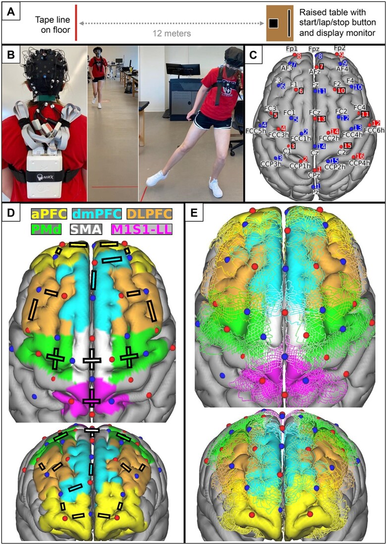
Methods: Adults without disability (N = 26; 65% women; 25 [standard deviation = 5] years old) performed a vigorous exercise experiment involving 20 trials of maximal speed overground walking. Half of the trials were randomized to include "extra-motivation" stimuli (lap timer, tracked best lap time, and verbal encouragement). Wearable near-infrared spectroscopy measured oxygenated hemoglobin responses from frontal lobe regions, including the dmPFC, primary sensorimotor, dorsolateral prefrontal, anterior prefrontal, supplementary motor, and dorsal premotor cortices.
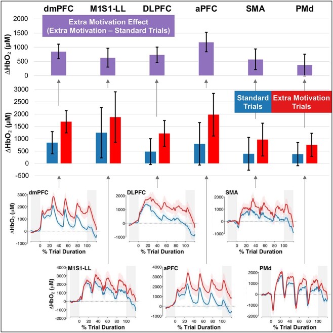
Results: Compared with standard trials, participants walked faster during extra-motivation trials (2.43 vs 2.67 m/s; P < .0001) and had higher oxygenated hemoglobin responses in all tested brain regions, including dmPFC (+842 vs +1694 μM; P < .0001). Greater dmPFC activity was correlated with more self-determined motivation for exercise between individuals (r = 0.55; P = .004) and faster walking speed between trials (r = 0.18; P = .0002). dmPFC was the only tested brain region that showed both of these associations.
Conclusion: Simple motivational stimuli during walking exercise seem to upregulate widespread brain regions. Results suggest that dmPFC may be a key brain region linking affective signaling to motor output.
Impact: These findings provide a potential biologic basis for the benefits of motivational stimuli, elicited with clinically feasible methods during walking exercise. Future clinical studies could build on this information to develop prognostic biomarkers and test novel brain stimulation targets for enhancing exercise motivation (eg, dmPFC).
Keywords: Aerobic Exercise; Behavior Regulation; Brain; Gait; Locomotion; fNIRS.
© The Author(s) 2023. Published by Oxford University Press on behalf of the American Physical Therapy Association. All rights reserved. For permissions, please email: journals.permissions@oup.com.
Figures
References
-
- Araújo D, Davids K, Bennett SJ, Button C, Chapman G. Emergence of sport skills under constraints. In: Williams AM, Hodges NJ. (eds), Skill Acquisition in Sport. Routledge; 2004: 433–458.
-
- Button C, Seifert L, Chow JY, Davids K, Araujo D. Dynamics of skill acquisition: an ecological dynamics approach. 2nd ed. Human Kinetics. 2020: 1–288. 10.5040/9781718214125. - DOI
-
- Davids K, Araújo D, Shuttleworth R, Button C. Acquiring skill in sport: a constraints-led perspective. Int J Comput Sci Sport. 2003;41:5–16.
-
- Davids K, Button C, Bennett S. Dynamics of skill acquisition: a constraints-led approach. 1st ed. Hum Kinet. 2008: 1–264.
-
- Guccione AA, Neville BT, George SZ. Optimization of movement: a dynamical systems approach to movement systems as emergent phenomena. Phys Ther. 2019;99:3–9. - PubMed
Publication types
MeSH terms
Substances
Grants and funding
LinkOut - more resources
Full Text Sources
Research Materials
