. 2024 Aug 1;109(8):2542-2554.
doi: 10.3324/haematol.2023.283917.

Germline bi-allelic SH2B3/LNK alteration predisposes to a neonatal juvenile myelomonocytic leukemia-like disorder

Affiliations

Germline bi-allelic SH2B3/LNK alteration predisposes to a neonatal juvenile myelomonocytic leukemia-like disorder

Chloé Arfeuille et al. Haematologica. .

Abstract

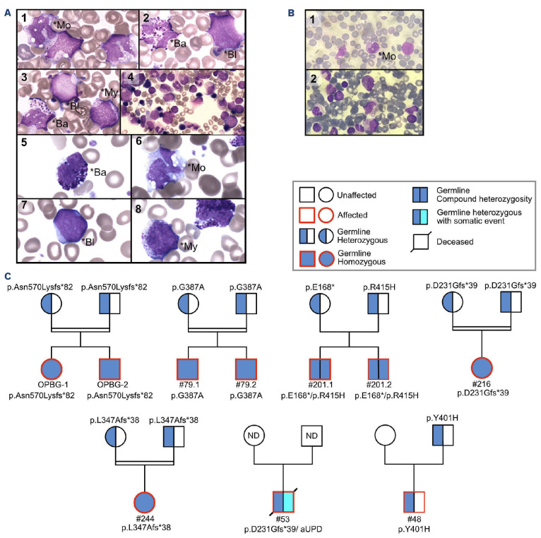
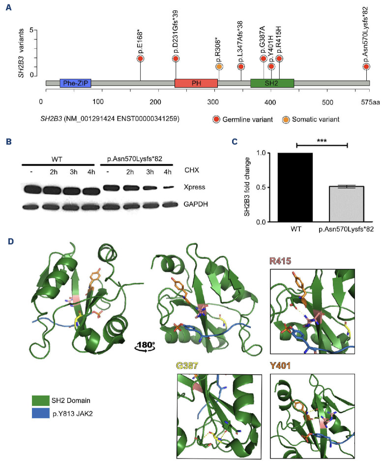
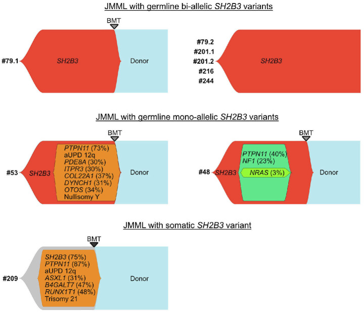
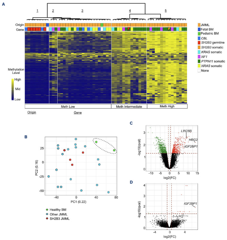
Juvenile myelomonocytic leukemia (JMML) is a rare, generally aggressive myeloproliferative neoplasm affecting young children. It is characterized by granulomonocytic expansion, with monocytosis infiltrating peripheral tissues. JMML is initiated by mutations upregulating RAS signaling. Approximately 10% of cases remain without an identified driver event. Exome sequencing of two unrelated cases of familial JMML of unknown genetics and analysis of the French JMML cohort identified 11 patients with variants in SH2B3, encoding LNK, a negative regulator of the JAK-STAT pathway. All variants were absent from healthy population databases, and the mutation spectrum was consistent with a loss of function of the LNK protein. A stoploss variant was shown to affect both protein synthesis and stability. The other variants were either truncating or missense, the latter affecting the SH2 domain that interacts with activated JAK. Of the 11 patients, eight from five families inherited pathogenic bi-allelic SH2B3 germline variants from their unaffected heterozygous parents. These children represent half of the cases with no identified causal mutation in the French cohort. They displayed typical clinical and hematologic features of JMML with neonatal onset and marked thrombocytopenia. They had a hypomethylated DNA profile with fetal characteristics and did not have additional genetic alterations. All patients showed partial or complete spontaneous clinical resolution. However, progression to thrombocythemia and immunity-related pathologies may be of concern later in life. Bi-allelic SH2B3 germline mutations thus define a new condition predisposing to a JMML-like disorder, suggesting that JAK pathway deregulation is capable of initiating JMML, and opening new therapeutic options.


Figures

Figure 1.
Figure 1.
Patients’ cytomorphological data and pedigrees. (A, B) May-Grünwald-Giemsa-stained smears showing cytomorphological features at diagnosis patients with of juvenile myelomonocytic leukemia (JMML). (A) Patients #79.1 (A1-4) and #79.2 (A5-8). (B) Patient #OPBG (B1, 2). Peripheral blood smears (A1-3; A5-8; B1) show monocytes (*Mo), myeloid precursors (*My), dysmorphic basophils (*Ba) and undifferentiated myeloid blasts (*Bl). Bone marrow smears (A4; B2) show hypercellularity with myeloid blasts and discrete signs of dysgranulopoiesis. The erythrocyte lineage is decreased and the megakaryocytic lineage is absent. (C) Pedigrees of patients with germline SH2B3 variants. All children, but none of the heterozygous parents, had JMML. Germline variants (dark blue) were either bi-allelic (full circle/square; families 1 to 4) or mono-allelic (half circle/square; families 5 and 6) in the affected child. Somatic alteration is indicated in light blue. aUPD: acquired uniparental isodisomy; JMML: juvenile myelomonocytic leukemia; ND: not done.
Figure 2.
Figure 2.
SH2B3 variants and consequences on the LNK protein. (A) Lollipop plot showing the distribution of SH2B3/LNK variants over a schematic representation of the LNK protein. The PH domain enables interaction with cell membrane phospholipids and the SH2 domain binds to the phosphorylated tyrosines of target proteins. (B, C) A disease-causing SH2B3 stoploss frameshift mutation affects RNA and protein stability. (B) Protein stability was assessed in 293T cells transiently transfected with wild-type Xpress-tagged SH2B3 and mutant carrying the c.1709dupA (p.Asp570Lysfs82) mutation. After transfection (48 h), cells were treated with cycloheximide (20 [ig/mL) for the indicated time or left untreated. Protein levels were assessed by immunoblotting, using an anti-Xpress monoclonal antibody. GAPDH levels are shown to document equal loading of total proteins from cell lysates. Western blot from a representative experiment of three performed is shown. (C) Quantitative polymerase chain reaction analysis performed on RNA extracted from fibroblasts taken from patients and controls shows a significant decrease of SH2B3 expression level indicating RNA decay of the allele carrying the SH2B3 variant. Data in the graph indicate fold change of SH2B3 in patients’ fibroblasts over that in control cells (WT), set as 1. GAPDH was used as an endogenous control. Histograms show mean values ± standard deviation of three independent experiments, each performed in triplicate. The analysis of expression was performed calculating the fold change using the 2-ΔΔCt formula and the results were statistically analyzed by PRISM7, using a two-tailed unpaired t test with the Bonferroni correction. ***P<0.001. (D) Three-dimensional structure of the SH2 domain of the LNK protein with the JAK2 phosphopeptide pY813 modeled from crystallographic data in the report from Morris et al .36 (PDB:7R8W). The position of variant amino acids was determined by structural homology between mouse and human SH2 domains. aa: amino acids; CHX: cycloheximide; WT: wild-type.
Figure 3.
Figure 3.
Clonal architecture of SH2B3 mutated juvenile myelomonocytic leukemia. Patients with a bi-allelic germline mutation of SH2B3 (red clones) (#79, #201, #216, #244) all have a monomorphic profile with no additional genetic alterations. Only patient #79.1 received a bone marrow transplant (upper panel). Patients with a germline mono-allelic SH2B3 mutation (#53, #48) and/or somatic SH2B3 mutation (orange clones) acquired several additional somatic mutations. Patient #53 had a somatic PTPN11 variant and patient #48 had somatic variants of PTPN11 and NF1, whose order of appearance could not be inferred from the allelic frequencies. Both patients underwent bone marrow transplantation (middle panel). In patient #209, the allelic frequency of the somatic SH2B3 variant was consistent with its presence in the full leukemia clone. Multiple other somatic alterations were acquired, including a driver somatic mutation in PTPN11. Here again, all potentially driver mutations were present at variant allele frequencies consistent with early co-occurrence during the course of the disease and order or appearance could not be determined (lower panel). JMML: juvenile myelomonocytic leukemia; BMT: bone marrow transplant; aUPD: acquired uniparental isodisomy.
Figure 4.
Figure 4.
DNA methylation pattern and gene expression profiling suggest persistence of fetal cues in juvenile myelomonocytic leukemia with bi-allelic germline SH2B3 variants. (A) DNA methylation analysis of patients with SH2B3 variants (N=8) and reference juvenile myelomonocytic leukemia (JMML) of known genetic groups (i.e., PTPN11 somatic, NRAS somatic, KRAS somatic, NF1 or CBL) (N=54). Unsupervised hierarchical clustering based on the 1,000 most variant 100 bp tiles identified three main DNA methylation subgroups (low, intermediate, high). JMML with bi-allelic SH2B3 germline mutations defines a subgroup of hypomethylated JMML (cluster 1), whereas JMML with either mono-allelic or somatic SH2B3 mutations cluster in the intermediate (cluster 4) or high (cluster 5) methylation groups. (B-D) Gene expression profile analyses of mononucleated cells of JMML with bi-allelic SH2B3 germline alteration (N=4) in comparison with other types of JMML (N=17) and bone marrow samples from healthy subjects. Principal component analysis (B) and volcano plots showing differential gene expression between JMML with bi-allelic SH2B3 germline alteration and healthy bone marrow (C) or other types of JMML (D). A qval threshold of ≤0.05 and a minimum fold-change of 1.5 were used to define differentially expressed genes. JMML: juvenile myelomonocytic leukemia; BM: bone marrow; Meth: methylation; PC: principal component; FC: fold change.

Comment in

References

    1. Chang TY, Dvorak CC, Loh ML. Bedside to bench in juvenile myelomonocytic leukemia: insights into leukemogenesis from a rare pediatric leukemia. Blood. 2014;124(16):2487-2497. - PubMed
    1. Locatelli F, Niemeyer CM. How I treat juvenile myelomonocytic leukemia. Blood. 2015;125(7):1083-1090. - PubMed
    1. Locatelli F, Nöllke P, Zecca M, et al. Hematopoietic stem cell transplantation (HSCT) in children with juvenile myelomonocytic leukemia (JMML): results of the EWOG-MDS/ EBMT trial. Blood. 2005;105(1):410-419. - PubMed
    1. Lapidot T, Grunberger T, Vormoor J, et al. Identification of human juvenile chronic myelogenous leukemia stem cells capable of initiating the disease in primary and secondary SCID mice. Blood. 1996;88(7):2655-2664. - PubMed
    1. Caye A, Rouault-Pierre K, Strullu M, et al. Despite mutation acquisition in hematopoietic stem cells, JMML-propagating cells are not always restricted to this compartment. Leukemia. 2020;34(6):1658-1668. - PMC - PubMed

Publication types

Substances

LinkOut - more resources