

Lingual morphology of domesticated Asian small-clawed otters in Yogyakarta, Indonesia
- PMID: 37981903
- PMCID: PMC10581529
- DOI: 10.17221/62/2022-VETMED
Lingual morphology of domesticated Asian small-clawed otters in Yogyakarta, Indonesia
Abstract
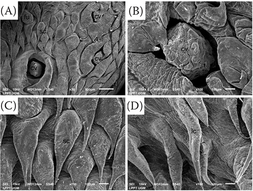
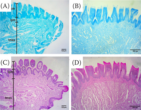
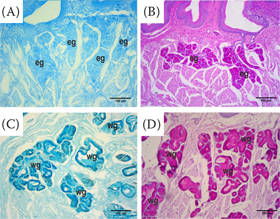
This study aimed to observe the lingual morphology of the domesticated Asian small-clawed otter, Aonyx cinereus (A. cinereus), from Yogyakarta, Indonesia. Six domesticated A. cinereus adults were obtained from a local otter breeder in Yogyakarta, with no regard to sex. The animals were acclimated to the laboratory for one day, following this, the animals underwent macroscopy identification and scanning electron microscopy (SEM) and light microscopy (LM) analysis. Macroscopically, the tongue of domesticated A. cinereus is divided into three parts: the apex, corpus, and radix. The apex is the shortest part and can move freely. A median groove is bent along the corpus. Typically, the radix contains circumvallate papillae and the epiglottic valleculae. The SEM and LM observations revealed that the lingual morphology of A. cinereus consisted of two types of papillae: mechanical papillae (horny filiform, leaf-like filiform, bifid filiform, trifid filiform, elongated leaf-like filiform, triangular filiform and conical papillae) and gustatory papillae (fungiform and circumvallate papillae). The lingual glands consisted of Weber's glands and von Ebner's glands secreting acid and neutral mucins. Collagen fibres are found in the lamina propria and muscular layer. In conclusion, the papillae of the Asian short-clawed otter have the same structure as those of other Mustelidae family members.
Keywords: Aonyx cinereus; electron and light microscopy; lingual glands; lingual papillae.
Copyright: © 2023 Anjani et al.
Conflict of interest statement
The authors declare no conflict of interest.
Figures


References
-
- Can M, Atalgin SH, Aydin MF. Scanning electron microscopic studies of the lingual papillae in the English horse. Acta Vet-Beograd. 2016 Jun;66(2):257-64.
-
- Damia U, Anjani AK, Wihadmadyatami H, Kusindarta DL. Identification of the lingual papillae in the sugar glider (Petaurus breviceps) by scanning electron microscopy and light microscopy. Anat Histol Embryol. 2021;50(6):918-30. - PubMed
-
- El-Bakary NER, Abumandour MMA. Morphological studies of the tongue of the Egyptian water buffalo (Bubalus bubalis) and their lingual papillae adaptation for its feeding habits. Anat Histol Embryol. 2017 Oct;46(5):474-86. - PubMed
-
- Emura S, Tamada A, Hayakawa D, Chen H, Shoumura S. Morphology of the dorsal lingual papillae in the bush dog (Speothos venaticus). Okajimas Folia Anat Jpn. 2000 Dec;77(5):137-41. - PubMed
-
- Emura S, Hayakawa D, Chen H, Shoumura S. Morphology of the dorsal lingual papillae in the newborn panther and Asian black bear. Okajimas Folia Anat Jpn. 2001 Dec;78(5):173-7. - PubMed
LinkOut - more resources
Full Text Sources
Miscellaneous