. 2023 Nov 13:3:88.
doi: 10.12688/openreseurope.15973.2. eCollection 2023.

Ergolide mediates anti-cancer effects on metastatic uveal melanoma cells and modulates their cellular and extracellular vesicle proteomes

Affiliations

Ergolide mediates anti-cancer effects on metastatic uveal melanoma cells and modulates their cellular and extracellular vesicle proteomes

Husvinee Sundaramurthi et al. Open Res Eur. .

Abstract

Background: Uveal melanoma is a poor prognosis cancer. Ergolide, a sesquiterpene lactone isolated from Inula Brittanica, exerts anti-cancer properties. The objective of this study was to 1) evaluate whether ergolide reduced metastatic uveal melanoma (MUM) cell survival/viability in vitro and in vivo; and 2) to understand the molecular mechanism of ergolide action.

Methods: Ergolide bioactivity was screened via long-term proliferation assay in UM/MUM cells and in zebrafish MUM xenograft models. Mass spectrometry profiled proteins modulated by ergolide within whole cell or extracellular vesicle (EVs) lysates of the OMM2.5 MUM cell line. Protein expression was analyzed by immunoblots and correlation analyses to UM patient survival used The Cancer Genome Atlas (TCGA) data.

Results: Ergolide treatment resulted in significant, dose-dependent reductions (48.5 to 99.9%; p<0.0001) in OMM2.5 cell survival in vitro and of normalized primary zebrafish xenograft fluorescence (56%; p<0.0001) in vivo, compared to vehicle controls. Proteome-profiling of ergolide-treated OMM2.5 cells, identified 5023 proteins, with 52 and 55 proteins significantly altered at 4 and 24 hours, respectively ( p<0.05; fold-change >1.2). Immunoblotting of heme oxygenase 1 (HMOX1) and growth/differentiation factor 15 (GDF15) corroborated the proteomic data. Additional proteomics of EVs isolated from OMM2.5 cells treated with ergolide, detected 2931 proteins. There was a large overlap with EV proteins annotated within the Vesiclepedia compendium. Within the differentially expressed proteins, the proteasomal pathway was primarily altered. Interestingly, BRCA2 and CDKN1A Interacting Protein (BCCIP) and Chitinase Domain Containing 1 (CHID1), were the only proteins significantly differentially expressed by ergolide in both the OMM2.5 cellular and EV isolates and they displayed inverse differential expression in the cells versus the EVs.

Conclusions: Ergolide is a novel, promising anti-proliferative agent for UM/MUM. Proteomic profiling of OMM2.5 cellular/EV lysates identified candidate pathways elucidating the action of ergolide and putative biomarkers of UM, that require further examination.

Keywords: BRCA2 and CDKN1A Interacting Protein; Chitinase Domain Containing 1; Metastatic uveal melanoma; ergolide; extracellular vesicles.

Plain language summary

The most common form of adult eye cancer is uveal melanoma (UM). Once UM cancer cells spread to organs in the rest of the body, metastatic UM (MUM), there is a poor prognosis for patients with only one approved drug treatment. Hence, it is vital to better understand the cellular and extracellular proteins that regulate UM pathology in order to uncover biomarkers of disease and therapeutic targets. In this original study, we demonstrate a compound called ergolide is capable of severely reducing the metabolic activity and growth of UM cancer cells, grown as isolated monolayers. Ergolide was also able to reduce the growth of human MUM cells growing as tumors in transplanted zebrafish larvae. We identify that ergolide alters specific proteins found in the human UM cells. These proteins once analyzed in detail offer opportunities to understand how new treatment strategies can be developed for UM.


Conflict of interest statement

No competing interests were disclosed.

Figures

Figure 1.
Figure 1.. Ergolide treatment significantly reduced UM/MUM cell viability.
( A, B) Dose-response analysis and determination of IC 50 of ergolide in OMM2.5 cells as 2.9 μM via the MTT assay. ( C, D) Long-term survival of Mel285, Mel270 and OMM2.5 clones were significantly (****, adjusted p=0.0001) reduced in ergolide treated samples compared to vehicle control (N = 3). Statistical analysis performed using One-way ANOVA with Dunnett's Test for Multiple Comparisons.
Figure 2.
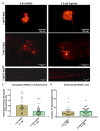
Figure 2.. Anti-cancer effects of ergolide observed in vivo in zebrafish OMM2.5 xenograft models.
( A, C) Representative images of zebrafish larvae transplanted with OMM2.5 Dil labelled cells at 2 days post fertilization (dpf, top panel); representative images of zebrafish larvae transplanted with OMM2.5 Dil labelled cells at 5 dpf, 3 days post treatment (dpt, bottom panel). A 56% (****, p<0.0001%) reduction, on average, in primary xenograft fluorescence observed in the 2.5 μM ergolide (n = 32) treated samples compared to 0.5% DMSO (n = 30) treated samples. ( B, D) A significant difference was not detected in the average number of disseminated cells. Statistical analysis performed using Student's T-test.
Figure 3.
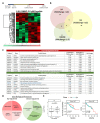
Figure 3.. Proteome profiling following 24 hours ergolide treatment of OMM2.5 cells.
( A) Schematic diagram depicting treatment regime and heatmap highlighting differentially expressed proteins. ( B) Venn diagram showing the number of proteins significantly differentially expressed (up/down) or differentially expressed (DE), given a cutoff of p≤0.05 and a ≥ 1.2 fold change. ( C) Table of top ten proteins that are significantly up- or down-regulated following 24 hours of ergolide treatment. ( D) Pathways enriched for GO: Biological processes. ( E) TCGA analysis of UM patient samples comparing MIF expression levels to chromosome 3 (Chr 3) status, overall survival and disease-free survival (DFS), (n = 80).
Figure 4.
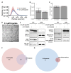
Figure 4.. Characterization of EV isolated from OMM2.5 cells.
( A) NTA analysis of isolated OMM2.5 EV particle size ranging from 50 nm to 250 nm in both 0.1% treated DMSO and 2.5 μM ergolide treated samples. ( B and C) A significant difference in the amount of particles and particle size was not observed between ergolide treated samples and vehicle control. ( D) TEM image of particles isolated from OMM2.5 cells. ( E) Immunoblot analysis of select proteins in OMM2.5 EV isolates and cell lysate (N = 1). ( F) Comparison between Vesiclepedia compendium and EV proteome. ( G) 88 proteins from the list of top 100 EV markers identified in OMM2.5 EV proteome.
Figure 5.
Figure 5.. Analysis of differentially expressed proteins in EVs isolated from OMM2.5 cells treated with ergolide.
( A) Heatmap of upregulated proteins in ergolide treatment group (left panel); list of top 20 significantly upregulated proteins in samples treated with ergolide (right panel). ( B) Heatmap of downregulated proteins in ergolide treatment group (left panel); list of top 20 significantly downregulated proteins in samples treated with ergolide (right panel). ( C) KEGG pathway analysis to determine pathways enriched in EVs following ergolide treatment.
Figure 6.
Figure 6.. Comparison of cellular lysate and EV proteome.
( A) Venn diagram depicting 2139 common proteins in the proteome of OMM2.5 cell lysate and OMM2.5 EV samples. ( B) Among the differentially expressed proteins, two common proteins were identified in the OMM2.5 cell lysate and EV samples. ( C) Table presenting CHID1 and BCCIP as significantly upregulated (red) in ergolide treated OMM2.5 cell lysate and downregulated (green) in EVs isolated from ergolide treated OMM2.5 cells. ( D) Higher BCCIP ( p = 0.005) expression levels noted in chromosome 3 (Chr 3) monosomy UM patients, with no difference in CHID1 expression level and Chr 3 status. ( E) High or low expression levels of BCCIP and CHID1 did not correlate to overall survival or disease-free survival (DFS) in UM TCGA patient samples.
Figure 7.
Figure 7.. Integrated Pathway overview of 407 proteins significantly altered by ergolide in OMM2.5 EVs or cell lysates analysed in Reactome’s hierarchical pathway structure.
( A) The Pathway Overview showing results from an overrepresentation analysis conducted using the “Analyse Gene list” tool with a list of Gene name identifiers. ( B) Voronoi diagram in the ReacFoam format representing an overview of altered pathways. The overrepresentation analysis calculates a probability score for each pathway, corrected for false discovery rate by the Benjamini-Hochberg method. Both visualisations represent pathway coverage in the set.

Similar articles

Cited by

References

    1. Bronkhorst IHG, Jager MJ: Inflammation in uveal melanoma. Eye (Lond). 2013;27(2):217–23. 10.1038/eye.2012.253 - DOI - PMC - PubMed
    1. Smit KN, Jager MJ, de Klein A, et al. : Uveal melanoma: Towards a molecular understanding. Prog Retin Eye Res. 2020;75: 100800. 10.1016/j.preteyeres.2019.100800 - DOI - PubMed
    1. Bustamante P, Piquet L, Landreville S, et al. : Uveal melanoma pathobiology: Metastasis to the liver. Semin Cancer Biol. 2021;71:65–85. 10.1016/j.semcancer.2020.05.003 - DOI - PubMed
    1. Carvajal RD, Schwartz GK, Tezel T, et al. : Metastatic disease from uveal melanoma: treatment options and future prospects. Br J Ophthalmol. 2017;101(1):38–44. 10.1136/bjophthalmol-2016-309034 - DOI - PMC - PubMed
    1. Mallone F, Sacchetti M, Lambiase A, et al. : Molecular Insights and Emerging Strategies for Treatment of Metastatic Uveal Melanoma. Cancers (Basel). 2020;12(10): 2761. 10.3390/cancers12102761 - DOI - PMC - PubMed