. 2023 Dec 12;17(23):23466-23477.
doi: 10.1021/acsnano.3c05341. Epub 2023 Nov 20.

Fusogenic Coiled-Coil Peptides Enhance Lipid Nanoparticle-Mediated mRNA Delivery upon Intramyocardial Administration

Affiliations

Fusogenic Coiled-Coil Peptides Enhance Lipid Nanoparticle-Mediated mRNA Delivery upon Intramyocardial Administration

Ye Zeng et al. ACS Nano. .

Abstract

Heart failure is a serious condition that results from the extensive loss of specialized cardiac muscle cells called cardiomyocytes (CMs), typically caused by myocardial infarction (MI). Messenger RNA (mRNA) therapeutics are emerging as a very promising gene medicine for regenerative cardiac therapy. To date, lipid nanoparticles (LNPs) represent the most clinically advanced mRNA delivery platform. Yet, their delivery efficiency has been limited by their endosomal entrapment after endocytosis. Previously, we demonstrated that a pair of complementary coiled-coil peptides (CPE4/CPK4) triggered efficient fusion between liposomes and cells, bypassing endosomal entrapment and resulting in efficient drug delivery. Here, we modified mRNA-LNPs with the fusogenic coiled-coil peptides and demonstrated efficient mRNA delivery to difficult-to-transfect induced pluripotent stem-cell-derived cardiomyocytes (iPSC-CMs). As proof of in vivo applicability of these fusogenic LNPs, local administration via intramyocardial injection led to significantly enhanced mRNA delivery and concomitant protein expression. This represents the successful application of the fusogenic coiled-coil peptides to improve mRNA-LNPs transfection in the heart and provides the potential for the advanced development of effective regenerative therapies for heart failure.

Keywords: fusogenic coiled-coil; iPSC-CM; intramyocardial delivery; lipid nanoparticles; mRNA delivery.


Conflict of interest statement

The authors declare no competing financial interest.

Figures
Scheme 1
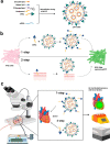
Scheme 1. Overview of LNP Formulation and Delivery in Vitro and in Vivo. (a) Schematic Illustration of mRNA Encapsulating LNP-CPE4. (b) Fusogenic Coiled-Coil Peptide Modified Lipid Nanoparticles (LNPs) for EGFP-mRNA delivery in iPSC-CMs. (c) Schematic Illustration of Intramyocardial Administration of LNPs Encapasulating Luciferase-mRNA
In the 1-step protocol, CPK4 and LNP-CPE4 are premixed and added to the cells. In the 2-step protocol, cells were first pretreated with CPK4 before incubation with LNP-CPE4.
Figure 1
Figure 1
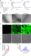
Premixing of CPK4 and CPE4 modified LNPs results in efficient transfection in cells. (a) Fluorescence intensity changes of fluorescein-labeled K4 peptide after addition to LNPs. (mean ± s.d., n = 5). (b) Hydrodynamic diameter changes over time after mixing LNP1-CPE4 with CPK4. (mean ± s.d., n = 3). (c) Hydrodynamic diameter changes over time after adding 10% FCS to the mixture of LNP1-CPE4 and CPK4. The nanoparticle diameter was monitored by DLS (mean ± s.d., n = 3). (d) Representative cryo-EM images of LNP1, and coiled-coil peptide modified LNP1-CPE4 before and after mixing with the complimentary peptide CPK4. The scale bar represents 50 nm. (e) Confocal microscopy images of EGFP-mRNA transfection of LNPs. CPK4+LNP1-CPE4 2-step: HeLa cells were pretreated with CPK4 (10 μM) for 2 h; then, the medium was removed, and LNP1-CPE4 was added (EGFP-mRNA, 1 μg/mL) and incubated for 24 h before imaging. LNP1-CPE4/CPK4 1-step: medium containing CPK4 (10 μM) and LNP1-CPE4 (EGFP-mRNA, 1 μg/mL) was added to HeLa cells and incubated for 24 h before imaging. GFP: green fluorescent protein; BF: bright field; scale bar is 20 μm. (f) Flow cytometry measurements of GFP expression intensity (GFP MFI) of LNPs. MFI was normalized to the LNP1 group. (mean ± s.d., n = 3, ****, P < 0.0001, ***, P < 0.001, **, P < 0.01, *, P < 0.05, ns, no significant difference). (g) CPK4:CPE4 ratio optimization upon a 1-step incubation protocol in HeLa cells (EGFP-mRNA, 1 μg/mL). (mean ± s.d., n = 3)
Figure 2
Figure 2
LNPs modified with fusogenic coiled-coil peptides display enhanced mRNA transfection efficiency in iPSC-CMs. (a) Characterization of iPSC-CMs: Characterization of iPSC-CMs: fluorescent immunostaining showing α-actinin positive iPSC-CMs (in green) and vimentin positive noncardiomyocytes (in red) present in 2D culture, and representative flow cytometry plot of percentage α-actinin positive cells after iPSC-CM differentiation showing high purity (98.4% α-actinin positive) of differentiation. (b) The transfection efficiency (GFP MFI) of LNPs in iPSC-CMs was measured by flow cytometry. 2-step 2h: iPSC-CMs were pretreated with CPK4 (10 μM) for 2 h and afterward incubated with LNP1-CPE4 (2 μg/mL) for 2 h. After 2 h, the supernatant was removed, and cells were cultured for another 24 h before flow cytometry measurements. 1-step 2h incubation: medium containing CPK4 and LNP1-CPE4 (2 μg/mL, CPK4/CPE4 = 5 μM) was added to the iPSC-CMs and incubated for 2 h. Next, the medium was removed, and cells were cultured for another 24 h before flow cytometry measurements. For the 2-step 24h and 1-step 24h groups: iPSC-CMs were incubated with LNPs for 24 h before measuring; all the other steps in the protocol remained the same. (****, P < 0.0001, ***, P < 0.001, **, P < 0.01, *, P < 0.05, ns, no significant difference) In all panels, error bars represent mean ± s.d. (n = 3). (c) The confocal microscopy images of LNP-mediated EGFP-mRNA transfection in iPSC-CM using a 1-step incubation protocol. Blue: DAPI; green: GFP, green fluorescent protein; scale bar represents 50 μm.
Figure 3
Figure 3
Coiled-coil fusogenic peptides enhance LNP-mediated mRNA delivery upon intramyocardial injection. Balb/c mice were intramyocardially injected with 5 μg (in 10 μL) of firefly luciferase mRNA encapsulated in (1) CPK4 and LNP1-CPE4 (final concentration of CPK4/CPE4, 125 μM), either premixed before injection or (2) injected in 2 sequential steps, (3) LNP1-CPE4 or (4) LNP1. As controls, free mRNA and PBS were also injected. Twenty-four h postadministration, organs were harvested, and the luminescence was measured by IVIS imaging. (a) Luminescence images of mice hearts. (b) Luciferase activity in heart lysates. Statistical significance was calculated with a one-way ANOVA (****, P < 0.0001, ***, P < 0.001, **, P < 0.01, *, P < 0.05, ns, no significant difference). Data are represented as the mean ± s.d. (n = 4 for LNP1-CPE4/CPK4 1-step and CPK4+LNP1-CPE4 2-step; n = 6 for LNP1-CPE4; n = 5 for LNP1; n = 3 for free mRNA; n = 2 for PBS.)
Figure 4
Figure 4
LNPs modified with fusogenic coiled-coil peptides display enhanced in vitro mRNA transfection efficiency for different ionizable lipids. (a–d) HeLa and Jurkat cells were transfected with LNPs containing different ionizable lipids (EGFP-mRNA, 1 μg/mL). LNP2 and LNP3 represent the Covid-19 mRNA-LNP formulations of Pfizer/BioNTech and Moderna, respectively. The transfection efficiency was analyzed 24 h after transfection utilizing flow cytometry (GFP MFI). (e) The transfection efficiency on iPSC-CMs of LNPs composed of different ionizable lipids was measured by flow cytometry after 24 h. Statistical significance was calculated by an unpaired student t test. (f) The cellular uptake efficiency of LNPs on iPSC-CMs using the 1-step incubation protocol and incubating for 24 h was measured by quantifying DiD fluorescence. 0.5 mol% of DiD was included in the lipid composition. Statistical significance was calculated by an unpaired student t test. (****, P < 0.0001, ***, P < 0.001, **, P < 0.01, *, P < 0.05, ns, no significant difference). In all panels, error bars represent mean ± s.d. (n = 3). (g) The cell uptake mechanism of fusogenic LNPs (LNP1) using 1-step incubation protocol on iPSC-CMs after 2 h incubation in the presence of endocytosis inhibitor or incubated at 4 °C. 0.5 mol% of DiD was included in the lipid composition and DiD intensity was normalized to 1-step of fusogenic LNPs in the absence of inhibitors. Error bars represent mean ± s.d. (n = 3).

References

    1. Bui A. L.; Horwich T. B.; Fonarow G. C. Epidemiology and risk profile of heart failure. Nat Rev Cardiol. 2011, 8 (1), 30–41. 10.1038/nrcardio.2010.165. - DOI - PMC - PubMed
    1. Senyo S. E.; Steinhauser M. L.; Pizzimenti C. L.; Yang V. K.; Cai L.; Wang M.; Wu T.-D.; Guerquin-Kern J.-L.; Lechene C. P.; Lee R. T. Mammalian heart renewal by pre-existing cardiomyocytes. Nature 2013, 493 (7432), 433–436. 10.1038/nature11682. - DOI - PMC - PubMed
    1. Whelan R. S.; Kaplinskiy V.; Kitsis R. N. Cell Death in the Pathogenesis of Heart Disease: Mechanisms and Significance. Annu. Rev. Physiol. 2010, 72 (1), 19–44. 10.1146/annurev.physiol.010908.163111. - DOI - PubMed
    1. Yang Q.; Fang J.; Lei Z.; Sluijter J. P. G.; Schiffelers R. Repairing the heart: State-of the art delivery strategies for biological therapeutics. Adv. Drug Deliv. Rev. 2020, 160, 1–18. 10.1016/j.addr.2020.10.003. - DOI - PubMed
    1. Laflamme M. A.; Murry C. E. Heart regeneration. Nature 2011, 473 (7347), 326–335. 10.1038/nature10147. - DOI - PMC - PubMed

Publication types