. 2024 Mar 1;13(3):bio060011.
doi: 10.1242/bio.060011. Epub 2024 Mar 1.

Deterministic nuclear reprogramming of mammalian nuclei to a totipotency-like state by Amphibian meiotic oocytes for stem cell therapy in humans

Affiliations

Deterministic nuclear reprogramming of mammalian nuclei to a totipotency-like state by Amphibian meiotic oocytes for stem cell therapy in humans

Ming-Hsuan Wen et al. Biol Open. .

Abstract

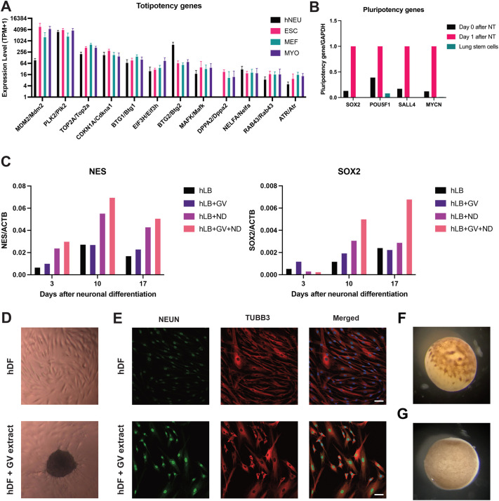
The ultimate aim of nuclear reprogramming is to provide stem cells or differentiated cells from unrelated cell types as a cell source for regenerative medicine. A popular route towards this is transcription factor induction, and an alternative way is an original procedure of transplanting a single somatic cell nucleus to an unfertilized egg. A third route is to transplant hundreds of cell nuclei into the germinal vesicle (GV) of a non-dividing Amphibian meiotic oocyte, which leads to the activation of silent genes in 24 h and robustly induces a totipotency-like state in almost all transplanted cells. We apply this third route for potential therapeutic use and describe a procedure by which the differentiated states of cells can be reversed so that totipotency and pluripotency gene expression are regained. Differentiated cells are exposed to GV extracts and are reprogrammed to form embryoid bodies, which shows the maintenance of stemness and could be induced to follow new directions of differentiation. We conclude that much of the reprogramming effect of eggs is already present in meiotic oocytes and does not require cell division or selection of dividing cells. Reprogrammed cells by oocytes could serve as replacements for defective adult cells in humans.

Keywords: Germinal vesicle; Oocyte; Reprogramming resistant gene; Somatic cell nuclear reprogramming; Stem cell therapy; Totipotency-like stem cell.


Figures

Fig. 1.
Fig. 1.
The chromatin changes and nuclear envelope breakdown in transplanted MEFs injected in GVs of non-dividing oocytes. (A) Schematic diagram shows the procedures of nuclear transfer to oocytes and following analyses. (B) Heatmap shows similar transcriptional changes for oocyte-induced genes in donor cell types and nuclear transplants at 48 h after nuclear transfer. Pluripotency genes are indicated at the right side of the heatmap. (C) Venn diagram shows similar gene induction by oocyte factors for three cell types at 48 h after nuclear transfer. (D,E) Bar charts show the different responses between cell types to the induction of pluripotency genes (D) and repression of myogenic genes (E) by oocyte factors at 48 h after nuclear transfer.
Fig. 2.
Fig. 2.
Completion of transcriptional reprogramming to a totipotency-like state by oocytes within 24 h after nuclear transfer. (A) Images show the dispersion of chromatin (in green) and loss of HP1α (in magenta) in MEF after nuclear transfer. Scale bars: 10 µm. (B) Bar chart shows that the chromatin in MEFs dispersed by twofold in area at 24 h after nuclear transfer. Error bars represent±SD. *P<0.05 by the Student's t-test, n=3. (C) Bar chart shows the loss of HP1α in three density levels of chromatin in MEFs from 4 h onwards. At all three time points, the more condensed the chromatin is, the more areas of chromatin occupied by HP1α. Error bars represent±SD. *P<0.05, **P<0.01 by the Student's t-test, n=3. (D) Images show the breakdown of nuclear envelope (in magenta) and burst of chromatin (in green) in nuclei of injected MEF after nuclear transfer. The breakdown of nuclear envelope where chromatin burst was observed (white arrowhead) at 4 h after nuclear transfer. Scale bars: 10 µm. (E) Dot plot shows the high correlation (r=0.78) of transcriptomes between day 1 and day 2 after MEF nuclei transplanted into Xenopus meiotic oocytes. (F,G) Pluripotency genes (F) and trophoblast genes (G) in transplanted MEFs were activated by oocyte factors on day 1 after nuclear transfer and remain at the same expression level on day 2. (H) Sox2 transcripts (white spots) were activated by oocyte factors in all transplanted MEFs at 24 h after nuclear transfer. The red color shows TO-PRO-1 stain for chromatin of MEFs. The areas outlined in blue show the absence of hybridized probes to the background areas between MEF nuclei.
Fig. 3.
Fig. 3.
Retention of gene resistance to oocyte reprogramming after the completion of oocyte reprogramming. (A) Venn diagram shows the differentiated MYO nuclei are more resistant to oocyte reprogramming than embryonic ESC and MEF. Among oocyte-inducible genes, the differentially expressed genes (fold change≥ 4) indicates the gene resistance in certain cell types with lower gene expression after nuclear transfer. Considering the transcriptional activity of donor cells may affect the results, genes that are downregulated by oocytes and shown in the oocyte-resistant genes are excluded. (B) Heatmap shows three cell types respond to oocyte reprogramming similarly for the top 1500 highly expressed oocyte-induced genes in MYO-NT. The top 1500 highly expressed genes in MYO-NT includes pluripotency genes, Jun, Klf4 and Myc. (C) Boxplot shows the transcriptional increase in numbers (TPM) for the top 1500 highly expressed oocyte-induced genes in MYO-NT. (D) Heatmap shows the different response among three cell types to oocyte reprogramming for the oocyte-resistant genes in MYO-NT. Oocyte-resistant genes in MYO-NT include six pluripotency genes, Klf2, Sox2, Pou5f1, Sall4, Utf1 and Mycn. (E) Boxplot shows the transcriptional changes in numbers (TPM) for the oocyte-resistant genes in MYO-NT.
Fig. 4.
Fig. 4.
Translational approach from nuclear transfer to in vitro culture. (A) Bar chart shows the induction of totipotency genes in mouse cells (ESC, MEF, and MYO) and human cells (hNEU) on day 2 after nuclear transfer. ESC, n=3; MEF, n=4; MYO, n=3; hNEU, n=3. (B) Bar chart shows the activation of pluripotency genes in human lung stem cells after nuclear transfer. (C) Bar chart shows the increase in gene expression of the neural progenitor markers from reprogrammed hLB by GV extracts after neuronal differentiation. GV, treatment of GV extracts; ND, treatment of neuronal differentiation medium. (D) hDF formed and maintained as embryoid bodies on day 15 after GV extract treatment. The dark area in the lower figure is an embryoid body. (E) GV-extract treated hDF shows the expression of neuronal differentiation markers on day 21 after neuronal differentiation. NEUN (in green) and TUBB3 (in red) are markers for neuronal differentiation. The nuclei are stained by DAPI (in blue). Scale bar: 50 µm. (F) Image shows the unfertilized egg (metaphase II) 8 h after injecting adult human lung stem cell nuclei. (G) Image shows the oocyte in meiotic prophase I 3 days after the injection of adult human lung stem cell nuclei.

References

    1. Alberio, R., Johnson, A. D., Stick, R. and Campbell, K. H. (2005). Differential nuclear remodeling of mammalian somatic cells by Xenopus laevis oocyte and egg cytoplasm. Exp. Cell Res. 307, 131-141. 10.1016/j.yexcr.2005.02.028 - DOI - PubMed
    1. Bannister, A. J., Zegerman, P., Partridge, J. F., Miska, E. A., Thomas, J. O., Allshire, R. C. and Kouzarides, T. (2001). Selective recognition of methylated lysine 9 on histone H3 by the HP1 chromo domain. Nature 410, 120-124. 10.1038/35065138 - DOI - PubMed
    1. Buganim, Y., Faddah, D. A., Cheng, A. W., Itskovich, E., Markoulaki, S., Ganz, K., Klemm, S. L., Van Oudenaarden, A. and Jaenisch, R. (2012). Single-cell expression analyses during cellular reprogramming reveal an early stochastic and a late hierarchic phase. Cell 150, 1209-1222. 10.1016/j.cell.2012.08.023 - DOI - PMC - PubMed
    1. Clift, D., Mcewan, W. A., Labzin, L. I., Konieczny, V., Mogessie, B., James, L. C. and Schuh, M. (2017). A method for the acute and rapid degradation of endogenous proteins. Cell 171, 1692-1706.e18. 10.1016/j.cell.2017.10.033 - DOI - PMC - PubMed
    1. Do, J. T. and Scholer, H. R. (2004). Nuclei of embryonic stem cells reprogram somatic cells. Stem Cells 22, 941-949. 10.1634/stemcells.22-6-941 - DOI - PubMed

Publication types