. 2023 Dec 5;3(12):2455-2467.
doi: 10.1158/2767-9764.CRC-23-0104.

Temporary Knockdown of p53 During Focal Limb Irradiation Increases the Development of Sarcomas

Affiliations

Temporary Knockdown of p53 During Focal Limb Irradiation Increases the Development of Sarcomas

Andrea R Daniel et al. Cancer Res Commun. .

Abstract

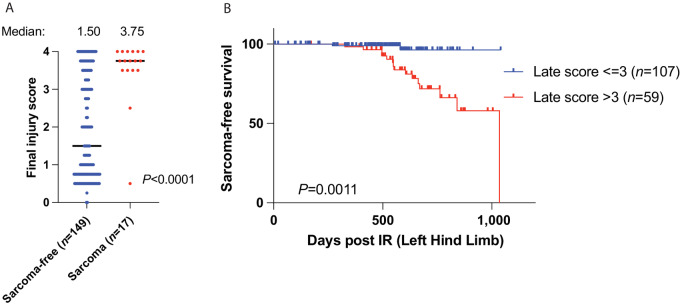
Approximately half of patients with cancer receive radiotherapy and, as cancer survivorship increases, the low rate of radiation-associated sarcomas is rising. Pharmacologic inhibition of p53 has been proposed as an approach to ameliorate acute injury of normal tissues from genotoxic therapies, but how this might impact the risk of therapy-induced cancer and normal tissue injuries remains unclear. We utilized mice that express a doxycycline (dox)-inducible p53 short hairpin RNA to reduce Trp53 expression temporarily during irradiation. Mice were placed on a dox diet 10 days prior to receiving 30 or 40 Gy hind limb irradiation in a single fraction and then returned to normal chow. Mice were examined weekly for sarcoma development and scored for radiation-induced normal tissue injuries. Radiation-induced sarcomas were subjected to RNA sequencing. Following single high-dose irradiation, 21% of animals with temporary p53 knockdown during irradiation developed a sarcoma in the radiation field compared with 2% of control animals. Following high-dose irradiation, p53 knockdown preserves muscle stem cells, and increases sarcoma development. Mice with severe acute radiation-induced injuries exhibit an increased risk of developing late persistent wounds, which were associated with sarcomagenesis. RNA sequencing revealed radiation-induced sarcomas upregulate genes related to translation, epithelial-mesenchymal transition (EMT), inflammation, and the cell cycle. Comparison of the transcriptomes of human and mouse sarcomas that arose in irradiated tissues revealed regulation of common gene programs, including elevated EMT pathway gene expression. These results suggest that blocking p53 during radiotherapy could minimize acute toxicity while exacerbating late effects including second cancers.

Significance: Strategies to prevent or mitigate acute radiation toxicities include pharmacologic inhibition of p53 and other cell death pathways. Our data show that temporarily reducing p53 during irradiation increases late effects including sarcomagenesis.


Figures

FIGURE 1
FIGURE 1
Temporary reduction in p53 expression during irradiation increases sarcomagenesis. A, Schematic showing mice fed dox diet for 10 days to drive expression of shRNA to knockdown p53, irradiated with 30 or 40 Gy to the hind limb, returned to normal chow, and followed for sarcoma development and normal tissue injury in the radiation field. B, Kaplan–Meier curves show radiation-induced sarcoma-free survival of control and p53KD mice irradiated with 30 or 40 Gy to the hind limb. P value is from a log-rank test. C, Representative images of a H&E-stained radiation-induced sarcoma subtypes and table with the number of tumors from each subtype. D, Quantitation and representative IHC images of mouse muscle tissue from control and p53KD mice 4 hours after 30 Gy irradiation stained with antibodies recognizing cleaved caspase 3. The number of positive cells per tissue section is graphed. E, Representative flow cytometry dot plots of GFP+ muscle satellite cells isolated from unirradiated (left) and irradiated (right) limbs from control (top) or p53KD (bottom) mice. F, The relative ratio of GFP+ cells in the irradiated limb (30 Gy) over the unirradiated limb is graphed (±SEM). Each dot represents one mouse. P value is from a t test. G, Schematic showing that cells with functional p53 undergo apoptosis or cell-cycle arrest and repair after irradiation, while damaged cells with impaired p53 function are protected from cell death and persist. Figure generated using BioRender.
FIGURE 2
FIGURE 2
Temporary reduction of p53 during irradiation increases chronic injuries in subsets of mice. A, Kaplan–Meier curves show acute injury-free survival (score 1+) of control and p53KD male and female mice irradiated with 40 Gy to the hind limb. P value is from a log-rank test. B, Kaplan–Meier curves show acute injury-free survival (score 1+) of control and p53KD male and female mice irradiated with 30 Gy to the hind limb. P value is from a log-rank test. Kaplan–Meier curves show chronic injury-free survival from scores 1+ (C), 2+ (D), 3+ (E), or 4 (F) of control and p53KD male and female mice irradiated with 30 Gy to the hind limb. P value is from a log-rank test.
FIGURE 3
FIGURE 3
Radiation-induced chronic injuries increase the risk of sarcomagenesis. A, The final injury scores of the control and p53KD mice that received 30 or 40 Gy are plotted. The final injury scores of mice that did not develop a radiation-induced sarcoma (blue) are compared with the scores of mice that did develop a radiation-induced sarcoma (red). P value is from a t test. B, Kaplan–Meier curves show radiation-induced sarcoma-free survival of the control and p53KD mice irradiated with 30 or 40 Gy to the hind limb. Mice with chronic injury scores equal to or less than 3 are compared with mice with chronic injury scores greater than 3. P value is from a cox proportional hazards model.
FIGURE 4
FIGURE 4
Radiation-induced sarcomas exhibit an increase in proliferative gene programs and a decrease in myogenic differentiation programs. A, Heat map of the top 75 differentially expressed genes in radiation-induced sarcomas (n = 16) versus normal muscle (n = 7). Genes (rows) are colored by scaled, normalized expression values. Both rows and columns are clustered. B, Volcano plot of log2 fold change for all genes with base mean greater than 50 in tumor (n = 16) versus normal (n = 7). Labeled genes have a nominal −log10P value greater than or equal to 30. C, Normalized enrichment scores for all FDR significant pathways for GSEA of Hallmark pathways. Points are colored by −log10 (nominal P value). D, Scatter plot comparing the differentially expressed pathways between tumors that arose in irradiated tissues (Rad) versus sporadic (Spor) tumors in mouse (y-axis) and human (x-axis) datasets. The EMT pathway is highlighted with a green dot. E, Boxplot showing gene expression of EMT pathway targets in mouse radiation-induced UPS compared with p53/RB tumors (top) and in human radiation-associated undifferentiated sarcomas compared with sporadic undifferentiated sarcomas.
FIGURE 5
FIGURE 5
Comparison of CNV and expression of specific oncogenes. Boxplot showing gene expression of Yap1 (A), Met (B), and Cdk4 (C) in radiation-induced sarcomas where the gene amplification status is known to be amplified (pink), not amplified (orange), or unknown (gray) compared with normal muscle (blue).

References

    1. Lee CL, Blum JM, Kirsch DG. Role of p53 in regulating tissue response to radiation by mechanisms independent of apoptosis. Transl Cancer Res 2013;2:412–21. - PMC - PubMed
    1. Gudkov AV, Komarova EA. Pathologies associated with the p53 response. Cold Spring Harb Perspect Biol 2010;2:a001180. - PMC - PubMed
    1. Singh VK, Seed TM, Olabisi AO. Drug discovery strategies for acute radiation syndrome. Expert Opin Drug Discov 2019;14:701–15. - PubMed
    1. Mito JK, Mitra D, Doyle LA. Radiation-associated sarcomas: an update on clinical, histologic, and molecular features. Surg Pathol Clin 2019;12:139–48. - PubMed
    1. Samartzis D, Nishi N, Cologne J, Funamoto S, Hayashi M, Kodama K, et al. . Ionizing radiation exposure and the development of soft-tissue sarcomas in atomic-bomb survivors. J Bone Joint Surg Am 2013;95:222–9. - PubMed

Publication types

Substances