

Immunoglobulin G is a natural oxytocin carrier which modulates oxytocin receptor signaling: relevance to aggressive behavior in humans
- PMID: 37983005
- PMCID: PMC10587035
- DOI: 10.1007/s44192-023-00048-z
Immunoglobulin G is a natural oxytocin carrier which modulates oxytocin receptor signaling: relevance to aggressive behavior in humans
Abstract
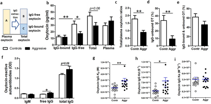
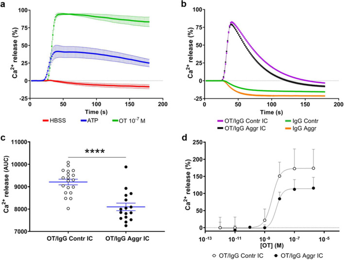
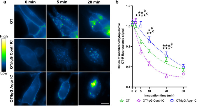
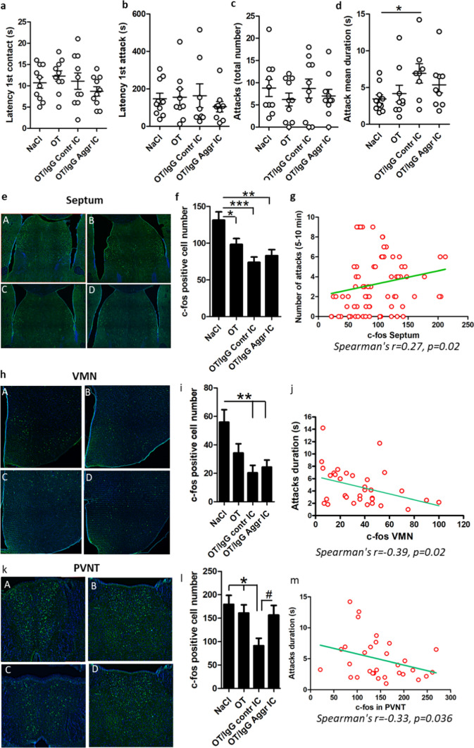
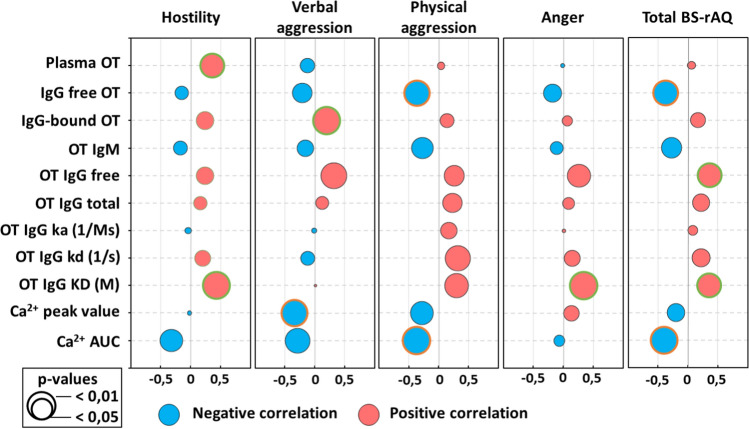
Oxytocin is a neuropeptide produced mainly in the hypothalamus and secreted in the CNS and blood. In the brain, it plays a major role in promoting social interactions. Here we show that in human plasma about 60% of oxytocin is naturally bound to IgG which modulates oxytocin receptor signaling. Further, we found that IgG of violent aggressive inmates were characterized by lower affinity for oxytocin, causing decreased oxytocin carrier capacity and reduced receptor activation as compared to men from the general population. Moreover, peripheral administration of oxytocin together with human oxytocin-reactive IgG to resident mice in a resident-intruder test, reduced c-fos activation in several brain regions involved in the regulation of aggressive/defensive behavior correlating with the attack number and duration. We conclude that IgG is a natural oxytocin carrier protein modulating oxytocin receptor signaling which can be relevant to the biological mechanisms of aggressive behavior.
Keywords: Aggressive behavior; Autoantibodies; Brain; Human; Intracellular signaling; Mice; Neuroendocrinology; Neuropeptides; Oxytocin; Oxytocin receptor; c-fos.
© 2023. The Author(s).
Conflict of interest statement
No direct conflict of interest to this study have been declared by any of the co-authors. SOF, EL and MN are co-inventors on patent applications related to the oxytocin signaling.
Figures
Similar articles
-
Adolescent oxytocin treatment affects resident behavior in aggressive but not tame adult rats.Physiol Behav. 2020 Oct 1;224:113046. doi: 10.1016/j.physbeh.2020.113046. Epub 2020 Jun 30. Physiol Behav. 2020. PMID: 32619528
-
Functional role of immunoglobulin G as an oxytocin-carrier protein.Peptides. 2024 Jul;177:171221. doi: 10.1016/j.peptides.2024.171221. Epub 2024 Apr 14. Peptides. 2024. PMID: 38626844 Review.
-
Medial amygdala lesions modify aggressive behavior and immediate early gene expression in oxytocin and vasopressin neurons during intermale exposure.Behav Brain Res. 2013 May 15;245:42-9. doi: 10.1016/j.bbr.2013.02.002. Epub 2013 Feb 10. Behav Brain Res. 2013. PMID: 23403283
-
Aggressive behavior and stress response after oxytocin administration in male Norway rats selected for different attitudes to humans.Physiol Behav. 2019 Feb 1;199:210-218. doi: 10.1016/j.physbeh.2018.11.030. Epub 2018 Nov 22. Physiol Behav. 2019. PMID: 30472394
-
Central nervous system effects of the neurohypophyseal hormones and related peptides.Front Neuroendocrinol. 1993 Oct;14(4):251-302. doi: 10.1006/frne.1993.1009. Front Neuroendocrinol. 1993. PMID: 8258377 Review.
Cited by
-
The Oxytocin System and Implications for Oxytocin Deficiency in Hypothalamic-Pituitary Disease.Endocr Rev. 2025 Jul 15;46(4):518-548. doi: 10.1210/endrev/bnaf008. Endocr Rev. 2025. PMID: 39985439 Free PMC article. Review.
-
Possible roles of neuropeptide/transmitter and autoantibody modulation in emotional problems and aggression.Front Psychiatry. 2024 Sep 24;15:1419574. doi: 10.3389/fpsyt.2024.1419574. eCollection 2024. Front Psychiatry. 2024. PMID: 39381606 Free PMC article. Review.
-
Navigating Central Oxytocin Transport: Known Realms and Uncharted Territories.Neuroscientist. 2025 Jun;31(3):234-261. doi: 10.1177/10738584241268754. Epub 2024 Aug 7. Neuroscientist. 2025. PMID: 39113465 Free PMC article. Review.
-
Long-Term Dynamics of Serum α-MSH and α-MSH-Binding Immunoglobulins with a Link to Gut Microbiota Composition in Patients with Anorexia Nervosa.Neuroendocrinology. 2024;114(10):907-920. doi: 10.1159/000539316. Epub 2024 Jun 8. Neuroendocrinology. 2024. PMID: 38852579 Free PMC article.
References
LinkOut - more resources
Full Text Sources
