. 2023 Nov 28;120(48):e2309506120.
doi: 10.1073/pnas.2309506120. Epub 2023 Nov 20.

Riding apoptotic bodies for cell-cell transmission by African swine fever virus

Affiliations

Riding apoptotic bodies for cell-cell transmission by African swine fever virus

Peng Gao et al. Proc Natl Acad Sci U S A. .

Abstract

African swine fever virus (ASFV), a devastating pathogen to the worldwide swine industry, mainly targets macrophage/monocyte lineage, but how the virus enters host cells has remained unclear. Here, we report that ASFV utilizes apoptotic bodies (ApoBDs) for infection and cell-cell transmission. We show that ASFV induces cell apoptosis of primary porcine alveolar macrophages (PAMs) at the late stage of infection to productively shed ApoBDs that are subsequently swallowed by neighboring PAMs to initiate a secondary infection as evidenced by electron microscopy and live-cell imaging. Interestingly, the virions loaded within ApoBDs are exclusively single-enveloped particles that are devoid of the outer layer of membrane and represent a predominant form produced during late infection. The in vitro purified ApoBD vesicles are capable of mediating virus infection of naive PAMs, but the transmission can be significantly inhibited by blocking the "eat-me" signal phosphatidyserine on the surface of ApoBDs via Annexin V or the efferocytosis receptor TIM4 on the recipient PAMs via anti-TIM4 antibody, whereas overexpression of TIM4 enhances virus infection. The same treatment however did not affect the infection by intracellular viruses. Importantly, the swine sera to ASFV exert no effect on the ApoBD-mediated transmission but can partially act on the virions lacking the outer layer of membrane. Thus, ASFV has evolved to hijack a normal cellular pathway for cell-cell spread to evade host responses.

Keywords: ASFV; apoptotic bodies; cell to cell transmission; immune evasion.


Conflict of interest statement

Competing interests statement:The authors declare no competing interest.

Figures

Fig. 1.
Fig. 1.
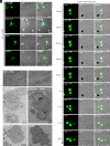
ASFV induces apoptosis of PAMs to shed ApoBDs. (A) Analysis of cellular apoptosis of ASFV-infected cells. PAMs were mock-infected or infected with ASFV strain HN09 at an MOI of 0.1 and harvested at indicated times post infection for Western blot with indicated antibodies to caspase-3, β-actin, and p30. β-actin served as a loading control, and ASFV p30 was used as an indicator of infection. (B) Growth kinetics of ASFV HN09 in PAMs at an MOI of 0.1. (C) Representative images showing the formation of ApoBDs. PAMs were mock-infected or infected with ASFV strain HN09 at an MOI of 0.1 and analyzed by DIC microscopy at 48 hpi. (D) Mock- and ASFV-infected PAMs (MOI = 0.1) were fixed at 48 hpi and analyzed by TEM. The characteristic ApoBDs (asterisk) and viral factory could be visualized only in ASFV-infected cells. (E) Size distribution of the vesicles around ASFV-infected PAMs (N = 100). The size of vesicles was measured via the scale bar under light microscope and electron microscope. (F) Immunostaining and 3D reconstruction of the ApoBDs. PAMs were infected with ASFV-GFP and stained with A5-Alexa568 at 48 hpi, and the images were reconstructed by Imaris software. Data information: The images were acquired by Nikon A1 confocal microscope and HITACHI HT7700 electron microscope.
Fig. 2.
Fig. 2.
ApoBDs derived from ASFV-infected PAMs can be swallowed by neighboring PAMs to establish a secondary infection. (A) Distribution pattern of the ASFV-positive ApoBDs. PAMs on coverslips in six-well plates were mock or infected with ASFV strain HN09 at an MOI of 0.1. The cells were fixed, permeablized, stained with indicated antibodies, and examined by confocal and DIC microscopy at 48 hpi. The arrows indicated the spreading trend of ASFV-positive ApoBDs. (B) ASFV-infected PAMs were fixed at 48 hpi and analyzed by TEM. (C) Live-cell imaging of the infection dynamics of ASFV-GFP-infected PAMs and GFP-positive ApoBDs. PAMs were seeded into 35-mm dishes and infected with ASFV-GFP at an MOI of 0.01. At 36 hpi, cells were imaged for time-lapse DIC and confocal microscopy. Representative images were collected from the taken video at the indicated time points. Data information: The images were acquired by Nikon A1 confocal microscope and HITACHI HT7700 electron microscope.
Fig. 3.
Fig. 3.
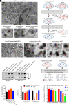
Purified ApoBDs from ASFV-infected PAMs contain infectious virions. (A) Presence of different forms of virions in ASFV-infected PAMs. (B) Schematic diagram of the procedure for purifying ApoBDs via differential centrifugation. PAMs seeded T-25 flask were infected with ASFV strain HN09 at an MOI of 0.1. At 60 hpi, the cells and culture were harvested for isolation of ApoBDs. (C) TEM analysis of the isolated ApoBDs, extracellular virions, and intracellular virions from ASFV-infected PAMs. (D) Dot blot analysis of PS of the three forms of virions. The purified virions were resuspended in phosphate buffered saline (PBS) (not to destroy the membrane) or lysed in Radio-Immunoprecipitation Assay (RIPA) buffer, and then spotted onto nitrocellulose membranes, followed by detection with indicated antibodies. PBS or RIPA was spotted as a negative control. (E) qPCR analysis of ASFV nucleic acid in different fractions. (F) Titration of ASFV and PRRSV load of different fractions in PAMs by TCID50. (G) Schematic presentation of the precipitation and dilution assay. The ApoBD-associated and extracellular virions were isolated same as (B) and centrifuged at 3,000 g for 20 min to collect the supernatant, and the pellet was then resuspended by RPMI-1640. (H) TCID50 analysis of virus titer after dilution. Data information: The images were acquired by HITACHI HT7700 electron microscope.
Fig. 4.
Fig. 4.
The purified ASFV-containing ApoBDs can transmit ASFV and establish a productive infection. PAMs grown on coverslips in six-well plates were incubated with the purified ApoBDs from WT ASFV-infected PAMs at 37 °C for 2 h, followed by cell fixation, permeablization, and staining with the antibodies to p30 (A) or swine serum to ASFV (B). (C) Live-cell imaging of the infection dynamics of the ApoBDs purified from ASFV-GFP-infected PAMs. Representative images were collected from the taken video at indicated time points. Data information: The images were acquired by Nikon A1 confocal microscope. (D) The dynamics of viral load in different fractions. PAMs were infected with WT ASFV or ASFV-GFP at an MOI of 0.1 or 0.01. At indicated time points, the three forms of virions were isolated and titrated by TCID50 in PAMs.
Fig. 5.
Fig. 5.
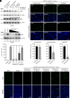
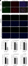
Blocking PS lipids suppresses ApoBD-mediated ASFV infection. Different forms of ASFV were isolated at 48 hpi from PAMs infected with ASFV strain HN09 at an MOI of 0.1 and then incubated, respectively, with Annexin V protein (5 µg/mL) prior to exposure to naive PAMs seeded in 24-well plates. The effect on viral replication was determined by Western blot (A) and IFA analysis (B). The infection efficiency was expressed as proportion of p30-positive cells via comparing Annexin V-treated group with control group, respectively. (C) Dose-dependent effect of Annexin V on ASFV infection by Western blot analysis. The relative band density of p30 was expressed as percentage compared to the untreated ASFV control (lane 2) after being normalized against β-actin in the corresponding lane. (D) Dose-dependent effect of Annexin V on the infection of ApoBD-associated virions by IFA analysis. Data information: Error bars indicate means ± SDs.
Fig. 6.
Fig. 6.
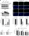
ApoBD-mediated ASFV infection is dependent on the efferocytosis receptor TIM4. (A) PAMs in 24-well plates were incubated with antibodies to TIM4 (10 µg/mL) or isotype IgG for 1 h and then exposed to three forms of virions before performing Western blot analysis at 12 hpi. (B) The same as (A), except by IFA analysis. The infection efficiency was expressed as proportion of p30-positive cells via comparing anti-TIM4 antibody treated group with that of isotype IgG-treated group, respectively. (C) Dose-dependent analysis. The same as (A) except that different doses of anti-TIM4 antibody was used. The relative band density of p30 was expressed as percentage compared to the ASFV infection control (lane 1) after being normalized against β-actin in the corresponding lane. (D and E) Effect of overexpression of TIM4 or MFG-E8 on the replication of ASFV in WSL-R4 cells. WSL-R4 cells in 12-well plates were transfected with pCMV-TIM4-HA, pCMV-MFG-E8-HA, or vector control (1.5 µg). At 24 h posttransfection, the cells were subjected to Western blot analysis with antibodies to HA tag (D) or infected with three forms of virions at an MOI of 1.0 for 24 h before being harvested for titration (E). Data information: Error bars indicate means ± SDs.
Fig. 7.
Fig. 7.
ApoBD-mediated viral transmission is fully resistant to swine sera to ASFV. (A) IFA analysis of the titer of swine anti-ASFV serum in PAMs with the antibody to p30 as a control. (BD) Intracellular virions, extracellular virions, and ASFV-containing ApoBDs were incubated with negative swine serum (−) or ASFV antibody positive swine serum (+) at a volume ratio of 1:1 at 37 °C for 1 h. The mixtures were then incubated with naive PAMs at 37 °C for 1 h followed by washes and addition of culture medium. (B) At 12 hpi, the cells were fixed and stained with antibodies to p30 for IFA. (C) The infection efficiency of three virion forms was expressed as proportion of p30-positive cells by comparing sera (+) group with sera (−) group, respectively. (D) The same as above, but the cells were harvested at 24 hpi for viral titration by TCID50. Data information: Error bars indicate means ± SDs.
Fig. 8.
Fig. 8.
A proposed model for ASFV transmission. ASFV induces cell apoptosis to shed the virion-loaded ApoBDs at late stage of infection via caspase-3 activation. The PS-positive ApoBDs carrying single-membrane virions are then phagocytosed by neighboring PAMs via PS interaction with efferocytosis receptors (e.g., TIM4, MFG-E8, etc.). Meanwhile, ASFV particles can directly bud from plasma membrane to acquire the PS-positive outer membrane. These extracellular virions can enter into recipient PAMs by means of either efferocytosis, clathrin-mediated endocytosis, or macropinocytosis.

Similar articles

Cited by

References

    1. Netherton C. L., Connell S., Benfield C. T. O., Dixon L. K., The genetics of life and death: Virus-host interactions underpinning resistance to African swine fever, a viral hemorrhagic disease. Front. Genet. 10, 402 (2019). - PMC - PubMed
    1. Zhou X., et al. , Emergence of African swine fever in China, 2018. Transbound. Emerg. Dis. 65, 1482–1484 (2018). - PubMed
    1. Zhao D., et al. , Replication and virulence in pigs of the first African swine fever virus isolated in China. Emerg. Microbes Infect. 8, 438–447 (2019). - PMC - PubMed
    1. Wen X. X., et al. , Genome sequences derived from pig and dried blood pig feed samples provide important insights into the transmission of African swine fever virus in China in 2018. Emerg. Microbes Infect. 8, 303–306 (2019). - PMC - PubMed
    1. Mighell E., Ward M. P., African Swine fever spread across Asia, 2018–2019. Transbound. Emerg. Dis. 68, 2722–2732 (2021). - PubMed

LinkOut - more resources